140
J
ournal of Epilepsy and ClinicalNeurophysiology
J Epilepsy Clin Neurophysiol 2011;17(4):140-143
The Boston Naming Test as a Predictor of Post-surgical
Naming Dysfunctions in Temporal Lobe Epilepsy
Sara Escorsi-Rosseta, Cecília Souza-Oliveiraa, Ana Carolina Gargaro-Silvaa,
Érica Regina Coimbraa, Eliane Correa Miottob, Marino Muxfelt Bianchina,c,
Vera Cristina Terraa, Américo Ceiki Sakamotoa
Centro de Cirurgia de Epilepsia (CIREP) – FMRP-USP
ABSTRACT
Objectives: Patients that undergo epilepsy surgery for temporal lobe epilepsy (TLE) in the dominant hemisphere are more susceptible to naming deficits. The aim of the present study was to perform an observational retrospective study comparing two groups of patients for naming performance, those with left and right temporal lobe resections regarding the performance in naming by Boston Naming Test (BNT). Methods: A total of 120 right-handed patients (52 right temporal lobe and 68 left temporal lobe), aged between 18 and 59, with pharmacoresistant mesial TLE were retrospectively analyzed. All patients underwent pre and postoperative neuropsychological assessment. Results and Conclusions: The BNT was a good predictor for possible post-surgical language deficits in patients submitted to left temporal lobectomy.
Keywords: epilepsy; temporal lobe; neuropsychology; language deficits.
RESUMO
O Boston Naming Test como marcador para disfunção nominativa no pós-operatório de epilepsia do lobo temporal
Objetivo: Pacientes submetidos a cirurgia de epilepsia portadores de epilepsia do lobo temporal (ELT) em hemisfério dominante são mais suscetíveis a apresentarem déficits de nomeação. O objetivo do presente estudo foi realizar um estudo retrospectivo observacional comparando dois grupos de pacientes sendo um grupo submetido a lobectomia temporal dominante e outro a lobectomia temporal não dominante em relação ao desempenho na tarefa de nomeação através do Boston Naming Test (BNT). Metódos: Um total de 120 pacientes destros foram retrospectivamente analisados (52 temporal direito e 68 temporal esquerdo) com idade entre 18 e 59 anos, com epilepsia do lobo temporal mesial fármaco resistente. Todos os pacientes foram submetidos a avaliação neuropsicológica pré e pós-operatória utilizando o BNT para medida de nomeação. Resultados e Conclusões: O BNT foi mostrou-se um bom instrumento para predizer possíveis déficits de linguagem em pacientes submetidos a lobectomia temporal esquerda.
Unitermos: epilepsia; lobo temporal; neuropsicologia; déficits de linguagem.
a Epilepsy Surgery Center (CIREP), Department of Neurology, Ribeirão Preto School of Medicine, University of São Paulo, Ribeirão Preto, Brazil b Division of Psychology and Department of Neurology, University of São Paulo, São Paulo, Brazil
c Division of Neurology, Department of Internal Medicine, Federal University of Rio Grande do Sul, UFRGS, Porto Alegre, Brazil.
141
The Boston Naming Test as a predictor of post-surgical naming dysfunctions in TLE
INTRODUCTION
Word finding difficulties are the most frequent complaints of patients with seizures originating from the language-dominant cerebral hemisphere1. Patients that undergo epilepsy surgery for temporal lobe epilepsy (TLE) in the dominant hemisphere are more susceptible to cognitive deficits related to verbal processing, specifically in the ability of naming and verbal memory2.
The main challenge of temporal lobe epilepsy surgery is to successfully remove sufficient amount of the epilepto- genic tissue to reach post-operative seizure-free status, without removing or damaging cortical areas responsible for language and memory function. In this context, pre- surgical neuropsychological assessments play a critical role in predicting cognitive impairments in postsurgical outcome3.
The Boston Naming Test (BNT) has been widely used to assess naming abilities in candidates for epilepsy surgery.4 In particular, patients presenting with dominant TLE have worse BNT performance in comparison to non-dominant TLE.5 This was already demonstrated for native English speakers, but rarely addressed in studies involving native Portuguese speakers. A translated version of BNT has been successfully applied to native Portuguese speakers. 6,7
The aim of the present study was to perform an observational retrospective study comparing two groups of native Portuguese speakers for naming performance, those with dominant and non-dominant temporal lobe resections.
PATIENTS AND METHODS
Patients
We retrospectively analyzed 120 right-handed patients (71 females, 49 males) with pharmacoresistant mesial TLE that underwent anterior-mesial temporal lobectomy at our Epilepsy Surgery Center. Patients’ age ranged from 18 to 59 years old. Fifty-two patients underwent operation on the non-dominant (right temporal lobe) and 68 patients in the dominant temporal lobe (left temporal lobe). All patients were submitted to standardized evaluation protocol that included presurgical and postsurgical neuropsychological extensive testing.
Methods
In addition to the neuropsychological assessment, all patients were submitted to a standardized presurgical evaluation protocol including medical history, neurological examination, long-term video-electroencephalographic monitoring, Magnetic Resonance Imaging (MRI), ictal and interictal SPECT, psychiatric and social assessments. In the present study we did not perform the Wada test in all cases. For this reason only right-handed patients were included, to avoid the possibility of atypical language representation.
Postsurgical neuropsychological reevaluation was performed within the first 5 to 8 months after surgery.
Inclusion criteria were hippocampal sclerosis on neuropathological examination and completely seizure-free status postoperatively (Engel class I). Patients with
IQ ≤69 (Wechsler Intelligence Scale)8 , psychiatric disease, bilateral mesial temporal lobe sclerosis and double pathology were excluded. The demographic data including gender, years of education, pre and postsurgical IQ, and naming performance (BNT) were first analyzed.
All patients underwent the same temporal lobe resection technique, extending 4.5 cm from the tip of both temporal lobes. After surgery, antiepileptic drugs were maintained equal to the presurgical regimen. Postsurgical outcome was determined based on Engel’s classification.9
Neuropsychological assessment
A number of tests were administered including the Edinburgh Handedness Inventory 10, IQ evaluation, memory (verbal and non-verbal), visuoperceptual/ spatial skills, attention, executive functions and language assessment (naming and fluency). The BNT was used to assess naming ability. This test consists of 60 figures, graded from lower to higher difficulty levels and patients are required to name the pictures. The subjects may receive a semantic cue if the object is misperceived or no answer is provided within 20 seconds, and then a phonemic cue is provided if the subject has not been able to name the picture after the semantic cue. The test is discontinued after six consecutive failures.
Statistical analysis
Firstly, a comparison between the two groups, left and right temporal lobe resections, was carried out using the SPSS 13.0 for Windows (SPSS Inc., 1989-2004). Demographic categorical variables were evaluated with the Chi-Square Test. Numeric variables were analyzed using the Independent-Samples T Test. The BNT scores were analyzed using Repeated Measures ANOVA. Pre- or Post- surgical BNT applications were used as within-subjects variable. The side of the surgery was used as a between-subject factor. Wilk’s Lambda was used as the multivariate test. All results were significant if p<0.05.
RESULTS
142
Escorsi-Rosset S, Souza-Oliveira C, Gargaro-Silva AC et al.
There were significant differences for pre- and post-surgical BNT scores according to the side of surgery (GLM,
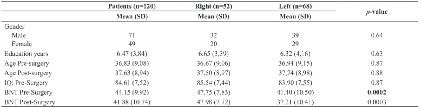
F(1,118)=18.00; p<0.0001; Wilk’s Lambda=0.84; partial eta squared=0.16; observed power 0.99). The analysis of within-subjects effects revealed that post-surgical BNT scores were significantly lower than pre-surgical scores (GLM, F(1,118)=22.44; p<0.0001; partial eta squared=0.16; observed power 0.99). The analysis of between-subjects effects revealed that BNT post-surgical scores were significantly lower only when considering the side of the surgery (GLM, F(1,118)=26.43; p<0.0001; partial eta squared=0.18; observed power 0.99). An inspection of the mean BNT scores (Table 1) indicates that left-dominant patients produced lower pre-surgical scores than right-non-dominant patients. In fact, surgery did not affect BNT performance on right-non-dominant temporal lobe epilepsy. However, already compromised patients with left-dominant mesial temporal lobe epilepsy presented an additional decrement of approximately 10% in BNT raw
scores after surgery. Figure 1 illustrates the magnitude of this effect, which can be considered important due to the scores of the partial eta squared observed.
DISCUSSION
The present study shows that the BNT can be a good predictor for post-surgical language deficits in patients submitted to left temporal lobectomy in a population of native Portuguese speakers. In addition, our study showed that BNT should be useful to provide default localization to the hemisphere that will be resected whereas left TLE patients showed a presurgical naming deficits. This is in line with previous studies with English language patients.11 A study was developed to investigate the utility of the BNT in a large group of TLE patients. Through a regression equation, they found that the BNT predicted the ultimate side of surgery in 69.5% of the sample, which 73.8% of patients were right TLE and 65.1% were left TLE. The authors highlighted that the prediction could be improved by using of a comprehensive neuropsychological battery that includes other tests sensitive to hemispheric lateralization.12 Similar results were found in study which, among several neuropsychological tests, the BNT has proved to be highly sensitive to lateralize the hemispheric lesions.5
In our study, we identified that despite the education may be one factor that has much influence on the performance of the BNT, it was similar in our two groups. Is highlighted that education is the factor that most influences the BNT performance.6 The authors said that the BNT performance should be viewed with caution, particularly in patients with low education and brain injury because it could be a real deficit or simply cultural deprivation. In our study we noted that the majority of patients in both groups had cultural deprivation, homogeneously influencing the performance of BNT.
There is a well-documented association between mesial temporal lobe epilepsy and naming impairments in visually-based tests, particularly in patients with epileptogenic zone in the language dominant hemisphere.13-16
Table 1. Characteristics of the sample according to side of surgery.
Patients (n=120) Right (n=52) Left (n=68)
p-value
Mean (SD) Mean (SD) Mean (SD)
Gender Male Female
71 49
32 20
39 29
0.64
Education years 6.47 (3,84) 6.65 (3,39) 6.32 (4,16) 0.63
Age Pre-surgery 36,83 (9,08) 36,67 (9,06) 36,94 (9,15) 0.87
Age Post-surgery 37,63 (8,94) 37,50 (8,97) 37,74 (8,98) 0.88
IQ: Pre-Surgery 84.61 (7,52) 85.54 (7,44) 83.90 (7,55) 0.87
BNT Pre-Surgery 44.15 (9.92) 47.75 (7.83) 41.40 (10.50) 0.0002
BNT Post-Surgery 41.88 (10.74) 47.98 (7.72) 37.21 (10.41) 0.0003
Abbreviations: SD: standarted deviation; IQ: intellectual quotient; BNT: Boston Naming Test, n: number.
143
The Boston Naming Test as a predictor of post-surgical naming dysfunctions in TLE
Confrontation naming performance reflects the capacity to access the precise name of objects on demand. As previously described, the temporal lobe mediates naming abilities beyond memory functions. Considering anatomical organization, the anterior temporal lobe areas are closely related to auditory naming, while middle and posterior areas are more related to visual confrontation naming. The consistency in the anterior/posterior distribution of auditory and visual naming sites is not surprising, due to the spatial relationship between auditory and visual association areas. This mapping was previously demonstrated by cortical stimulation, positron emission tomography (PET) and functional magnetic resonance image (fMRI) studies.17 Even when the resection is performed in anterior areas, it seems that dysfunction in posterior temporal lobe areas could be observed, showing that language dominant hemisphere is much susceptible to cognitive impairments.
Despite the fact that the BNT is still not standardized and validated to our population, and currently only a translated version is available for use, we nevertheless observed the same results previously described for English language patients.
The identification that patients of native Portuguese speakers maintains the performance in the BNT, as described in international literature noted that this test permits to be a useful tool to suggest evocation deficits before surgery. Furthermore, it allows to patients to have a precise orientation before surgery with respect to possible cognitive deficits that could be present after surgery.
ACKNOWLEDGEMENTS
Fundação de Amparo a Pesquisa do Estado de São Paulo – FAPESP (Grant 04/14004-CInAPCe program).
REFERENCES
Hamberger MJ, McClelland S 3rd, McKhann GM 2nd, Williams AC, 1.
Goodman RR. Distribution of auditory and visual naming sites in nonlesional temporal lobe epilepsy patients and patients with space-occupying temporal lobe lesions. Epilepsia 2007; 48(3):531-8. Davies KG, Bell BD, Bush AJ, Hermann BP, Dohan Jr FC, Jaap AS. 2.
Naming decline after left anterior temporal lobectomy correlates with pathological status of resected hippocampus. Epilepsia 1998; 39(4):407-19.
Sawrie SM, Martin RC, Gilliam FG, Faught RE, Maton B, Hugg JW, 3.
Bush N, Sinclair K, Kuzniecky RI. Visual confrontation naming and
hippocampal function: A neural network study using quantitative (1) H magnetic resonance spectroscopy. Brain 2000; 123(Pt 4):770-80. Sanyal SK, Chandra PS, Gupta S, Tripathi M, Singh VP, Jain S, 4.
Padma MV, Mehta VS. Memory and intelligence outcome following surgery for intractable temporal lobe epilepsy: relationship to seizure outcome and evaluation using a customized neuropsychological battery. Epilepsy Behav 2005; 6(2):147-55.
Raspall T, Donate M, Boget T, Carreno M, Donaire A, Agudo R, 5.
Bargallo N, Rumia J, Setoain X, Pintor L and others. Neuropsychological tests with lateralizing value in patients with temporal lobe epilepsy: reconsidering material-specific theory. Seizure 2005; 14(8):569-76. Mansur LL, Radanovic M, Taquemori L, Greco L, Araujo GC. A 6.
study of the abilities in oral language comprehension of the Boston Diagnostic Aphasia Examination – Portuguese version: a reference guide for the Brazilian population. Braz J Med Biol Res 2005; 38(2):277-92.
Miotto EC, Sato J, Lucia MC, Camargo CH, Scaff M Development of 7.
an adapted version of the Boston Naming Test for Portuguese speaker. Rev Bras Psiquiatr 2010; 32(3):279-82.
Wechsler D. Wechsler Adult Intelligence Scale – Revised Manual 8.
San Antonio-TX; 1981.
Engel J, Jr. ILAE classification of epilepsy syndromes. Epilepsy Res 9.
2006; 70(Suppl 1):S5-10.
Oldfield R. Assessment and Analysis of Handness – Edinburgh 10.
Inventory Neuropsychologia 1971; 9:97-100.
Stafiniak P, Saykin AJ, Sperling MR, Kester DB, Robinson LJ, 11.
O’Connor MJ, Gur RC. Acute naming deficits following dominant temporal lobectomy: prediction by age at 1st risk for seizures. Neurology 1990; 40(10):1509-12.
Busch RM, Frazier TW, Haggerty KA, Kubu CS. Utility of the 12.
Boston naming test in predicting ultimate side of surgery in patients with medically intractable temporal lobe epilepsy. Epilepsia 2005; 46(11):1773-9.
Chelune GJ, Naugle RI, Luders H, Awad IA. Prediction of cognitive 13.
change as a function of preoperative ability status among temporal lobectomy patients seen at 6-month follow-up. Neurology 1991; 41(3):399-404.
Schwarz M, Pauli E, Stefan H. Model based prognosis of postoperative 14.
object naming in left temporal lobe epilepsy. Seizure 2005; 14(8):562-8.
Schwarz M, Pauli E. Postoperative speech processing in temporal lobe 15.
epilepsy: Functional relationship between object naming, semantics and phonology. Epilepsy Behav 2009; 16(4):629-3.
Sherman EM, Wiebe S, Fay-McClymont TB, Tellez-Zenteno J, Metcalfe 16.
A, Herandez-Ronquillo L, Hader WJ, Jette N. Neuropsychological outcomes after epilepsy surgery: systematic review and pooled estimates. Epilepsia 2011; 52(5):857-69.
Votaw JR, Faber TL, Popp CA, Henry TR, Trudeau JD, Woodard 17.
JL, Mao H, Hoffman JM, Song AW. A confrontational naming task produces congruent increases and decreases in PET and fMRI. Neuroimage 1999; 10(4):347-56.
Address Correspondence to: Sara Rosset
Centro de Cirurgia de Epilepsia (CIREP) – Hospital das Clínicas Faculdade de Medicina de Ribeirão Preto – USP
Av. Bandeirantes, 3900 – Bairro Monte Alegre CEP 14048-900, Ribeirão Preto, SP, Brazil