Epilepsy and Clinical
Neurophysiology
J Epilepsy Clin Neurophysiol 2011;17(3):93-99
The Usefulness of Neuronavigation in
Functional Hemispherectomy
Murilo Sousa de Menesesa, Charles Kondageskia, Heraldo Ney Larocca dos Santosd,
Pedro André Kowacsd, Giselle Caselatto Coelhob, Giovana Gadensc,
Samanta Blattes da Rochac, Cristiane Simãoc
Neurological Institute of Curitiba (Instituto de Neurologia de Curitiba)
ABSTRACT
Catastrophic epileptic encephalopathies in children comprise devastating conditions that features cerebral dysfunction in association with refractory epileptic seizures. The diagnosis is based on the clinical findings, on magnetic resonance imaging (MRI) of the brain and on electroencephalographic findings. For these conditions, surgery remains essential for attaining seizure control. We report two cases of 5-year-old girls. The first one had a diagnosis of Rasmussen’s syndrome. The second one had a large porencephalic cyst secondary to perinatal cerebral ischemia. Despite trials of anticonvulsants, both patients deteriorated, and a functional hemispherectomy guided by neuronavigation was indicated and performed, with low morbidity and excellent seizure control. The neuronavigation proved to be a valuable guidance tool in performing the functional hemispherectomy, making the disconnections more accurate, and thus decreasing the surgical time and blood loss.
Keywords: Rasmussen’s syndrome; hemispherectomy; hemispherotomy; neuronavigation; epilepsy surgery.
RESUMO
Aplicabilidade da neuronavegação em hemisferectomia funcional
As encefalopatias epilépticas catastróficas da infância compreendem condições graves que associam disfunção cerebral e crises epilépticas refratárias. Seu diagnóstico é firmado com base nos dados clínicos e nos achados de ressonância magnética e eletrencefalográficos. Para algumas destas condições o tratamento cirúrgico continua sendo essencial para o controle das crises. Relatamos dois casos de pacientes de 5 anos. A primeira teve diagnóstico de síndrome de Rasmussen. A segunda tinha antecedentes de encefalopatia hipóxico-isquêmica perinatal. Ambas apresentaram epilepsia parcial refratária em associação com rápida deterioração neurológica, e foram submetidas à hemisferectomia funcional com auxílio da neuronavegação, com baixa morbidade e excelente controle das crises. A neuronavegação se mostrou como uma valiosa ferramenta na realização da hemisferectomia funcional, possibilitando desconexões mais precisas, menor tempo de cirurgia e menor perda sanguínea.
Palavras-chave: síndrome de Rasmussen; hemisferotomia; hemisferectomia; neuronavegação; cirurgia de epilepsia.
a Neurosurgical Department, Neurological Institute of Curitiba.
b Neurosurgical Department, Residency Program, Neurological Institute of Curitiba. c Psychology Department, Neurological Institute of Curitiba.
INTRODUCTION
Rasmussen’s syndrome is a well-known cause of refractory epilepsy in the young patient. Its early recognition and treatment are crucial for a better prognosis in terms of seizure control and neurological performance.1-8 Anatomical hemispherectomy and functional hemispherotomy and its variants have been employed on the management of this condition with good results.9-17 Other refractory epileptic diseases may also be treated by these procedures.22-25 Nonetheless, these surgeries are challenging in terms of their anatomical complexities and associated intra operative and post-operative risks. The introduction of modern neuronavigation guidance in neurosurgery has improved surgical results by increasing accuracy, safety and decreasing surgical time.26-31 The aim of this article is to discuss the usefulness of the neuronavigation in the functional hemispherectomy for refractory epilepsy.
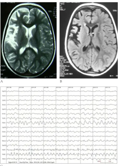
Figure 1.A) T2-weighted MRI imaging demonstrating atrophic changes in the right hemisphere, more evident around the Sylvian fissure and insular cortex. B) Subtle Flair-sequence signal intensities can be seen in the right insular cortex. C) Telemetry shows sharp waves and spikes on the anterior and medium right temporal projections against a slowed background.
Case One
A 5-year-old girl presented to our Epilepsy Unit with a history of seizures since the age of 3 years and 8 months. The first tonic-clonic seizure occurred during sleep, followed by left sided clonic seizures that progressively increased in frequency (1 to 2 events daily). At the age of 4 years and 6 months, she went on to develop complex partial seizures, preceded by an epigastric discomfort and autonomic symptoms. Initial EEG study was normal, and brain magnetic resonance imaging (MRI) disclosed mild hippocampal asymmetry, left hippocampus being smaller than the right hippocampus. Her fits were partially controlled with lamotrigine. Nonetheless, at the age of 4 years, the seizures became refractory. Further investigation with video-EEG telemetry showed a highly active epileptic zone on the anterior temporal projection of the right hemisphere (Fig. 1A and 1B). New MRI displayed atrophic changes in
A B
C
the right insular cortex, as well as enlargement of
the Sylvian fissure and cortical sulcii in the same
side (Fig. 1C). Neuropsycological evaluation revealed visual memory and executive planning impairment, in association with left sided fine motor dysfunction.
After the diagnosis of Rasmussen encephalitis was made, the treatment options were discussed with the parents, and surgery (Image-guided Right Functional Hemispherectomy) was offered, accepted and consented by the patient’s parents.
Case Two
A 5-year-old girl presented to our unit with a history of perinatal cerebral ischemia with a severe disruption of gray and white matter architecture with a cystic lesion in the left hemisphere. She had seizures since her birth. Disturbances on cognition, behavior and motor development were present. She had a right hemiparesis, but was able to walk independently. She was presenting an increasing number of partial epileptic seizures, becoming refractory to different antiepileptic drugs. MRI revealed severe atrophy with infarction and cyst lesion in the left cerebral hemisphere. Prolonged video-EEG recording disclosed continuous epileptogenic activity originated from the whole left hemisphere. Neuropsychological evaluation demonstrated severe cognitive impairment.
Surgical procedure
Along with routine pre-operative work up, MRI-3D reconstructed images were obtained for neuronavigation pre-operative planning and calibration. The neuronavigation system used was the Vector Vision, Brain Lab.
At the time of anesthesia induction, IV antibiotics were given, the reconstructed images were loaded into the navigation machine and the 3-pin head fixation was put in place. The navigation 3-spheres reference device was attached to the head clamp, after which the system was calibrated for use through the z-touch® technique. The accuracy given by the system was appropriate for the pro- cedure proposed. With the help of the navigation wand, the appropriate skin incision was marked (Fig. 2A and Fig. 2B). After prepping and marking of the skin, the surgical field was draped and the incision performed. The next step
consisted of a tailored fronto-temporal-parietal cranial flap followed by dural opening performed under microscopic view. With the help of the neuronavigation, a 4cm x 3cm corticotomy window was made on the supra insular region to gain access to the ipsilateral body of the lateral ventricle (Fig. 2C and Fig. 2D). In the second case there was a cystic brain lesion communicating with the lateral ventricle. After that, a right temporal lobectomy was performed, followed by resection of the mesial temporal lobe structures (amigdalo-hipocampectomy). Through the previously made ventricular window, and with the navigation guidance, the
corpus callosum was clearly identified, in the first case, and
sectioned widthwise from its right lateral extension (as also confirmed by the position of the pericallosal arteries) and lengthwise from the rostrum through the splenium
(Fig 2E and 2F). In the second case, callosotomy was only completed, because there was already an extended
Figure 2.A) and B) Marking of the incision. C) Right Supra-Insular resection window for access to the lateral ventricle. D) Navigation view: Supra-insular approach to the lateral ventricule. E) Navigation view: Callosotomy, genu of the corpus callosum. F) Navigation view: white matter fibers on the right lateral aspect of the splenium of the corpus callosum.
A B C
D
E
lesion of this structure. After this step, the remaining occipital and frontal intra-hemispheric white matter fibers were sectioned, completing the disconnection. Careful haemostasis was performed and no intra-dural drains were used. The bone flap was repositioned; muscles reinserted and a subcutaneous suction drainage system was left in place. The skin was closed with unabsorbable sutures. Total surgical time was about 6 hours in the first case and 4 hours in the second case and the estimated blood loss was 600ml for both.
Post operatory period
The first patient remained in the intensive care unit for 36 hours, and was extubated on the fifth post-operative hour after regaining full consciousness. The
second patient remained in the intensive care unit for 24 hours. Neurological examination depicted left hand side hemiplegia with no other deficits in the first case. The second patient remained clinically unchanged with a right hemiparesis. The subcutaneous drain was removed on the first post-operative day. During their hospital stay they did not present any seizures. Postoperative CT scans were performed for each patient in the post-operative period, and no evidence of hemorrhagic or hydrocephalic complications were found. The usual anticonvulsant regimen was maintained at the time of discharge. Post operative MRI obtained 3 months after surgery revealed total disconnection of the hemisphere in both cases (Fig. 3).
At last follow ups (12 months and 2 months, respectively, after surgery) they were seizure free.
Figure 3. A) and B) Post-operative T2-weighted MRI showing a residual cyst and general aspect of the right hemisphere after the functional hemispherectomy. C and D) Tractography dysplaing the discontinued white matter fibers within the the right hemisphere and corpus callosum.
A B
C D
DISCUSSION
Medically intractable epilepsy in children (MIE) is defined by the persistence of seizures despite the use of 2 or more anticonvulsants at optimal doses, though this diagnosis is very often established early after the onset of seizures, especially when the epilepsy is disabling or infantile spasms occur.19 Up to 30% of children bearing epilepsy will develop refractory fits.19 Rasmussen’s syndrome is currently understood as a chronic and progressive disease, which presents with neurological deterioration (speech, cognition and motor impairment) and continuous partial seizures. Auto immune response appears to play an important role in the pathogenesis of the disease.1-8 In Rasmussen’s syndrome, MRI of the brain generally depicts unilateral hemispheric atrophy of different degrees and electroencephalographic analysis demonstrates virtually whole hemispheric spikes and abnormal waves.5,8,19 The epileptic seizures are seldom responsive to anticonvulsants and some patients may respond to a course of steroids, intravenous immunoglobulin or plasmapheresis.5,20 Surgery remains the main treatment option, and should be considered in the early stages of the disease, when the best outcomes can be achieved.12 Surgical options include anatomical hemispherectomies and the various techniques of functional hemispherotomies. These techniques are also currently employed on the management of refractory epilepsy caused by other conditions, like hemimegaencephaly, ischemia related seizures and Sturge-Weber syndrome.9-12,14
McKenzie performed anatomical hemispherectomy in the 30’s and Krynauw in the 50’s, and it still is the technique of choice in some epilepsy surgery centers.21,24 It involves the removal of brain tissue in one hemisphere after ligation of major supratentorial main arteries, but sparing the basal ganglia. Despite the good seizure control rates, the anatomical techniques carry a higher morbidity and mortality risks, mainly related to the prolonged operative time, induced coagulation disorders, hemosiderosis, hydrocephalus and infection.9,10,14,15,20,22 Nonetheless, more recent hemispherectomy series achieve a lesser degree of complications and mortality rates in comparison with earlier reports.10,12,15,20,21,24
Other approaches developed in the 70’s and 80’s included functional hemispherectomy and functional hemidecortication, which aimed at decreasing the amount of brain tissue removal. Both techniques achieve good seizure control rates with decreased morbidity and mortality in comparison with anatomical resections.11,21
In the 90’s, new techniques, namely functional hemis- pherotomies, emerged with the purpose of disconnecting the brain hemisphere from the contra-lateral side and intra- hemispheric white matter fibers while preserving the vessels and removing the minimal amount of brain tissue.9,23 Various
modified techniques have arisen, but the vertical and peri-insular hemispherotomies introduced by Delalande and Villemure are the most popular. The first consists of disconnecting the targeted hemisphere from a parasagital corticectomy, accessing the ipsilateral lateral ventricle, performing a callosotomy and then contouring the basal ganglia down and anteriorly to the hippocampus. Then the remaining occipital and frontal white matter is divided to complete the disconnection. The second implies on the creation of a supra and infra-insular window, access to the lateral ventricle, callosotomy and frontal and occipital dis- connections.14-17,20,21 The lower rate of complications described following a functional hemispherotomy appears to result from the lesser degree of tissue resection. Also, the surgical time is reduced in comparison to anatomical techniques.25
In the cases presented in this article, we chose to perform a modified functional hemispherectomy, under neuronavigation guidance, aiming at a least necessary brain tissue removal.
Neuronavigation has been employed in neurosurgical procedures since the mid eighties, when Roberts put forward the concept of the frameless stereotactic system and Watanabe and Reinhardt developed arm-assisted systems.30 Ever since, several technologies have emerged, making it possible for the surgeon to be guided intra-operatively.27-31 All major steps of a given neurosurgical procedure potentially benefit from navigation techniques: pre operative planning, skin incision marking and selection of the approach, localization of the lesion, definition of its boundaries and relevant regional anatomy. Currently, the most widespread navigation devices rely on optical systems, where reflective spheres attached to the patient’s head clamp are identified by infra-red cameras. The position of the patient’s head is worked out through the three dimensional situation of the spheres after calibration of the system. A new set of spheres attached to a mobile surgical tool or a pointer, is brought in proximity to the head, which in turn gets automatically perceived by the cameras. The three dimensional location of the mobile spheres is determined with reference to the fixed spheres through complex mathematical algorithms, and displayed in dedicated screen in a chosen sequence of MRI.29 The pointer is then used at any desired time to pinpoint the region of interest. Nonetheless, it´s main limitation and other navigation technologies is brain shift. As the system is based solely on pre-operative images, whenever a considerable amount of brain tissue or fluid is removed, the computer cannot accurately compensate the brain shift, and the surgeon can be mislead. To update the system, another set of images needs to be obtained (intra-operative MRI, CT-scan or ultrasound).
Considering that in many cases the anatomy is distorted, several epilepsy surgery centers currently use neuronavigation as an important aid on performing hemispherectomies/ hemispherotomies.17,23,25,27,28 The reported advantages in employing the navigation techniques include better orientation during the craniotomy and gaining access to the ventricles, on performing the tractotomies and callosotomy. Surgical time is reduced, and as a consequence blood loss is decreased. However, as already mentioned, brain shift is a common situation on hemispherotomies/ hemispherectomies, since a considerable amount of CSF or brain tissue is expected to be removed.
In our experience, the neuronavigation guidance was helpful during the several steps of the procedure: pre-operative surgical planning, definition of the skin incision, craniotomy location and its size. But at the time of gaining access to the lateral ventricle, performing the callosotomy and the frontal and occipital white matter sectioning the navigation was essential. One of the main aspects of the procedure, in our opinion, is to initially gain access to the ipsilateral lateral ventricle, through a supra-insular window. The navigation guidance was very useful on determining the extension of the parietal cortex resection and direction of the white matter dissection, so that the approach for the callosotomy and later frontal/occipital disconnections could be established accurately. Moreover, to minimize CSF loss after the ventricular opening, and to secure its access, cotton patties are always placed at the aperture. With regards to the callosotomy, since it is performed from a lateral intraventricular approach, one needs to have complete control of its anterior-posterior extension. Again, using the neuronavigator, we were able to disconnect the crossing fibers of the corpus callosum from the rostrum through the splenium, as shown in the post-operative MRI of the first patient. Furthermore, another particular challenging step of the procedure is to complete the disconnection within the frontal lobe medial white matter fibers. Neuronavigation provided us with useful guidance to accomplish it.
When an image-guided neurosurgical procedure is performed, as in the cases of stereotaxis or neuronavigation, brain-shift may occur. This is due to the fact that the prior determined coordinates may lose accuracy during surgery. This phenomenon is related to the displacement of brain structures due to surgical manipulation, tissue resection and the aspiration of cerebrospinal fluid.
The possibility of brain-shift increases when the surgery is more aggressive. The only way to avoid this problem is the use of intraoperative neuroimaging. We have employed intraoperative magnetic resonance imaging (MRI) in cases of brain tumors microsurgical resections. However, this procedure increases morbidity, particularly the postoperative infections.
MRI has excellent image resolution and is the best option to demonstrate details of brain structures. Image-guided MRI, as with the stereotaxis and neuronavigation, has great accuracy. However, it is accepted that a possibility of distortion in the proportion of few millimeters may exist.
It is important to understand that functional hemispherectomy is already a well established surgery and it has been performed routinely in many centers with microsurgical methods but without image-guided techniques. The use of this new method enables the functional hemispherectomy to be performed with the same safety of microsurgical conventional operation plus all the advantages of neuronavigation.
The vision of the brain anatomy with the operative microscope is allied to the sophisticated location by an image-guided technique allowing, for example, the penetration of the brain tissue from the superficial cortex into the lateral ventricle faster and safer than with only the conventional way. The same advantages are used in the resection of the corpus callosum from the lateral ventricle up until the visualization of the anterior cerebral arteries, as well as through the brain fascicles of white matter for a complete disconnection.
Neuronavigation proved to be an effective tool in guiding the surgeon through the major steps of the functional hemispherectomy performed in the present cases, with emphasis in the more demanding phases of the procedure, like the callosotomy and frontal/occipital disconnection. Considering the growing experience with navigation techniques and the emergence of new technologies that could overcome brain shift, it is our belief that these technologies should be employed routinely in all complex neurosurgical procedures such as hemispherectomies.
ACKNOwLEDGEMENT
We acknowledge Dr. Ronaldo Vosgerau M.D. for providing us with the MRI images.
REFERENCES
1. Rasmussen T, Olszewski J, Lloyd-Smith D. Focal seizures due to chronic localized encephalitis. Neurology 1958;8:435-45.
2. Watson R, Jepson JEC, Bermudez I, Alexander S, Hart Y, McKnight K, Roubertie A, Fecto F, Valmier J, Sattelle DB, Beeson D, Vincent A, Lang B. 7-Acetylcholine receptor antibodies in two patients with Rasmussen encephalitis. Neurology 2005;65:1802-4.
3. Larionov S, König R, Urbach H, Sassen R, Elger CE, Bien CG. MRI brain volumetry in Rasmussen encephalitis: the fate of affected and “unaffected” hemispheres. Neurology 2005;64(5): 885-7.
4. Bien CG, Bauer J, Deckwerth TL, et al. Destruction of neurons by citotoxic T cells: a new pathogenic mechanism in Rasmussen’s encephalitis. Ann Neurol 2002;51:311-8.
6. Korn-Lubetzki I, Bien CG, Bauer J, Gomori M, Wiendl H, Trajo L, Ovadia H, Wilken B, Hans VH, Elger CE, Hurvitz H, Steiner I. Rasmussen encephalitis with active inflammation and delayed seizures onset. Neurology 2004;62:984-6.
7. Thomas P, Zifkin B, Ghetâu G, Delalande O. Persistence of ictal activity after functional hemispherectomy in Rasmussen syndrome. Neurology. 2003;60:140-2.
8. Pardo CA, Vining EP, Guo L, Skolasky RL, Carson BS, Freeman JM. The pathology of Rasmussen syndrome: stages of cortical involvement and neuropathological studies in 45 hemispherectomies. Epilepsia 2004;45(5):516-26.
9. Villemure JG, Mascott, C. Peri-insular hemispherotomy: surgical principles and anatomy. Neurosurgery 1995;37(5):975-81. 10. Fonseca LF, Melo RP, Cukiert A, Burattini JA, Mariani PP, Brandão
R, Ceda L, Baldauf CM, Argentoni M, Forster C, Baise C. Early functional hemispherectomy in hemimegalencephaly associated to refractory epilepsy. Arq Neuropsiquiatr 2004;62(4):1063-7. 11. Devlin AM, Cross JH, Harkness W, Chong WK, Harding B,
Vargha-Khadem F, Neville BG. Clinical outcomes of hemispherectomy for epilepsy in childhood and adolescence. Brain 2003;126(Pt 3): 556-66.
12. Lettori D, Battaglia D, Sacco A, Veredice C, Chieffo D, Massimi L, Tartaglione T, Chiricozzi F, Staccioli S, Mittica A, Di Rocco C, Guzzetta F. Early hemispherectomy in catastrophic epilepsy: a neuro-cognitive and epileptic long-term follow-up. Seizure 2008;17(1): 49-63.
13. Basheer SN, Connolly MB, Lautzenhiser A, Sherman EMS, Hendson G, Steinbok P. Hemispheric surgery in children with refractory epilepsy: seizure outcome, complications, and adaptive function. Epilepsia 2007;48(1):133-44.
14. Marras CE, Granata T, Franzini A, Freri E, Villani F, Casazza M, De Curtis M, Ragona F, Ferroli P, D’Incerti L, Pincherle A, Spreafico R, Broggi G. Hemispherotomy and functional hemispherectomy: indications and outcome. Epilepsy Res 2010;89(1):104-12. 15. Morino M, Shimizu H, Ohata K. Anatomical analysis of different
hemispherotomy procedures based on dissection of cadaveric brains. J Neurosurg 2002;97:423-31.
16. Wen HT, Rhoton AL Jr, Marino R Jr. Anatomical landmarks for hemispherotomy and their clinical application. J Neurosurg 2004;101(5):747-55.
17. Chandra PS, Padma VM, Shailesh G, Chandreshekar B, Sarkar C, Tripathi M. Hemispherotomy for intractable epilepsy. Neurol India 2008;56(2):127-32.
18. Go C, Snead OC 3rd. Pharmacologically intractable epilepsy in children: diagnosis and preoperative evaluation. Neurosurg Focus 2008;25(3):E2.
19. Faria AV, Reis F, Dabus GC, Zanardi VA, Guerreiro MM, Cendes F. MRI findings in the diagnosis and monitoring of Rasmussen’s encephalitis. Arq Neuropsiquiatr 2009;67(3B):792-7.
20. Muto A, Oguni H, Takahashi Y, Shirasaka Y, Sawaishi Y, Yano T, Hoshida T, Osaka H, Nakasu S, Akasaka N, Sugai K, Miyamoto A, Takahashi S, Suzuki M, Ohmori I, Nabatame S, Osawa M. Nationwide survey (incidence, clinical course, prognosis) of Rasmussen’s encephalitis. Brain Dev 2010;32(6):445-53.
21. Kossoff EH, Vining EP, Pillas DJ, Pyzik PL, Avellino AM, Carson BS, Freeman JM. Hemispherectomy for intractable unihemispheric epilepsy etiology vs outcome. Neurology 2003;61(7):887-90. 22. Limbrick DD, Narayan P, Powers AK, Ojemann JG, Park TS, Bertrand
M, Smyth MD. Hemispherotomy: efficacy and analysis of seizure recurrence. J Neurosurg Pediatr 2009;4(4):323-32.
23. De Ribaupierre S, Delalande O. Hemispherotomy and other disconnective techniques. Neurosurg Focus 2008;25(3):E14. 24. Fountas KN, Smith JR, Robinson JS, Tamburrini G, Pietrini D,
Di Rocco C. Anatomical hemispherectomy. Childs Nerv Syst 2006;22(8):982-91.
25. Pollo C, Debatisse D, Pralong E, Levivier M. Periinsular hemis- pherotomy: surgical technique, intraoperative EEG monitoring and results on seizure outcome. Neurochirurgie 2008;54(3):303-10. 26. Benifla M, Sala F Jr, Jane J, Otsubo H, Ochi A, Drake J, Weiss S,
Donner E, Fujimoto A, Holowka S, Widjaja E, Snead OC 3rd, Smith ML, Tamber MS, Rutka JT. Neurosurgical management of intractable rolandic epilepsy in children: role of resection in eloquent cortex. Clinical article. J Neurosurg Pediatr 2009;4(3):199-216.
27. Johnson RD, Stacey RJ. The impact of new imaging technologies in neurosurgery. Surgeon 2008;6(6):344-9.
28. Stone SS, Rutka JT. Utility of neuronavigation and neuromonitoring in epilepsy surgery. Neurosurg Focus 2008;25(3):E17.
29. Gumprecht HK, Widenka DC, Lumenta CB. BrainLab VectorVision Neuronavigation System: technology and clinical experiences in 131 cases. Neurosurgery 1999;44(1):97-104; discussion 104-5. 30. Enchev Y. Neuronavigation: geneology, reality, and prospects.
Neurosurg Focus 2009;27(3):E11.
31. Hayhurst C, Byrne P, Eldridge PR, Mallucci CL. Application of electromagnetic technology to neuronavigation: a revolution in image-guided neurosurgery. J Neurosurg 2009;111(6):1179-84.
Corresponding author:
Murilo Sousa de Meneses Street Jeremias Maciel Perretto, 300 81210-310, Curitiba, PR, Brazil Phone: (55-41)3028-8534