Peroral esophageal segmentectomy and anastomosis
with single transthoracic trocar: a step forward in
thoracic NOTES
Authors C. Rolanda1,2, D. Silva1,3, C. Branco1, I. Moreira4, G. Macedo5, J. Correia-Pinto1,6
Institutions Institutions are listed at the end of article.
submitted 2 January 2010 accepted after revision 12 August 2010
Bibliography
DOIhttp://dx.doi.org/
10.1055/s-0030-1256012 Endoscopy 2011; 43:
14–20 © Georg Thieme
Verlag KG Stuttgart · New York ISSN 0013-726X
Corresponding author
J. Correia-Pinto, MD PhD
Instituto de Ciências da Vida e Saúde
Escola de Ciências da Saúde Universidade do Minho, Campus de Gualtar
4709-057 Braga Portugal
Fax: +351-253-604831 [email protected]
Introduction
!
A minimally invasive esophageal approach is being used more frequently for both benign and malignant esophageal disease [1]. Since the first thoracoscopic mobilization of the esophagus de-scribed by Cuschieri in 1992, different surgical techniques have been introduced [2, 3]. However, it should be emphasized that thoracic incisions even if small are painful [4].
With natural orifice transluminal endoscopic sur-gery (NOTES) [5–8], a new approach to the thor-ax and mediastinum has been proposed: transe-sophageal access [9]. Although there are descrip-tions of transvesical transdiaphragmatic thoraco-scopy [10] and transgastric transdiaphragmatic pericardial fenestration, the most reasonable re-ported thoracic access is the transesophageal. If the esophagus was used as an entry site into the chest, a direct access to the thorax and posterior mediastinum could reasonably be established [11]. In fact, the technical feasibility of a
transeso-phageal approach has been described in porcine models for several simple thoracic procedures [11–16]. Even if the general concept is appealing, the transesophageal approach seems the one that most stretches the limits, even in the NOTES world. Access by the transesophageal approach may be highly risky, and mechanical abrasion and trauma of surrounding structures is higher when compared with the other visceral access routes. Moreover, the potential hemodynamic compromise due to an uncontrolled pneumome-diastinum, and the consequences of an ineffective esophagotomy closure can be devastating for the patient, because of the morbid consequences of mediastinitis. In this sequence, the well-known problems of NOTES [17], such as safe enterotomy creation, infection prevention, tissue manipula-tion, and, primarily, suturing and the establish-ment of anastomosis, seem to be even more limit-ing in the transesophageal approach.
Mirroring what is being done in abdominal sur-gery, where the hybrid approach became a transi-Background and study aims: A transesophageal
natural orifice transluminal endoscopic surgery (NOTES) approach has been proposed for thoracic and mediastinal access. Similarly to transgastric surgery, serious limitations remain related to creating an esophagotomy and its safe closure. A hybrid approach in thoracic NOTES could work as an intermediate step before pure transesophageal NOTES. We assessed the benefit of hybrid thoracic NOTES for peroral segmental esophagectomy and subsequent complete esophageal anastomosis with a single transthoracic port.
Methods: Two protocols were used to attempt esophago-esophageal anastomosis: ex vivo using a phantom model (n = 5), and in vivo after esoph-ageal mobilization, and segmental esophagect-omy achieved using either a gastroscope (flexible) (n = 5) or thoracoscope (rigid) instruments (n = 5). A forward-viewing double-channel endoscope
and a transthoracic operative thoracoscope with a working channel were coordinated in order to create a complete single-layer, end-to-end esoph-ageal anastomosis ex vivo as well as in vivo. Feasi-bility and anastomosis quality were evaluated by inside and outside assessment of: patency, the in-corporation of mucosa in all stitches, and a leak test.
Results: Anastomosis was achieved in all ex vivo experiments and thoracoscopically-led in vivo procedures. All anastomoses were patent, allow-ing distal passage of the endoscope, with mucosa incorporation. In in vivo experiments, a leak was detected in three animals and corrected with ad-ditional stitching.
Conclusions: Peroral esophageal anastomosis with a single transthoracic trocar is feasible, which may represent a step forward in thoracic NOTES.
tional step for humans in reducing the number of transabdomin-al ports and enabling a naturtransabdomin-al orifice approach without losing safety [18, 19], we hypothesized that the use of a peroral ap-proach in a hybrid mode can significantly reduce the number of transthoracic ports.
Aiming to eliminate some of the current limitations of transeso-phageal NOTES, we designed this study to carry out peroral esophageal mobilization, segmental esophagectomy and esopha-go-esophageal anastomosis with a single trocar, to assess the re-liability of this strategy for opening, resection and suturing of the esophagus.
This project was funded by the following grants: Bolsa de Investi-gação da Sociedade Portuguesa de Endoscopia Digestiva 2007 and the FCT project–PTDC/SAU-OSM/105578/2008.
Materials and methods
!
Study design
This study was approved by ethical review boards of Minho Uni-versity (Braga, Portugal).
The study was divided in two main branches: ex vivo studies in which esophago-esophageal anastomoses were carried out in a phantom model (five porcine esophaguses), and in vivo studies in anesthetized pigs. The in vivo studies involved esophageal mo-bilization and segmental esophagectomy using either a flexible gastroscope (five animals) or rigid instruments introduced through the working channel of an operative thoracoscope (five animals). Esophago-esophageal anastomoses were then carried out in the subset group in which esophagectomy had been car-ried out using rigid instruments introduced through the working channel of an operative thoracoscope. In all conditions, we used a forward-viewing double-channel gastroscope (G28/34; Karl Storz, Tuttlingen, Germany) and a transthoracic 10-mm operative thoracoscope with a 5-mm working channel (26036AA; Karl Storz).
Ex vivo studies
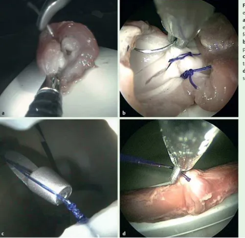
Phantom modelA three-dimensional structure consisting of syn-thetic polyurethane foam with an attached PVC container was mounted to simulate the thoracic cavity, and a tube 20 cm long was used to reproduce the oropharynx. Esophaguses harvested from adult pigs were positioned, fixed and sectioned in the mid-dle segment, creating a gap 4 cm long between the ends (
●
" Fig. 1 a). The model had a black, nontransparent removablecover with an opening on the right side to position the trocar (
●
" Fig. 1 b).Surgical techniqueThe operative thoracoscope was inserted into the phantom model through a 12-mm trocar (Excel port; Ethicon Endo-Surgery, Cincinnati, USA), whereas the gastroscope was in-troduced through the proximal esophagus; both instruments were coordinated to carry out an esophago-esophageal anasto-mosis (
●
" Fig. 2).First, a suturing needle with a 3–0 absorbable PDS stitch (Poly-dioxanone; Ethicon Endo-Surgery) was mounted on a needle-holder (26178KPL; Karl Storz), previously passed through the op-erative thoracoscope working channel and introduced into the phantom model. Then the gastroscope was moved towards the free lower extremity of the esophagus to grasp it on its posterior wall, including always muscle and mucosa, making proximal traction for the first stitch passage (inside to outside). The needle was then repositioned by the needle-holder for approaching the
esophageal upper extremity, and the gastroscope was used to promote esophageal stump alignment and esophageal wall trac-tion for outside-to-in proximal puncture. The knot-tying was achieved by extracorporeal knot techniques using a knot-pusher (26596D; Karl Storz) under gastroscopic vision. Then scissors (34410MW; Karl Storz) were introduced through the operative thoracoscope working channel and the excess suture lengths were cut. This procedure was repeated until 10 single-layer inter-rupted sutures had been completed, the posterior stitches with internal knotting and the later ones with external knotting. The duration of the procedure was recorded.
When the sutures were complete, the gastroscope was used to verify the immediate endoluminal reliability of the anastomosis, confirming that mucosa from both tips were touching each other, and identifying possible sites for supplementary stitches.
In vivo studies
Pig preparation Male 40–50 kg pigs (Sus scrofus domesticus) were fed liquids for 1 day and not allowed food and water for 8 hours before the surgery. All procedures were performed under general anesthesia with endotracheal intubation and mechanical ventilation, according to previous descriptions [20, 21]. Before the procedure, 1 mg of intravenous atropine was administered. Surgical technique The pig was placed in the prone position (
●
" Fig. 3). A 12-mm trocar was positioned around the eighthin-tercostal space in the right posterior axillary line, through which the operative thoracoscope was introduced; CO2insufflation was maintained with a pressure up to 6 mmHg. The gastroscope was advanced through an oropharyngeal overtube (US Endoscopy, Mentor, Ohio, USA) into the esophagus. Esophageal mobilization and segmental esophagectomy were carried out in the proximal third using either rigid instruments through an operative thora-coscope (five animals) or flexible instruments through a gastro-scope (five animals), as detailed schematically in
●
" Fig. 4.Fig. 1 Phantom model built to simulate the thoracic cavity:aporcine esophagus sectioned in its middle segment and positioned inside the
phantom model;bphantom model with its cover.
In those animals where esophagectomy was performed using flexible instruments, the proximal section was carried out with an endoscopic submucosal dissection (ESD) knife. This procedure was enhanced by clamping the esophageal lumen with an exter-nal grasper introduced through the working channel of the op-erative thoracoscope, which helped also in mobilizing the esoph-agus and providing traction while the gastroscope knife sec-tioned the esophagus circumferentially with cautery. Subse-quently, the gastroscope was introduced into the mediastinum, and the esophagus was released from its vessels and attachments using a flexible hemostatic grasper. Finally, the esophagectomy was completed using an endoscopic snare.
In those animals where esophagectomy was performed with transthoracic instruments, scissors were used through the opera-tive thoracoscope working channel. Under endoluminal traction, the proximal esophagus was dissected, mobilized, freed from its attachments and sectioned.
In both approaches, approximately 4 cm of esophagus was sec-tioned and removed perorally.
In those animals where the esophagectomy was accomplished with the rigid instruments, the gastroscope was reintroduced into the thoracic cavity providing traction and alignment of both esophageal stumps for suturing. The creation of the anastomosis followed the same steps as described in the ex vivo studies (
●
" Fig. 2). At the end of the procedure, the gastroscopecon-a b c
d e f
g h i
Fig. 2 Steps in esophageal anastomosis:afirst stitch with the gastroscope grasper pulling the
esophageal wall distally;bfirst stitch with the
gas-troscope grasper pushing the esophageal wall
proximally;cfirst stitch crossed;dknot-pusher
closing the first stitch under gastroscope image
control;eoperative thoracoscope scissors cutting
the suture;fposterior esophageal wall anastomosis
complete with inside knotting;g–hstitching the
anterior wall;ianterior esophageal wall
anastomo-sis complete with outside knotting.
Anesthetist
Gastroscope operator
Nurse Thoracoscope
operator
A B C
D
Fig. 3 Layout of the room: A, thoracoscope monitor; B, gastroscope monitor; C, ventilator; D, back table for equipment.
1 2
3 4
b
Fig. 4 aPeroral esophagectomy led by the operative thoracoscope. 1 and 2: gastroscope transillumination, thoracoscope positioning and transverse section with rigid scissors. 3 and 4: gastroscopic traction of the distal stump and again transverse section and esophageal segment release by the
thoracoscope.bPeroral esophagectomy led by the gastroscope. 1:
thora-coscope fixing, and use of the flexible endoscopic submucosal dissection
(ESD) knife to cut the esophagus. 2–4: thoracoscopic traction of the distal
stump through a flexible snare; snare section with cautery and esophageal segment release.
a
1 2
3 4
firmed the patency of the anastomosis and the incorporation of mucosa in all stitches; the thoracoscopic view was used to check the reliability of the anastomosis by looking for bubbles released under saline when the gastroscope was insufflating air inside the esophagus (air leak test).
Results
!
Ex vivo studies
The phantom model was easy to operate and simulated reason-ably well the steps of the the in vivo anastomosis, although the lung, other thoracic structures, and the cardio-respiratory move-ments were not present.
After some initial difficulties, we verified progressive coordina-tion, making possible the construction of a complete single-layer end-to-end esophageal anastomosis in all experiments, with at least 10 interrupted sutures having five knots each. The crucial step was always the first stitch. Gastroscopic grasping of the mus-cular and mucosal layers of the distal stump was easily achieved and useful as it provided positioning and traction for the needle passage (
●
" Fig. 5 a).Similarly, the gastroscope was useful in exposing the proximal extremity and for countertraction. Once the needle had crossed both extremities, the gastroscope view was also effective in mon-itoring the knotting. The subsequent stitches were progressively easier to apply (
●
" Fig. 5 b). The entrance of the knot-pusher inthe thorax was well monitored by the gastroscope image, allow-ing the operator to recommend pressure adjustment or even knot removal when the knot was not correctly applied (
●
" Fig. 5 c). The knots’edges were easily cut with the scissors(
●
" Fig. 5 d). As the anastomosis evolved, the benefit of the imageprovided by the gastroscope was gradually reduced, but the need for support and exposure was also less as the anastomosis was reaching completion. Although the aim was single-layer sutures involving both mucosa and muscle, we found that mucosa easily dislodged from the muscle layer and could be involuntarily miss-ed by the nemiss-edle; the gastroscope was very useful in monitoring the correct transmural involvement and in providing better ex-posure whenever necessary. The mean (standard deviation [SD]) time to perform complete anastomoses was 65.5 (10.9) minutes. The anastomoses were finally checked by their endoluminal as-pect for lumen patency, intersuturing space and stitch mucosal misplacement. It was found in a few sutures that some stitches did not include the mucosal layer.
In vivo studies
The prone approach and the CO2insufflation provided good ex-posure of the intrathoracic esophagus without the need for addi-tional retraction instruments. This and the endoluminal transil-lumination of the esophagus allowed rapid access of the opera-tive thoracoscope to select the esophageal segment of interest. In this study, we tested esophageal mobilization and esophagect-omy using either a flexible gastroscope (
●
" Video 1) or rigidin-struments (
●
" Video 2), and both approaches were feasible.How-Fig. 5 Ex vivo steps of a complete single-layer, end-to-end esophago-esophageal anastomosis:
agrasping, positioning and traction of the distal
esophageal extremity provided by the gastroscope for the first stitch passage (gastroscope view);
bsequential sutures and needle position for
proximal extremity puncture (thoracoscope view);
cknot-tying with the knot-pusher entrance
moni-tored by the gastroscope image (gastroscope view);
dcutting of excess suture material by the telescopic
scissors (thoracoscope view).
Video 1
Gastroscopic esophagectomy. Steps of proximal section done with endo-scopic submucosal dissection (ESD) knife. Esophageal dissection with a coagulation grasper. Once esophageal dissection was completed the distal esophagus was sectioned with a snare. The specimen was removed pero-rally.
ever, the margin of the proximal esophageal stump was left more irregular when esophagectomy was performed using a flexible instrument (the gastroscope) than using a rigid instrument (the operative thoracoscope).
With both strategies, segmental esophagectomy was done and the specimen was removed perorally without complications (
●
" Fig. 6 aandb).In animals where esophagectomy was performed using rigid in-struments (with a similar regularity of proximal and distal bor-der), we performed esophageal anastomosis following the same principles as in the ex vivo training. Apart from some movement interference and some additional caution because of adjacent structures, the anastomoses were feasible and reproducible in all cases (
●
" Video 3).An interesting aspect of these experiments was that the thoraco-scope view was sometimes not enough to see whether the needle position was the most appropriate for a specific suture orienta-tion. However, combining the thoracoscope view with the
gas-troscope view, it was always possible to correct the needle-hold-er position. The total mean opneedle-hold-erative time was 101.8 (32.9) min-utes, including dissection, segmentectomy and anastomosis. At the end of the procedure, the internal and external appearance of the anastomoses was checked (
●
" Video 4).Externally, the thoracoscope was used to inspect the surface of the anastomosis by using endoluminal transillumination; the gastroscope was used to make small rotations of the anastomosis (
●
" Fig. 6 c). Inside, all anastomoses were patent allowing distalpassage of the endoscope, and almost all sutures incorporated the mucosa (
●
" Fig. 6 d). Air leak (bubbling) was detected in threeanimals. This most commonly occurred when the mucosa was not included in the stitch or when the distance between two stit-ches was too long. In these cases, we were able to correct it with additional stitching.
Discussion
!
The esophagus is a fragile organ given its specific morphological characteristics and anatomical location, and it has been consid-ered by surgeons something of a special zone [4]. This probably contributes to the slower dissemination of video-assisted surgery in the thorax when compared with laparoscopy [22, 23].
Similar-Fig. 6 In vivo segmental esophagectomy and esophago-esophageal anastomosis procedure:
aesophageal segmentectomy (thoracoscope view);
besophageal segment being sectioned distally by
the gastroscope snare (gastroscope view);c
exter-nal fiexter-nal examination under endoscopic
transillumi-nation (thoracoscope view);dendoluminal
exami-nation of the anastomosis (gastroscope view).
Video 4
Final checking of the esophageal anastomosis. The inside and outside ap-pearance of the esophageal anastomosis.
online content including video sequences viewable at: www.thieme-connect.de/ejournals/abstract/endoscopy/ doi/10.1055/s-0030-1256012
Video 3
Esophageal anastomosis. Some steps of a complete single-layer, end-to-end esophago-esophageal anastomomosis in vivo, with images from a gastro-scope and an operative thoracogastro-scope sequentially intercalated.
Video 2
Operative thoracoscope esophagectomy. Using scissors through the op-erative thoracoscope working channel, and with slight movements of the gastroscope for esophageal traction, a safe blunt dissection of esophagus was carried out. The steps for esophagectomy are shown. The specimen was removed perorally.
ly, transesophageal thoracic NOTES inspires less enthusiasm than abdominal NOTES.
Transesophageal NOTES endorses the possible absence of trans-thoracic incisions. Avoiding intercostal neuralgia, it brings the ex-pectation of a potentially greater patient benefit in the thorax than in the abdomen [12]. Descriptions in a porcine model of transesophageal mediastinoscopy and thoracoscopy [9, 13], lung and pleura biopsy [13], lymphadenectomy [12, 13], pericardial fenestration [12], vagotomy and esophagomyotomy [14], Heller myotomy [15], esophageal wall resection [16], with or without the help of endoscopic ultrasound (EUS), can be found in the lit-erature. Besides the possible benefits, the procedure’s main risks are always discussed: the‘blind’creation of the esophagotomy, the unpredictable thoracic side exit without EUS or fluoroscopic assistance, and finally the possible devastating consequences of leaking from an incomplete esophageal closure.
We hypothesized that a hybrid thoracic approach could achieve the benefits of reducing the number of transthoracic ports and si-multaneously minimizing the limitations and risks of a pure trans-esophageal approach. This study explores the combination of the peroral route with a single transthoracic port for a complex intra-thoracic procedure–segmental esophagectomy with esophago-esophageal anastomosis. The reduction of the usual four or five trocars used to a single transthoracic instrument was obtained as a result of three main factors: the use of an operative thoracoscope with a working channel for 5-mm instruments, the prone position instead of the regular left-lateral decubitus, and the coordinated beneficial use of the peroral gastroscope instruments.
The prone position is being promoted because it allows gravity to provide exposure with minimal handling, giving good esopha-geal visualization, simplifying dissection and reducing the opera-tive times, without sacrificing patient safety [1]. In our in vivo ex-periments, we also confirmed these benefits with no need of any accessory port for lung retraction, and the single transthoracic in-strument was totally focused on esophageal surgery.
The great improvement explored in this study is the help provid-ed by the peroral/transesophageal port. The benefits start with the endoscopic transillumination of the esophagus by the thora-coscope; this step may be particularly helpful for direct visualiza-tion of a possible lesion area, orienting a more precise resecvisualiza-tion [24]. In this study, esophageal dissection and mobilization were carried out through two distinct strategies led either by a flexible gastroscope or by rigid thoracoscope instruments. Both strate-gies were successful in mobilizing, sectioning and dissecting the esophageal segment. When these processes were led by the gas-troscope instruments, esophageal dissection and distal section were quite reliable, but the proximal esophageal section often left the stump margin too irregular. In contrast, esophageal dis-section and dis-section carried out by the thoracoscope instruments was reliable and rapid, and both esophageal margins were similar and quite regular. This was our main reason for testing the feasi-bility of the esophageal anastomosis in those animals where esophageal dissection and sectioning were carried out by the op-erative thoracoscope. It should be emphasized that even in this strategy, coordinated movements of the endoscope and traction were important for the operative thoracoscope-oriented work. In minimally invasive esophagectomy, specimen retrieval is cur-rently performed via a separated neck incision, but in our experi-ments the specimen was always removed perorally; depending on its size, we predict this can be done in humans too.
The more impressive contribution of the gastroscope was during the creation of the anastomosis. The first stitch, which in a
con-ventional thoracoscopic procedure is frequently the most diffi-cult, was in our experience more straightforward because of the posterior esophageal wall grasping and proximal traction provid-ed by the gastroscope instruments. Furthermore, the exposure of tissue layers, the countertraction and the capability for needle re-trieval from and presentation to the operative thoracoscope whenever necessary were (apart from some inadequacy of the endoscopic forceps to grasp the needle securely) good technical potentialities revealed during experimentation. All in vivo ana-stomoses had at least 10 sutures, and each stitch was tied with five knots, with total control of the knotting by the gastroscope, from the thoracic entrance until its tissue adjustment. We found episodically that mucosa retracted and was missed during the stitch passage, but we could ameliorate this with increasing skills in coordination of tissue exposure and thoracoscope movements. Another advantage of a peroral access is that it is possibile imme-diately to check the luminal patency of the anastomosis and iden-tify suspected areas of weakness needing additional stitches. In addition, endoscopic delivery of conditioning agents at this time can facilitate healing, as has been proposed previously [25]. As potential indications for this approach we would imagine be-nign lesions such as leiomyomas, resistant strictures, gastrointes-tinal stromal tumors, esophageal ruptures, complicated foreign bodies, esophageal atresia and even some selected nonadvanced malignant esophageal diseases. Initial series proposed minimally invasive methods, essentially for selected patients with early-stage esophageal cancer (early-stage II and below) [26]. Excluding high grade dysplasia and intramucosal cancer where organ pre-servation by endoscopic mucosal resection might be preferred, and excluding tumors of the esophagogastric junction that should have an abdominal transhiatal approach, we envision the possible application of hybrid thoracic NOTES in T1sm–T2 le-sions even with thoracic lymphadenectomy. In this matter, re-cent reports support that sentinel node mapping, pre- or peri-tervention, can not only offer proper staging but also tailor an in-dividualized surgical strategy and help to plan the field of irradia-tion [4, 27]. Thus we predict the usefulness of our strategy for en-doluminal lesion location, endoscopic submucosal injection of an agent with real-time thoracoscopic monitoring for sentinel node, followed by a posterior combined dissection lymphadenectomy with peroral removal.
Moreover, this approach could contribute to eliminating most of the current limitations of pure transesophageal NOTES that are potentially applicable to other thoracic extraesophageal condi-tions, namely in all those procedures previously attempted by a thoracic NOTES approach. The combination of the peroral ap-proach with one transthoracic access seems advantageous be-cause of the availability of two distinct images of the same proce-dure, resulting in a more accurate and safe assessment of the in-tervention, mainly an anastomosis, in such a delicate space as the thoracic cavity. We also believe that a reduction of the number of transthoracic ports to one is the maximum that we can expect in minimally invasive thoracic surgery for now. In fact, convention-ally it is required to leave in place an external drain after any thoracic intervention in humans; this highlights the pertinence of our transthoracic access.
We would like to point out that our pig model has a healthy esophagus with no adhesions, and it is more elastic than human esophagus. Also, as a limitation, it might be emphasized that this was not a survival study. Thus, it is not possible to verify the safe-ty of the approach, although we believe this approach has a good chance of being translated to humans in a stepwise reduction of
transthoracic trocars, once there is secure endoscope sterility and the use of an oropharyngeal overtube.
In conclusion, this study confirms the feasibility of a hybrid per-oral transesophageal approach with a single transthoracic trocar to carry out segmental esophagectomy with complete single-lay-er, end-to-end intrathoracic esophageal anastomosis in a porcine model. Moreover, this study reinforces the logic and pertinence of using a transesophageal approach to the thoracic cavity.
Competing interests:Jorge Correia Pinto is a consultant to Karl Storz.
Institutions
1Surgical Sciences Research Domain, Life and Health Sciences Research
Institute (ICVS), School of Health Sciences, University of Minho, Braga, Portugal
2Department of Gastroenterology, Hospital de Braga, Braga, Portugal
3Department of Internal Medicine, Centro Hospitalar do Médio Ave, Vila Nova
de Famalicão, Portugal
4Department of Oncology, Centro Hospitalar do Alto Ave, Guimarães,
Portugal
5Department of Gastroenterology, Hospital de Sao Joao, Porto, Portugal
6Department of Pediatric Surgery, Hospital de Sao Joao, Porto, Portugal
References
1 Fabian T, Martin J, Katigbak M et al.Thoracoscopic esophageal mobili-zation during minimally invasive esophagectomy: a head-to-head comparison of prone versus decubitus positions. Surg Endosc 2008; 22: 2485–2491
2 Cuschieri A, Shimi S, Banting S.Endoscopic oesophagectomy through a right thoracoscopic approach. J R Coll Surg Edinb 1992; 37: 7–11 3 Shichinohe T, Hirano S, Kondo S.Video-assisted esophagectomy for
esophageal cancer. Surg Today 2008; 38: 206–213
4 Perretta S, Allemann P, Dallemagne B et al.Natural orifice transluminal orifice surgery (NOTES) for neoplasia of the chest and mediastinum. Surg Oncol 2009; 18: 177–180
5 Kalloo AN, Singh VK, Jagannath SB et al.Flexible transgastric peritoneo-scopy: a novel approach to diagnostic and therapeutic interventions in the peritoneal cavity. Gastrointest Endosc 2004; 60: 114–117 6 Lima E, Rolanda C, Pego JM et al.Transvesical endoscopic
peritoneosco-py: a novel 5 mm port for intra-abdominal scarless surgery. J Urol 2006; 176: 802–805
7 Decker A.Culdoscopy a method for visual diagnosis of gynecologic dis-ease. Clin Symp 1952; 4: 201–210
8 Fong DG, Pai RD, Thompson CC.Transcolonic endoscopic abdominal ex-ploration: a NOTES survival study in a porcine model. Gastrointest En-dosc 2007; 65: 312–318
9 Sumiyama K, Gostout CJ, Rajan E et al.Transesophageal mediastinosco-py by submucosal endoscomediastinosco-py with mucosal flap safety valve technique. Gastrointest Endosc 2007; 65: 679–683
10 Lima E, Henriques-Coelho T, Rolanda C et al.Transvesical thoracoscopy: A natural orifice translumenal endoscopic approach for thoracic sur-gery. Surg Endosc 2007; 21: 854–858
11 Willingham FF, Gee DW, Lauwers GY et al.Natural orifice transesopha-geal mediastinoscopy and thoracoscopy. Surg Endosc 2008; 22: 1042– 1047
12 Fritscher-Ravens A, Patel K, Ghanbarri A et al.Natural orifice translum-inal endoscopic surgery (NOTES) in the mediastinum: long-term sur-vival animal experiments in transesophageal access, including minor surgical procedures. Endoscopy 2007; 39: 870–875
13 Gee DW, Willingham FF, Lauwers GY et al.Natural orifice transesopha-geal mediastinoscopy and thoracoscopy: a survival series in swine. Surg Endosc 2008; 22: 2117–2122
14 Woodward T, McCluskey D, Wallace MB et al.Pilot study of tranesopha-geal endoscopic surgery: NOTES esophagomyotomy, vagotomy, lym-phadenectomy. J Laparoendosc Adv Surg Tech 2008; 18: 743–745 15 Pauli EM, Mathew A, Haluck RS et al.Technique for transesophageal
en-doscopic cardiomyotomy (Heller myotomy): video presentation at the Society of American Gastrointestinal and Endoscopic Surgeons (SA-GES). Surg Endosc 2008; 22: 2279–2280
16 Fritscher-Ravens A, Cuming T, Jacobsen B et al.Feasibility and safety of endoscopic full-thickness esophageal wall resection and defect clo-sure: a prospective long-term survival animal study. Gastrointest En-dosc 2009; 69: 1314–1320
17 ASGE, SAGES.ASGE/SAGES Working Group on Natural Orifice Translu-menal Endoscopic Surgery White Paper. Gastrointest Endosc 2005; 63: 199–203
18 Asakuma M, Perretta S, Alleman P et al.Challenges and lessons learned from NOTES cholecystectomy initial experience: a stepwise approach from the laboratory to clinical application. J Hepatobiliary Pancreat Surg 2009; 16: 249–254
19 Sodergren MH, Clark J, Athanasiou T et al.Natural orifice transluminal endoscopic surgery: critical appraisal of applications in clinical prac-tice. Surg Endosc 2009; 23: 680–687
20 Rolanda C, Lima E, Pego JM et al.Third-generation cholecystectomy by natural orifices: transgastric and transvesical combined approach. Gastrointest Endosc 2007; 65: 111–117
21 Rolanda C, Lima E, Silva D et al.In vivo assessment of gastrotomy clo-sure by over-the-scope-clips in an experimental model for varicoce-lectomy. Gastrointest Endosc 2009; 70: 1137–1145
22 Song SY, Na KJ, Oh SG et al.Learning curves of minimally invasive esophageal cancer surgery. Eur J Cardiothorac Surg 2009; 35: 689–693 23 Luketich JD, Alvelo-Rivera M, Buenaventura PO et al.Minimally invasive esophagectomy: outcomes in 222 patients. Ann Surg 2003; 238: 486– 495
24 Malek MM, Shah SR, Katz AL et al.Endoscopically guided thoracoscopic esophagectomy for stricture in a child. Surg Endosc 2009; [Epub ahead of print]:
25 Reavis MK.The esophageal anastomosis: how improving blood supply affects leak rate. J Gastrointest Surg 2009; 13: 1558–1560
26 Law S.Minimally invasive techniques for esophageal cancer surgery. Best Pract Res Clin Gastroenterol 2006; 20: 925–940
27 Cense HA, Van Eijck CH, Tilanus HW.New insights in lymphatic spread of esophageal cancer and its implications for the extent of surgical re-section. Best Pract Res Clin Gastroenterol 2006; 20: 893–906