RBCCV 44205-1625 DOI 10.5935/1678-9741.20150009
Simpliied method for esophagus protection
during radiofrequency catheter ablation of atrial
ibrillation - prospective study of 704 cases
Método simpliicado para proteção do esôfago durante a ablação por radiofrequência da ibrilação atrial
- estudo prospectivo de 704 casos
José Carlos Pachón Mateos
1, PhD; Enrique I Pachón Mateos
2, MD; Tomas G Santillana Peña
3, MD;
Tasso Julio Lobo
3, MD; Juán Carlos Pachón Mateos
4, PhD; Remy Nelson A Vargas
5, MD; Carlos
Thiene C Pachón
3, MD; Juán Carlos Zerpa Acosta
3; MD
1São Paulo Cardiology Institute of the University of São Paulo (USP), São Paulo
Heart Hospital (Director of Pacemaker Service at IDPC), São Paulo, SP, Brazil.
2Cardiology Institute of São Paulo Heart Hospital Dante Pazzanese, São
Paulo, SP, Brazil.
3São Paulo Heart Hospital, São Paulo, SP, Brazil.
4São Paulo Cardiology Institute of the University of São Paulo (USP), São
Paulo Heart Hospital, São Paulo, SP, Brazil.
5São Paulo Cardiology Institute São Paulo Heart Hospital, São Paulo, SP, Brazil.
This study was carried out at Instituto Dante Pazzanese (IDPC), Hospital do Coração Hcor, University of São Paulo Medical School, São Paulo, SP, Brazil.
No inancial support.
Correspondence address: José Carlos Pachón Mateos
Desembargador Eliseu Guilherme St, 123 – Paraíso - São Paulo, SP, Brazil Zip code: 04004-030
E-mail: [email protected]
Article received on January 26th, 2015 Article accepted on February 2nd, 2015
Abstract
Introduction: Although rare, the atrioesophageal istula is
one of the most feared complications in radiofrequency catheter
ablation of atrial ibrillation due to the high risk of mortality.
Objective: This is a prospective controlled study, performed
during regular radiofrequency catheter ablation of atrial ibril -lation, to test whether esophageal displacement by handling the transesophageal echocardiography transducer could be used for
esophageal protection.
Methods: Seven hundred and four patients (158 F/546M
[22.4%/77.6%]; 52.8±14 [17-84] years old), with mean EF of 0.66±0.8 and drug-refractory atrial ibrillation were submitted
to hybrid radiofrequency catheter ablation (conventional
pulmo-nary vein isolation plus AF-Nests and background tachycardia ablation) with displacement of the esophagus as far as possible
from the radiofrequency target by transesophageal
echocardiog-raphy transducer handling. The esophageal luminal temperature was monitored without and with displacement in 25 patients.
Results: The mean esophageal displacement was 4 to 9.1cm (5.9±0.8 cm). In 680 of the 704 patients (96.6%), it was enough to
allow complete and safe radiofrequency delivery (30W/40o
C/irri-gated catheter or 50W/60oC/8 mm catheter) without esophagus
overlapping. The mean esophageal luminal temperature changes
with versus without esophageal displacement were 0.11±0.13oC
versus 1.1±0.4oC respectively,
P<0.01. The radiofrequency had to
be halted in 68% of the patients without esophageal displacement because of esophageal luminal temperature increase. There was no incidence of atrioesophageal istula suspected or conirmed.
Only two supericial bleeding caused by transesophageal echo
-cardiography transducer insertion were observed.
Conclusion: Mechanical esophageal displacement by transe-sophageal echocardiography transducer during radiofrequency catheter ablation was able to prevent a rise in esophageal luminal
temperature, helping to avoid esophageal thermal lesion. In most cases, the esophageal displacement was suficient to allow safe ra -diofrequency application without esophagus overlapping, being a
convenient alternative in reducing the risk of atrioesophageal istula.
INTRODUCTION
Radiofrequency (RF) catheter ablation of atrial ibrillation (AF) has been the most widely used method to retrieve sinus rhythm when AF is refractory to drug therapy. During abla -tion there is a risk of thermal damage of the esophagus due to its proximity and contact with the left atrium[1] (Figure 1). The most feared complication is atrioesophageal istula[2-4], whose low but worrisome occurrence has been estimated to be <1%[5,6]. However, its true incidence is certainly unknown since there is no systematic report of this complication. A re -cent study of esophagogastroscopy performed in 28 patients 24 hours after catheter ablation without control of the esoph -agus position showed that 47% and 18% of the patients had esophageal lesions compatible with supericial thermal injury and necrosis or ulcer, respectively[7].
Technical developments in catheter ablation of AF have seen increasing use of high power large surface catheters (8 mm) and high power transfer systems (irrigated catheters) in
Resumo
Introdução: Apesar de rara, a fístula átrio-esofágica é uma das complicações mais temidas na ablação por radiofrequência
da ibrilação atrial pelo alto risco de mortalidade.
Objetivo: Este é um estudo prospectivo controlado, realizado
durante a ablação por radiofrequência da ibrilação atrial re
-gular, para testar se o deslocamento do esôfago ao manipular o
transdutor de ecocardiograia transesofágica poderia ser usado para a proteção de esôfago.
Métodos: Setecentos e quatro pacientes (158 mulheres e 546
homens [22,4%/77,6%]; 52,8±14 [17-84] anos), com EF média igual a 0,66±0,8 e com ibrilação atrial refratária ao tratamento
medicamentoso, foram submetidos à terapia híbrida com ablação por radiofrequência (isolamento convencional das veias
pulmo-nares e ninhos de ibrilação atrial e ablação de taquicardia de
background) com deslocamento do esôfago o mais longe possível
do alvo da radiofrequência por manuseio do transdutor de
eco-cardiograia transesofágica. A temperatura luminal esofágica foi monitorada com e sem deslocamento em 25 pacientes.
Resultados: O deslocamento esofágico signiicativo foi de 4 a 9,1 centímetros (5,9±0,8 cm). Em 680 dos 704 pacientes (96,6%), isso foi o suiciente para permitir a entrega completa e segura de
radiofrequência (30W/40°C/cateter irrigado ou
50W/60°C/cate-ter de 8 milímetros) sem sobreposição do esôfago. As al50W/60°C/cate-terações
médias de temperatura luminal esofágica com e sem
desloca-mento de esôfago foram de 0,11±0,13oC versus 1,1±0,4oC,
respec-tivamente, P<0,01. A radiofrequência teve que ser interrompida
em 68% dos pacientes sem deslocamento de esôfago devido ao aumento da temperatura luminal esofágica. Não houve nenhum caso, suspeito ou conirmado, de fístula átrio-esofágica. Foram observados apenas dois sangramentos supericiais causados por inserção do transdutor de ecocardiograia transesofágica.
Conclusão: O deslocamento mecânico do esôfago pelo
trans-dutor de ecocardiograia transesofágico durante a ablação com
radiofrequência foi capaz de impedir o aumento da temperatu
-ra luminal esofágica, ajudando a evitar lesão térmica. Na maio
-ria dos casos, o deslocamento esofágico foi suiciente para per
-mitir a aplicação segura de radiofrequência sem sobreposição
do esôfago, sendo uma alternativa conveniente para reduzir o risco de fístula átrio-esofágica.
Descritores: Ablação por Cateter. Esôfago. Fístula Esofágica. Fibrilação Atrial.
Abbreviations, acronyms & symbols
AF Atrial ibrillation
AEF Atrioesophageal istula
ED Esophageal displacement
EF Ejection fraction
ELT Esophageal luminal temperature
LA Left atrium
LAO Left anterior oblique
PA Postero-anterior
PV Pulmonary vein
RAO Right anterior oblique
RF Radiofrequency
RFA Radiofrequency catheter ablation
TET Transesophageal echocardiography transducer
left atrium regions that have great contact with the esophagus (pulmonary veins antrum and LA posterior wall). In addi -tion, the use of long and even conluent LA block lines has been a common practice[8]. These aspects make the risk of esophageal injury highly prevalent. The main problem is that atrioesophageal istula, in spite of being rare, is extremely serious with a high risk of mortality from stroke, mediastini -tis, sepsis, and endocarditis. It is caused by conductive heat transfer to the esophagus with trans-mural necrosis[9,10] and possible participation of ischemia because of circulation and damage to esophageal innervation.
other hand, as the esophagus has great spontaneous motili -ty, its pre-procedure location has little value and continuous monitoring is required[13,14].
As we regularly use transesophageal echocardiogra -phy (TEE) throughout catheter ablation of AF, we pro -posed that the TEE transducer could be regularly em -ployed to divert the esophagus out of the ablation area and, in January 2005, we started this prospective study to test its possible benefits (Figure 2).
Objective
The objective of this study was to test the hypothesis that controlled delection of the TEE transducer could meet two major aims:
1. Divert the esophagus out of the area to be ablated in order to avoid heating; and
2. Keep the esophagus stable in a well-known ixed posi -tion, avoiding an undesirable and unexpected interposition in the ablation area due to its natural motility.
This is a prospective study of regular clinical application. The purpose of this article is to show the results and discuss
details and limitations of this technique after 6 years of sys -tematic employment and follow-up.
METHODS
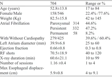
Seven hundred and four patients (158 female and 546 male [22.4%/77.6%]; mean±SD age, 52.8±14 [17 to 84] years) with drug-refractory AF (314 paroxysmal [44.6%], 332 persistent [47.2%], and 58 permanent [8.2%]) and treated by catheter RF endocardial ablation were included. Most patients (425, 60.4%) had no signiicant heart disease, with mean EF of 0.66±0.8 (0.3 to 0.8). In most cases, the LA diameter was ei -ther normal or slightly increased (39.9±6.8 mm) (Table 1). Fig. 1 - Cardiac computed tomography with esophagus visualization in RAO, PA and LAO, respectively, from left to right. There is a large contact area between the esophagus and the left atrium. This anatomical relationship can easily explain the risk of thermal injury of the esophagus during endocardial left atrial ablations. RAO=right anterior oblique; PA=postero-anterior; LAO=left anterior oblique
Fig. 2 - Lateral displacement of the esophagus usually obtained during transesophageal echocardiography. In this case the total displacement was 6.5 cm, allowing isolation of the the pulmonary vein (white circles) on each side with a good distance from the esophagus.
Table 1. Main features of 704 patients included in this study. N
Age (years) Female/Male Weight (Kg) Atrial Fibrillation
With/Without Cardiopathy Left Atrium diameter (mm) Ejection fraction
RF shots
X-ray duration (min) Number of sessions Max Esophageal displace
-ment (cm)
704
52.8±13.8
158/546
82.5±15.8 Paroxysmal 314 Persistent 332 Permanent 58
279/425
39.9±6.8 0.66±0.8 70.5±18.9 60.6±21.1 1.16 ±0.4
5.9±0.8
Range or % 17 to 84 22.4% /77.6% 42 to 145 44.6% 47.2% 8.2%
39.6% / 60.4% 25 to 60 0.3 to 0.8 40 to 120 10 to 99 1 to 4
4 to 9.1
Methodology
Patients using oral anticoagulants had the prothrombin time adjusted before the procedure (target INR ≤ 1.6). Con -ventional surface ECG monitoring, adhesive deibrillation patches, mechanical ventilation with intravenous or inhala -tion general anesthesia in addi-tion to the placement of TEE transducer were used. After having conirmed absence of intracardiac thrombus, four right femoral vein punctures were performed and a duodecapolar catheter was placed into coronary sinus. Trans-septal puncture was used in or -der to introduce both an ablation and a circular catheter in the left atrium. The following additional equipment was used: Cicero anaesthesia system (Dräger); multiparameter HP/Philips M1026A monitor; HP/Philips Sonos-2500 echo -cardiograph; 32 channels TEB polygraph with software for spectral analysis (Pachón-TEB); computerized spectrometer (Pachón®) for real-time spectral analysis, Siemens digital ra -dioscopy; Medtronic, Biotronik and Irvine RF generator; Philips Heartstart biphasic XL deibrillator with trans-cutaneous pace -maker and cerebral activity spectrometer (BIS). The ablations were carried out by using 8 mm catheter Blazer EPT, Medtronic Conductor and Johnson Irrigated. Activated clotting time was attained between 300 and 400 s by IV Heparin infusion. All the patients included in the study accepted the procedure being made aware of the methodology and potential complications, having signed the written informed consent.
Esophageal Displacement and Ablation
After obtaining a three-dimensional LA model with Navx system by handling the TEE, the esophagus was shifted and kept into the rightmost position before the ablation of the left half of LA (left pulmonary veins isolation and AF-Nests ab -lation). Afterwards, the TEE transducer was handled again in order to shift the esophagus to the leftmost location. Ablation of the half right of the LA was then performed. All positions were photographed in order to get accurate esophageal dis -placement measurements. In the inal ablation phase, in cases with LA background tachycardia, the esophagus was again shifted far from the ablation sites. At any time, at operator discretion before turning the RF on, the esophagus was ma -nipulated by the TEE transducer in order to keep it as far as possible from the RF delivery point.
Esophageal Temperature Monitoring versus Displacement
A group of 25 patients were also studied to see the effects of displacement in the esophageal luminal temperature (ELT). They had an esophageal probe and thermometer additional -ly inserted. In these cases the esophagus was contrasted with barium and ELT was monitored during the ablations, before and after displacements. With the esophagus in the natural po -sition, since the esophageal thermometer was in a good site (Figure 3) and whenever a ≥ 1oC ELT was observed, the ablation was immediately halted and the esophagus was quickly shifted.
Esophageal Endoscopy after Ablation
After ablation the patients were kept in the hospital under strict clinical monitoring for two days. Esophageal endosco -py was indicated whenever there was any symptom or sign of esophageal discomfort or lesion.
Medication after Ablation
Proton pump blockers were not used unless the patient was taking it prior to the ablation. During the irst 3 months, in all cases, antiarrhythmic medication (amiodarone, propafenone or beta blockers) was used depending on the patient’s toler -ance. Anticoagulation was strictly established for at least 2-3 months using warfarin (INR = 2 to 3) or dabigatran.
RESULTS
The anatomical course of the esophagus was quite vari -able. In 22.2% of the patients, it was centralized; however, in 57.5% and 20.3% of the patients, it was diverted near or su -perimposed onto the left or right pulmonary veins (Figures 4 and 5), respectively. In all cases, it was possible to achieve me -chanical esophageal displacement. For safety reasons, the dis -placement was applied even in cases with centered esophagus. Displacement ranged from 4 to 9.1 cm (5.9±0.8 cm). In 680 of the 704 patients (96.6%), the displacement was large enough to allow RF delivery with reasonable safety, even in the LA posterior wall (30W/40oC/irrigated catheter or 50W/60oC/8 mm catheter). For ablation of the LA posterior wall, the TEE transducer depth was modiied as needed for each case. In 24 cases (3.4%), the esophagus had reduced and dificult mobility or allowed only one-way displacement, as seen in Figures 4 and 5. However, with stepwise handling, it was possible to ob -tain reasonable segmental displacement to get safe RF delivery in most areas of the pulmonary veins, although not completely suficient for extensive treatment of the posterior LA wall. Fig. 3 - Barium esophagography showing esophageal displacement during RF catheter ablation of AF. In A, the thermometer position is satisfactory; however, in B, the thermometer is misplaced and should not be considered for temperature control of the ablation. The yellow dotted lines show the esophageal lumen contour. Enough bilateral esophageal displacement can also be observed, allowing safe ablation.
In the whole group there was neither a case nor a suspi -cion of atrioesophageal istula (mean follow-up of 37.9±81.9 months). Eight patients underwent esophageal endoscopy due to symptoms, with two of them showing slight bleed -ing. The investigation revealed normal esophagus in six of the eigh patients. The two cases with bleeding had superi -cial linear lesions in the high esophagus portion (above the atrial level), without signs of burning or necrosis. They were related to mechanical injury caused by introduction of the TEE transducer. Although small and supericial, the bleeding was certainly increased by anticoagulation. In both cases, the bleeding was promptly stopped with topic 1:1000 adrenaline solution. No cases presented symptoms compatible with in -jury to the periesophageal nerve plexus. There was no occur -rence of ileus palsy or pylori spasm.
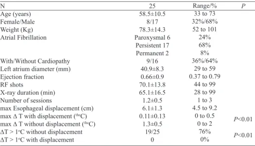
Study of Esophageal temperature versus Displacement
The results of 25 patients having ELT monitored without and with mechanical esophageal displacement are displayed in Table 2.
DISCUSSION
Several techniques have been proposed for esophageal protection during catheter ablation of AF. Any alternative limiting the amount or the sites of RF delivery increases the rate of ablation failure. Techniques have been used for:
1. Locating the esophagus before and/or during ablation; 2. Monitoring esophageal temperature;
3. Limitating RF energy or sites; 4. Esophageal cooling; and 5. Esophageal displacement. Fig. 4 - Esophagus positioned on the left at rest showing some
restriction to be moved. Nevertheless, there was enough displacement for treating left pulmonary vein. In such cases, the barium
esophagography is helpful to deine the real esophagus boundaries
and to show the extension of the displaced segment.
Fig. 5 - This patient had an old phrenic palsy and the esophagus was adhered to the same side showing restriction to be displaced to the left. However, enough displacement was achieved for right pulmonary vein ablation. Again, barium esophagography was helpful
to deine the real esophagus borders and to show the extension of
the displaced segment.
Table 2. ELT changes with and without esophageal displacement. In 19/25 (76%) patients the RF was interrupted due to ELT increase > 1oC during one RF delivery at least.
N
Age (years) Female/Male Weight (Kg) Atrial Fibrillation
With/Without Cardiopathy Left atrium diameter (mm) Ejection fraction
RF shots
X-ray duration (min) Number of sessions
max Esophageal displacement (cm) max ∆ T with displacement (theC)
max ∆ T without displacement (theC)
∆T > 1oC without displacement
∆T > 1oC with displacement
25
58.5±10.5
8/17
78.3±14.3 Paroxysmal 6
Persistent 17 Permanent 2
9/16
40.9±8.3 0.66±0.9 70.1±13.8 65.1±16.5 1.2±0.5 6.1±1.3 0.11±0.13
1.3±0.5
19/25 0
Range/% 33 to 73 32%/68% 52 to 101
24% 68% 8% 36%/64%
29 to 59 0.37 to 0.79
44 to 99 28 to 99 1 to 3 4.5 to 9.2
0 to 0.5 0 to 2
76% 0%
P
P<0.01
P<0.01
The esophagus location before ablation has been held with barium contrast radiography, computed tomography (Figure 1), MRI or electroanatomical 3D mapping[21]. These techniques allow a fairly accurate location, but with more or less infor -mation on the extent of the contact between the LA and the esophagus. The main disadvantages are spontaneous change of esophagus position and the need of reducing the RF energy in overlapping regions that can lead to incomplete ablation.
The esophagus location during ablation has been deter -minated through contrast with barium, intracardiac echocar -diography[22],and electroanatomical 3D mapping[23]. These methods have the great advantage of showing the actual position of the esophagus during ablation, but also limit RF delivery in overlapping areas. Therefore, if a AF relapse oc -curs due to restriction of RF energy release, the problem will persist in the same area in case of reablation[24].
Esophageal temperature monitoring during ablation[12] is another option, but it does not completely prevent compli -cations because the thermometer can be located outside the point of greatest heat and, in addition, there is the problem of thermal latency of the esophagus[25]. Studies have shown low correlation between the total energy delivered in LA and LET increase[25,26]. Furthermore, heating of the esophagus restricts RF, delivery bringing out the problem of incomplete ablation.
A quite creative technique is placing a cooled balloon irrigated with saline solution inside the esophagus[27,28]. However, more detailed studies need to be conducted to verify whether anterior displacement caused by esophageal balloon insuflation could be an additional problem, thereby reducing its potential beneits.
Several techniques have advocated esophagus protection by reducing the RF power in overlapping areas. So far, there is no evidence based on guidelines deining the limits of RF energy. Speculative recommendations suggest that RF ener -gy in overlapping areas of the esophagus should be <20W for less than 20 seconds and there should be at least 180 sec between two successive RF applications. However, the dis -advantage is that even when well-implemented, incomplete and insuficient ablation may predispose to relapse.
In this study, it was possible to demonstrate that the esoph -agus has a large motility. If on the one hand this feature is a disadvantage due to the risk of unexpected esophageal inter -position in the RF focus, on the other hand, it allows it to be mechanically displaced away from the point of RF application reducing the risk of thermal injury and atrioesophageal istula (Figure 2). Additionally, mechanical esophageal displacement by using the TEE transducer, allows it to keep it stable and far from the RF application site. Since January 2005, we have reg -ularly used this feature in any LA ablation as an indispensable requirement for ablation (Figure 6). Similar experience has been published supporting our observations and strengthening the idea that controlled esophageal displacement could be em -ployed as a protection against thermal lesions[29,30].
Fig. 6 - Good esophageal displacement allowing large bilateral antrum ablation.
The extent of displacement depends on several factors such as constitutional characteristics, thorax size, patient age, presence of adhesions and/or esophageal pathologies, operator experience, transducer mobility, etc. In this study, the displacement of the esophagus ranged from 4 to 9.1 cm (5.9±0.8 cm). This allowed for secured visualized RF ap -plication (keeping a safe distance from the RF ap-plication site) in 690 patients (98.1%). Only 14 cases (1.9%) had very low esophageal mobility and a careful displacement for each pulmonary vein ablation was needed. In these cases, despite having enough displacement to ablate the pulmonary veins antrum, it was not possible to safely apply RF to the posterior LA wall.
Besides visual control of the esophageal displacement, 25 patients underwent concomitant monitoring of ELT (Fig -ure 3). The RF was then applied with and without esopha -geal displacement. With displacement, it was not necessary to halt the RF energy whereas without displacement, the RF energy had to be stopped in 17 patients (68%) due to ELT increase of ≥1oC. In addition, it was found that the esoph -ageal displacement was able to reduce 10.9 times the ELT range (∆ELT = 0.11±0.13oC with versus 1.2±0.5oC without displacement, P<0.01) (Table 2). We decided to stop the RF
Authors’ roles & responsibilities
JCPM Analysis and/or interpretation of data; statistical analysis; inal approval of the manuscript; design and study design; conduct of the operations and/or experiments; writing of the manuscript or critical review of its content
EIPM analysis and/or interpretation of data; statistical analysis; inal approval of the manuscript; design and study design; conduct of the operations and/or experiments; writing of the manuscript or critical review of its content
TGSP Conduct of operations and/or experiments TJL Conduct of operations and/or experiments JCPM Conduct of operations and/or experiments RNAV Conduct of operations and/or experiments
CTCP Analysis and/or interpretation of data; conduct of the opera -tions and/or experiments
JCZA Conduct of operations and/or experiments
REFERENCES
1. Tsao HM, Wu MH, Higa S, Lee KT, Tai CT, Hsu NW, Chang CY, et al. Anatomic relationship of the esophagus and left atrium: implication for catheter ablation of atrial ibrillation. Chest. 2005;128(4):2581-7.
2. Gillinov AM, Pettersson G, Rice TW. Esophageal injury during radiofrequency ablation for atrial ibrillation. J Thorac Cardiovasc Surg. 2001;122(6):1239-40.
One problem observed in this study was the potential risk of mechanical trauma of the oropharynx and upper esophagus during the introduction of the TEE transducer, causing bleed -ing that was intensiied by anticoagulation. These observed cases were easily treated with topic application of adrena -line solution during diagnostic endoscopy. This complica -tion occurred at the beginning of the learning curve. Based on this experience, the TEE transducer insertion procedure was changed, with more appropriate lubrication and careful handling being applied. As a result, this complication was no longer observed.
The TEE transducer should never be advanced inside the esophagus with pronounced angulation. It is important to move it with extreme care. Its position must be changed often to avoid forcing it in a single point for a long time. It could cause an ischemic injury to the esophageal mucosa.
Study and Method Limitations
Active and controlled esophagus displacement during catheter RF of AF ablation seems to be able to prevent esoph -ageal temperature increase as well as esoph-ageal thermal le -sions. However, it depends on the use of the TEE transducer throughout the procedure. This may be considered a limita -tion for some services, but in our methodology, it becomes an advantage as we regularly use the TEE to replace the useful but more expensive intracardiac echocardiogram.
The insertion of TEE transducer depends on good seda -tion or general anesthesia, thus many services that perform AF ablation with a conscious patient or with supericial seda -tion may have dificulty employing this technique. The inser -tion process must be performed with addi-tional care because since the patient will be anti-coagulated any mucosal trauma may cause signiicant bleeding.
Due to its signiicant diameter (11 mm), the TEE trans -ducer may be considered a disadvantage as it forces the esophageal wall to the atrium, reducing the postero-anterior dimension of the LA. This could reduce the space for abla -tion, favoring esophageal heating. However, the proposal is to bend the transducer in order to maintain the esophagus as far as possible from the ablation site with minimal overlap -ping of the transducer and the LA.
One limitation of this study is that endoscopy was per -formed only in a few symptomatic cases. Since this is a prospective study of regular clinical application, though, it would be inconvenient and ethically questionable to per -form an additional semi-invasive procedure in asymptom -atic patients. This must be achieved through a randomized study. Nevertheless, the high number of cases treated with -out any occurrence of clinical esophageal lesion is a high -ly positive inding. Furthermore, the study of 25 patients undergoing ablation with ELT monitoring showed the high eficacy of this method for preventing esophageal tempera -ture increase.
Sometimes, malposition and low mobility of the esopha -gus may limit the application of this technique; however, the experience of this study shows that these cases are rare.The presence of the TEE transducer and, especially, the barium in the esophagus may reduce the radiological visibility in some degree. Currently, we have used barium only in cases with dificult esophagus displacement.
CONCLUSION
3. Scanavacca MI, D’ávila A, Parga J, Sosa E. Left atrial-esophageal fistula following radiofrequency catheter ablation of atrial ibrillation. J Cardiovasc Electrophysiol. 2004;15(8):960-2.
4. Pappone C, Oral H, Santinelli V, Lang CC, Manguso F, Torracca L, et al. Atrio-esophageal istula as a complication of percutaneous transcatheter ablation of atrial fibrillation. Circulation. 2004;109(22):2724-6.
5. Ghia KK, Chugh A, Good E, Pelosi F, Jongnarangsin K, Bogun F, et al. A nationwide survey on the prevalence of atrioesophageal istula after left atrial radiofrequency catheter ablation. J Interv Card Electrophysiol. 2009;24(1):33-6.
6. Cummings JE, Schweikert RA, Saliba WI, Burkhardt JD, Kilikaslan F, Saad E, et al. Brief communication: atrial-esophageal istulas after radiofrequency ablation. Ann Intern Med. 2006;144(8):572-4.
7. Schmidt M, Nölker G, Marschang H, Gutleben KJ, Schibgilla V, Rittger H, et al. Incidence of oesophageal wall injury post-pulmonary vein antrum isolation for treatment of patients with atrial ibrillation. Europace. 2008;10(2):205-9.
8. Pappone C, Rosanio S, Oreto G, Tocchi M, Gugliotta F, Vicedomini G, et al. Circunferential radiofrequency ablation of pulmonary vein ostia: A new anatomic approach for curing atrial ibrillation. Circulation. 2000;102(21):2619-28.
9. Teplitsky L, Hegland DD, Bahnson TD. Catheter based cryoablation and radiofrequency ablation for atrial ibrillation results in conductive heat transfer from and to the esophagus. Heart Rhythm. 2006;3(5):S242.
10. Ripley KL, Gage AA, Olsen DB, Van Vleet JF, Lau CP, Tse HF. Time course of esophageal lesions after catheter ablation with cryothermal and radiofrequency ablation: implication for atrio-esophageal istula formation after catheter ablation for atrial ibrillation. J Cardiovasc Electrophysiol. 2007;18(6):642-6.
11. Arruda MS. Pre clinical “in vivo” evaluation of an esophageal protective system: Implications on esophageal thermal injury during AF ablation. Heart Rhythm. 2008;5:S16.
12. Redfearn DP, Trim GM, Skanes AC, Petrellis B, Krahn AD, Yee R, et al. Esophageal temperature monitoring during radio frequency ablation of atrial fibrillation. J Cardiovasc Electrophysiol. 2005;16(6):589-93.
13. Good E, Oral H, Lemola K, Han J, Tamirisa K, Igic P, et al. Movement of the esophagus during left atrial catheter ablation for atrial ibrillation. J Am Coll Cardiol. 2005;46(11):2107-10.
14. Helms A, West JJ, Patel A, Mounsey JP, DiMarco JP, Mangrum JM, et al. Real-time rotational ICE imaging of the relationship of the ablation catheter tip and the esophagus during atrial ibrillation ablation. J Cardiovasc Electrophysiol. 2009;20(2):130-7.
15. Haıssaguerre M, Jaıs P, Shah DC, Garrigue S, Takahashi A, Lavergne T, et al. Electrophysiological end point for catheter
ablation of atrial ibrillation initiated from multiple pulmonary venous foci. Circulation. 2000;101(12):1409-17.
16. Morady F. Treatment of paroxysmal atrial fibrillation by pulmonary vein isolation. Circ J. 2003;67(7):567-71.
17. Pachón M JC, Pachón M EI Pachon M JC, Lobo TJ, Pachon MZ, Vargas RN, et al. A new treatment for atrial ibrillation based on spectral analysis to guide the catheter RF-ablation. Europace. 2004;6(6):590-601.
18. Arruda M, Natale A. Ablation of permanent AF: adjunctive strategies to pulmonary veins isolation: targeting AF NEST in sinus rhythm and CFAE in AF. J Interv Card Electrophysiol. 2008;23(1):51-7.
19. Huang SY, Lin YJ, Tsao HM, Chang SL, Lo LW, Hu HF, et al. The biatrial substrate properties in different types of paroxysmal atrial ibrillation. Heart Rhythm. 2011;8(7):961-7.
20. Mateos JC, Mateos EI, Lobo TJ, Pachón MZ, Mateos JC, Pachón DQ, et al. Radiofrequency catheter ablation of atrial ibrillation guided by spectral mapping of atrial ibrillation nests in sinus rhythm. Arq Bras Cardiol. 2007;89(3):124-34, 140-50.
21. Good E, Oral H, Lemola K, Han J, Tamirisa K, Igic P, et al. Movement of the esophagus during left atrial catheter ablation for atrial fibrillation. J Am Coll Cardiol. 2005;46(11):2107-10.
22. Kenigsberg DN, Lee BP, Grizzard JD, Ellenbogen KA, Wood MA. Accuracy of intracardiac echocardiography for assessing the esophageal course along the posterior left atrium: a comparison to magnetic resonance imaging. J Cardiovasc Electrophysiol. 2007;18(2):169-73.
23. Sherzer AI, Feigenblum DI, Kulkarni S, Pina JW, Casey JL, Salka KA, et al. Continuous nonluoroscopic localization of the esophagus during radiofrequency catheter ablation of atrial ibrillation. J Cardiovasc Electrophysiol. 2007;18(2):157-60.
24. Kennedy R, Good E, Oral H, Huether E, Bogun F, Pelosi F, et al. Temporal stability of the location of the esophagus in patients undergoing the repeat left atrial ablation procedure for atrial ibrillation or lutter. J Cardiovasc Electrophysiol. 2008;19(4):351-5.
25. Teplitsky L, Perzanowski C, Durrani S, Berman AE, Hranitzky P, Bahnson TD. Radio frequency catheter ablation for atrial ibrillation produces delayed and long lasting elevation of luminal esophageal temperature independent of lesion duration and power. Heart Rhythm. 2005;2(Suppl.):S8-S9.
26. Cummings JE, Schweikert RA, Saliba WI, Burkhardt JD, Brachmann J, Gunther J, et al. Assessment of temperature, proximity, and course of the esophagus during radiofrequency ablation within the left atrium. Circulation. 2005;112(4):459-64.
prevent thermal injury during endocardial radiofrequency surgical ablation of the left atrium: a inite element study. Phys Med Biol. 2005;50(20):N269-79.
28. Tsuchiya T, Ashikaga K, Nakagawa S, Hayashida K, Kugimiya H. Atrial ibrillation ablation with esophageal cooling with the cooled water-irrigated intraesophageal balloon: a pilot study. J Cardiovasc Electrophysiol. 2007;18(2):145-50.
29. Herweg B, Johnson N, Postler G, Curtis AB, Barold SS, Ilercil A. Mechanical esophageal delection during ablation of atrial
ibrillation. Pacing Clin Electrophysiol. 2006;29(9):957-61.
30. Chugh A, Rubenstein J, Good E, Ebinger M, Jongnarangsin K, Fortino J, et al. Mechanical displacement of the esophagus in patients undergoing left atrial ablation of atrial ibrillation. Heart Rhythm. 2009;6(3):319-22.