O T O L O G Y
Predictive factors for the appearance of myringosclerosis
after myringotomy with ventilation tube placement: randomized
study
Carla Branco1• Daniel Monteiro1•Joa˜o Pac¸o2
Received: 15 April 2016 / Accepted: 29 June 2016 ÓSpringer-Verlag Berlin Heidelberg 2016
Abstract Myringotomy with the insertion of ventilation tubes is the most frequent surgical procedure performed in children, and the appearance of myringosclerosis is one of its most frequent long-term complications. The objective of this study is to identify clinical factors and technique vari-ations that may have a relation with the appearance of myringosclerosis, after tube insertion. Patients submitted to myringotomy with transtympanic short-term tube insertion were studied in a longitudinal prospective and analytical cohort study with the prospective randomized open, blinded endpoint (PROBE) methodology, to study the influence of the location of myringotomy (anterior–inferior quadrant or posterior–inferior), directions of the incision (radial or non-radial) and aspiration or not of the middle ear. Our study included 156 patients (297 ears). Myringosclerosis was observed in 35.7 % of the operated ears. It appeared more often in patients with greater number of otitis (p=.001) and with greater number of otorrhea episodes (p =.029) and in patients in whom the tympanogram after the tube extraction was type A (according to Jerger´s classification) (p =0.016). We identified myringosclerosis in less patients, if the tube was in the tympanic membrane for less than 12 months (p =.009). Myringosclerosis was present more extensively if the tympanic incision was located in the anterior–inferior quadrant, with tympanic involvement superior to 25 % (p =.015). The results observed prove that, underlying the appearance of myringosclerosis, there exists an early
inflammatory or infectious process and a final cicatricial process. It was also found that when myringotomy is made in the anterior–inferior quadrant, myringosclerosis appears in a higher percentage of the tympanic membrane; therefore, it is not recommended to do the incision in this quadrant, because it may lead to a reduction of the tympanic mem-brane vibration.
Keywords MyringosclerosisMyringotomySclerotic
plaques
Introduction
Tympanosclerosis is a pathology that may appear when tissue repair occurs, in which high quantities of collagenic fibrosis tissue are deposited in the lamina propria which covers the ossicles, the walls of the tympanic cavity and the medial layer of the tympanic membrane [1, 2]. The thickness due to collagen gives way to the formation of a homogenous and hyaline substance, that is later subject to the deposition of crystals of calcium and phosphate [3–6]. When the deposition of crystals occurs only on the tympanic membrane, which is the most frequent location, it is named myringosclerosis [1, 7–9]. This process may assume clinical importance if it is able to interfere with the transmission of the sound vibrations through the structures of the middle ear [1,10].
Nowadays, myringotomy with ventilation tube place-ment is one of the most common surgical procedures per-formed in children [8, 11, 12]. Although considered a simple procedure with considerable benefits, it can have complications [13]. Several studies indicate myringoscle-rosis as the most frequent complication of this procedure [1,8,14–16].
& Carla Branco
1 ENT Department, Hospital de Vila Franca de Xira,
Lisbon, Portugal
2 ENT Department, Hospital CUF Infante Santo,
Lisbon, Portugal
The present study aims to identify predictive factors for the appearance of myringosclerosis after short-term tube insertion and, therefore, to identify risk groups and tech-niques that can potentially be responsible for the devel-opment of this complication.
The specific objectives are, to identify clinical factors that can predict the development of myringosclerosis; determine if the location of the myringotomy influences the development of the pathology; determine if the type of incision (radial or non-radial) influences the development of myringosclerosis; verify if the aspiration of the tympanic cavity impacts the development of myringosclerosis.
Materials and methods
Patients, with chronic otitis media with effusion, submitted to myringotomy with transtympanic short-term tube insertion, were studied in an analytical and longitudinal prospective cohort study, to evaluate criteria variables, either, pre, intra or post-operative. These patients were also submitted to a controlled clinical study with the prospec-tive randomized open, blinded endpoint (PROBE) methodology.
In the study, all the patients submitted to myringotomy with unilateral or bilateral tube insertion between February 2013 and May 2014, in the Vila Franca de Xira Hospital, were enrolled. A signed informed consent was mandatory to all the patients included in the study.
The exclusion criteria were the presence of tym-panosclerosis prior to surgery, any chronic pathology in the opposite ear of tube placement (excluding otitis media with effusion), missed attendance of the patient in any of the follow-up appointments and the refusal of the patient or their legal representative to participate in the study.
In the clinical study, the opposite ear was used as con-trol, comparing the type of incision (radial or non-radial), the location of the incision (anterior–inferior quadrant or posterior–inferior quadrant) and the aspiration or not of the middle ear.
The randomization of the patients was performed as follows: in patients born in the first 6 months of the year, a radial incision was performed in the right ear and a non-radial incision in the left ear; in patients, born in the last 6 months of the year, the opposite was performed. In patients, born on even months (February, April, June, August, October and December), the ventilation tube was inserted in the anterior–inferior quadrant on the right ear and in the posterior–inferior quadrant on the left ear; in patients, who were born in odd months, the opposite was performed. In patients, who were born in the first and third trimester of the year, the tympanic cavity was aspirated in the right ear and not in the left ear; in patients, who were
born on the other trimesters, the left ear was aspirated. In the sporadic cases, it was necessary to aspirate the middle ear, independent of randomization, due to hemorrhage.
The surgical procedure was performed under general anesthesia. In all cases, a fluoroplastic ventilation Shepard tube, from Xomed, with 1.14 nm, was inserted.
In post-operative period, the patients were followed every 6 months until the complete extrusion of the venti-lation tube. In each examination, a micro-otoscopy was performed to identify myringosclerosis, and the involve-ment, in percentage, of the tympanic membrane was noted. It was considered positive when white plaques could be identified in the tympanic membrane.
Computer software SPSS for Windows (version 21) was used for statistical analysis. To compare the different variables, Kendall’s correlation coefficient, Pearson’s cor-relation coefficient and Chi-squared test were used. It was considered statistically significant if a value of p\0.05 was obtained.
The protocol of this study was approved by the Ethical Committee of the Vila Franca de Xira Hospital and the Ethical Committee of The Faculdade de Cieˆncias Me´dicas de Lisboa da Universidade Nova de Lisboa.
Results
At our hospital, and during the time of this study, we performed myringotomy with tympanic tube placement on 173 patients. Of these patients, twenty-three were excluded either because they refused to participate in the study, or due to the presence of myringosclerosis prior to surgery or for not showing up for the follow-up appointments. The total number of patients included in the study was 156, on which myringotomy was unilateral in 15 patients, and bilateral in the remaining, in a total of 297 ears analyzed. The patients’ ages, at the time of surgery, ranged from 2 to 36 years (mean 5.5±3.65 years), and 89 were male and 67 were female.
The calcium values ranged between 7.6 and 10.2 mg/dl (mean 9.26±.52 mg/dl).
Concerning all the patients included in the study, 55.8 % stated not having any ear infection in the previous year, 19.9 % had had until 3 occurrences, 16.7 % between 4 and 6 and 7.7 % more than 6 occurrences. Regarding the number of antibiotic treatments in the previous year to surgery, 33.3 % of the patients were treated with less than 3 antibiotics, 29.5 % between 4 and 6 and 14.1 % took more than 6. Only 22.4 % did not take any antibiotic.
The pre-operative tympanogram was type B in 262 ears and type C in 35 (according to Jerger’s classification).
antero-inferior quadrant was the location for myringotomy in 52.9 % (n=157) of the cases, and the remaining cases had a posterior–inferior incision. Aspiration was performed in 60.3 % of the cases.
Bleeding during myringotomy occurred in 35.7 % of the cases. Effusion in the middle ear was detected in 62.2 % of the ears, 80 % was mucoid and the remaining was serous. Myringosclerosis was identified in 35.4 % of the oper-ated ears (n =105) from which, 48 exhibited more than 25 % of myringosclerosis involvement on the tympanic membrane.
Concerning tube extrusion, 21.9 % of the operated ears showed a complete extrusion in less than 6 months. In 49 %, the tube was present for 6 months to 12 months; in 20.5 %, from 13 to 18 months; in 7.9 %, from 19 to 24 months; and in 0.7 %, for more than 24 months.
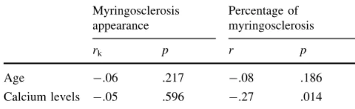
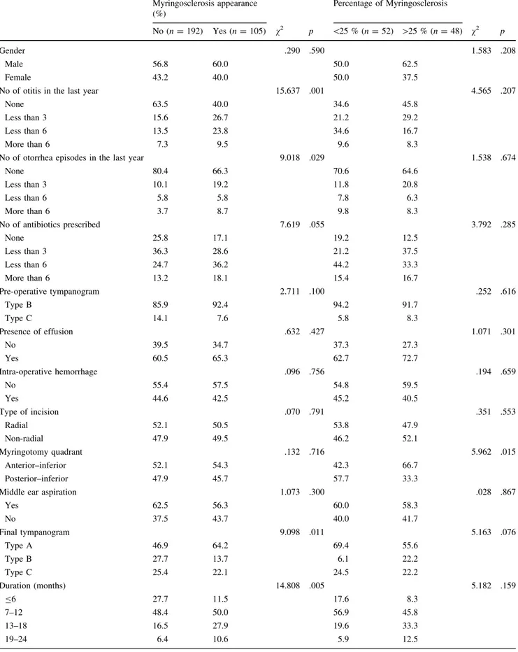
Regarding the socio-demographic, pre-operative, intra-operative and post-intra-operative variables (Tables1,2) there was no statistically significant association between the presence of myringosclerosis and the age of the patient (p=.217), the calcium levels (p =.596), gender (p=590), number of antibiotics taken (p=.055), pre-operative tympanogram (p =.100), pre-operative otoscopy (p=.058), presence of effusion (p=.427), type of effu-sion (p=.362), intra-operative hemorrhage (p=.756), incision type (p =.791), myringotomy quadrant (p=.716) and aspiration of the middle ear (p=.300). There were statistically significant associations between the presence of myringosclerosis and the number of ear infections in the previous year [v2(3) =15.637;p =.001]
and between the presence of myringosclerosis and the number of episodes with otorrhea in the previous years [v2
(3)=9.018; p=.029]. These results demonstrate that patients with myringosclerosis have, in their clinical his-tory, a higher number of otitis and otorrhea.
It was also possible to confirm statistically significant associations between the presence of myringosclerosis and the final tympanogram [v2 (4)=12.222; p=.016]
and the presence of myringosclerosis and the retention time of tympanic tube [v2(5)=15.382;p =.009]. These
results reveal that patients with myringosclerosis have, more frequently, a final tympanogram type A, and a
retention time of 7–18 months. Although patients without myringosclerosis also have a higher propensity to tym-panograms type A, the number of tymtym-panograms type B and type C is higher than in patients with myringoscle-rosis. On the other hand, these patients have a lower retention time of the tube; in most cases, inferior to 12 months.
Comparing the percentage of tympanic involvement by myringosclerosis and the pre-operative, intra-operative and post-operative variables (Tables 1,2), in the patients with myringosclerosis, it is possible to confirm a statistically significant correlation between the percentage of myringosclerosis and the calcemia levels (r=.27; p =.014). Therefore, it can be concluded that lower values of calcemia correspond to higher percentages of myringosclerosis.
Our study found statistically significant associations between the percentage of myringosclerosis and the quadrant of the myringotomy [v2(1)=5.962;p =.015].
The results reveal that the anterior–inferior quadrant was the most frequent location of incision, in patients with percentage of tympanic involvement higher than 25 %. The posterior–inferior quadrant was the most frequent location of incision, in patients with percentage of tympanic involvement lower than 25 %.
The only complication verified after the procedure was two perforations of the tympanic membrane; in both these cases, the myringotomy incision was in the anterior–infe-rior quadrant.
In all procedures, we did not identify any lesion of the ossicular chain.
Relatively to intra-operative hemorrhage (p =.113), the presence of the myringotomy tube (p=.959), and the result of the final tympanogram (p=.070), we did not identify statistical difference when the myringotomy tube was placed in the anterior–inferior or posterior–inferior quadrant.
Discussion
Myringotomy with tube insertion, for the treatment of otitis media with effusion, increases myringosclerosis incidence [1,17] between 17.1 % [18] and 56 % [19]. In the present study, the incidence was 35.4 %.
According to Koc and Uneri [4], there is a statistically significant difference between the development of myringosclerosis in children from the masculine and fem-inine gender, being the incidence in the masculine gender higher. From the results obtained in this study, it was not possible to establish any statistically significant association among gender and the development of myringosclerosis, as occurred with Kaur et al. [17] and Tos et al. [20]. Table 1 Correlation between appearance of myringosclerosis, the
percentage of tympanic membrane involved by myringosclerosis, age and calcium levels (correlation coefficient of Kendall)
Myringosclerosis appearance
Percentage of myringosclerosis
rk p r p
Age -.06 .217 -.08 .186
Table 2 Association between appearance of myringosclerosis and percentage of tympanic membrane involved by myringosclerosis with socio-demographic, pre, intra and post-operative variables
Myringosclerosis appearance (%)
Percentage of Myringosclerosis
No (n=192) Yes (n=105) v2 p \25 % (n=52) [25 % (n=48) v2 p
Gender .290 .590 1.583 .208
Male 56.8 60.0 50.0 62.5
Female 43.2 40.0 50.0 37.5
No of otitis in the last year 15.637 .001 4.565 .207
None 63.5 40.0 34.6 45.8
Less than 3 15.6 26.7 21.2 29.2
Less than 6 13.5 23.8 34.6 16.7
More than 6 7.3 9.5 9.6 8.3
No of otorrhea episodes in the last year 9.018 .029 1.538 .674
None 80.4 66.3 70.6 64.6
Less than 3 10.1 19.2 11.8 20.8
Less than 6 5.8 5.8 7.8 6.3
More than 6 3.7 8.7 9.8 8.3
No of antibiotics prescribed 7.619 .055 3.792 .285
None 25.8 17.1 19.2 12.5
Less than 3 36.3 28.6 21.2 37.5
Less than 6 24.7 36.2 44.2 33.3
More than 6 13.2 18.1 15.4 16.7
Pre-operative tympanogram 2.711 .100 .252 .616
Type B 85.9 92.4 94.2 91.7
Type C 14.1 7.6 5.8 8.3
Presence of effusion .632 .427 1.071 .301
No 39.5 34.7 37.3 27.3
Yes 60.5 65.3 62.7 72.7
Intra-operative hemorrhage .096 .756 .194 .659
No 55.4 57.5 54.8 59.5
Yes 44.6 42.5 45.2 40.5
Type of incision .070 .791 .351 .553
Radial 52.1 50.5 53.8 47.9
Non-radial 47.9 49.5 46.2 52.1
Myringotomy quadrant .132 .716 5.962 .015
Anterior–inferior 52.1 54.3 42.3 66.7
Posterior–inferior 47.9 45.7 57.7 33.3
Middle ear aspiration 1.073 .300 .028 .867
Yes 62.5 56.3 60.0 58.3
No 37.5 43.7 40.0 41.7
Final tympanogram 9.098 .011 5.163 .076
Type A 46.9 64.2 69.4 55.6
Type B 27.7 13.7 6.1 22.2
Type C 25.4 22.1 24.5 22.2
Duration (months) 14.808 .005 5.182 .159
B6 27.7 11.5 17.6 8.3
7–12 48.4 50.0 56.9 45.8
13–18 16.5 27.9 19.6 33.3
In this study, it was not possible to find an association between elevated calcemia levels and the development of myringosclerosis, as described by Leal et al. [13], who pointed this as one of the factors responsible for the development of myringosclerosis. In contrary, in our study, patients with lower levels of calcemia presented larger myringosclerosis plaques, contradicting Leal et al.
Several studies acknowledge that the inflammatory pro-cess is responsible for the myringosclerosis pathology [2,14,21]. In the results obtained in our study, there is a similar and strong, statistically significant association between the development of myringosclerosis and the number of otitis media and otorrhea. Several factors seem to be involved in the inflammation process, related with the onset and the progression of myringosclerosis [7,22]. These factors include macrophages, B cells and other inflamma-tory mediators such as interleukin 6 (IL-6) and inducible nitric oxide synthase (iNOS), although their mechanism is not completely understood and defined [7,22].
It was not possible to verify any association between the development of myringosclerosis and the presence and the type of effusion (mucous or serous); the intra-operative hemorrhage (as described by Spree et al. [23] and Parker et al. [16]); the myringotomy incision orientation or the middle ear aspiration (discordant with the conclusions of Liana et al. [24]).
In our study, although there was no significant difference regarding the place of myringotomy and the incidence of myringosclerosis, it was more extensive when the incision location was in the anterior–inferior quadrant, compared with the incision performed on the posterior–inferior quad-rant. Apparently, the pathological process of myringoscle-rosis is more noticeable when the tube is inserted in the anterior–inferior quadrant. This can be explained by the anatomic differences of the lamina propria in the anterior and posterior quadrants, described by Pac¸o [25]. According to this author, in the posterior quadrants, the circular fibrous layer is less dense which can represent less tissue damage and an inferior healing response. Another explanation is related with the proximity of the Eustaquian tube to the anterior–inferior quadrant. This proximity can increase the oxygen concentration in this quadrant, compared with the posterior, and, therefore, augment the formation of free oxygen radicals and myringosclerosis. The different vascu-larization of the two quadrants, higher in the anterior35, can also justify an increased formation of free oxygen radicals when the incision is made in this quadrant. This fact leads us to recommend the placement of the myringotomy incision and tube in the posterior–inferior quadrant, which is tech-nically easier in narrow ear canals and with protruding anterior wall and is not associated with any increase in intra or post-operative complications and has similar surgical outcomes.
According to Yaman et al. [3], the myringosclerosis rate was higher in patients who maintained the ventilation tubes for more than 12 months. In the present study, the myringosclerosis rate was higher in the patients who retained the ventilation tubes for more than 6 months, similarly as described by Maw [26]. Contrary to what was expected, higher rates of myringosclerosis did not appear in the ears which had longer tube retention time.
Finally, it was also possible to determine an association between the development of tympanosclerosis and the recovery of otitis with effusion, as demonstrated by the type A tympanogram obtained after the extraction of the ventilation tube. It is, therefore, possible to infer that the healing process, initiated by the insertion of the ventilation tubes, is important for the recovery of the ear pathology, though it can also be responsible for the development of myringosclerosis. From these results, it is also plausible to infer that, in contrary to what would be expected, patients with more severe ear pathology are not more prone to have myringosclerosis.
Conclusion
Myringosclerosis frequently develops in patients submitted to myringotomy with insertion of ventilation tubes. Myringosclerosis is closely related with the inflammatory process (patients with more otitis have a higher incidence of myringosclerosis) which may be enhanced by the insertion of the ventilation tube. Furthermore, myringosclerosis is also associated with the healing process responsible for the recovery of the otological pathology in the origin of surgery (a higher incidence of myringoscle-rosis is associated with a final tympanogram type A).
In addition, it was found that when myringotomy is made in the anterior–inferior quadrant, myringosclerosis appears in a higher percentage of the tympanic membrane; therefore, it is not recommended to do the incision in this quadrant, because it may lead to a reduction of the tym-panic membrane vibration.
Compliance with ethical standards
Conflict of interest The authors declare that they have no conflict of interest.
References
1. Gibb AG, Pang YT (1994) Current considerations in the etiology and diagnosis of tympanosclerosis. Eur Arch Otorhinolaryngol 251(8):439–451
prevention of myringosclerosis. Eur Arch Oto Rhino Laryngol 269(11):2335–2341
3. Yaman H, Guclu E, Yilmaz S, Ozturk O (2010) Myringosclerosis after tympanostomy tube insertion: relation with tube retention time and gender. Auris Nasus Larynx (Internet) 37(6):676–679 4. Koc A, Uneri C (2001) Sex distribution in children with
tym-panosclerosis after insertion of a tympanostomy tube. Eur Arch Otorhinolaryngol 258(1):16–19
5. Tympanosclerosis Chang IW (1969) Electron microscopic study. Acta Otolaryngol (Internet) 68(1):62–72
6. Wielinga EWJ, Kerr AG (1993) Tympanosclerosis. Clin Oto-laryngol 18:341–349
7. Forse´ni M, Bagger-Sjo¨ba¨ck D, Hultcrantz M (2001) A study of inflammatory mediators in the human tympanosclerotic middle ear. Arch Otolaryngol Head Neck Surg 127(5):559–564 8. Uneri C, Bagˇlam T, Yazici M (2006) The effect of vitamin E
treatment on the development of myringosclerosis after ventila-tion tube insertion. Int J Pediatr Otorhinolaryngol 70(6):1045–1048
9. Leal MC, Bento RF, Neto SD et al (2006) Influence of hyper-calcemia in the formation of tympanosclerosis in rats. Otol Neurotol (Internet) 27(1):27–32
10. Helms J, Steinbach E (1973) Tympanosclerosis. Ind J Otol XXV(1):25–27
11. Khodaverdi M, Jørgensen G, Lange T et al (2013) Hearing 25 years after surgical treatment of otitis media with effusion in early childhood. Int J Pediatr Otorhinolaryngol (Internet) 77(2):241–247
12. Kinis V, Ozbay M, Alabalik U et al (2015) Effect of caffeic acid phenethyl ester on myringosclerosis development in the tympanic membrane of rat. Eur Arch Oto Rhino Laryngol (Internet) 272(1):29–34
13. de Carvalho Leal M, Ferreira Bento R, da Silva Caldas Neto S et al (2006) Influence of hypercalcemia in the formation of tympanosclerosis in rats. Otol Neurotol 27(1):27–32
14. Friedman EM, Sprecher RC, Simon S, Dunn JK (2001) Quanti-tation and prevalence of tympanosclerosis in a pediatric oto-laryngology clinic. Int J Pediatr Otorhinolaryngol 60(3):205–211
15. O¨ zcan C, Go¨ru¨r K, Cinel L, Talas DU, U¨nal M, Cinel I (2002) The inhibitory effect of topical N-acetylcysteine application on myringosclerosis in perforated rat tympanic membrane. Int J Pediatr Otorhinolaryngol 63(3):179–184
16. Parker A, Maw A, Powell J (1990) Intra-tympanic membrane bleeding after grommet insertion and tympanosclerosis. Clin Otolaryngol Allied Sci 15(3):203–207
17. Kaur K, Sonkhya N, Bapna AS (2006) Tympanosclerosis revis-ited. Indian J Otolaryngol Head Neck Surg 58(2):128–132 18. Barati B, Hashemi SM, Tabrizi AG (2012) Otological findings
10 years after myringotomy with tympanostomy tube insertion. Iran J Otorhinolaryngol 24(4):181–186
19. De Beer BA, Schilder AGM, Zielhuis GA, Graamans K (2005) Natural course of tympanic membrane pathology related to otitis media and ventilation tubes between ages 8 and 18 years. Otol Neurotol 26(5):1016–1021
20. Tos M, Bonding P, Poulsen G (1983) Tympanosclerosis of the drum in secretory otitis after insertion of grommets. A prospec-tive, comparative study. J Laryngol Otol 97(6):489–496 21. Schiff M, Yoo TJ (1985) Immunologic aspects of otologic
dis-ease: an overview. Laryngoscope 95(3):259–269
22. Forse´ni Flodin M, Hultcrantz M (2002) Possible inflammatory mediators in tympanosclerosis development. Int J Pediatr Otorhinolaryngol 63(2):149–154
23. Sprem N, Branica S, Dawidowsky K (2002) Experimental hematotympanum–aspects to the tympanosclerosis development. Coll Antropol 26(1):267–272
24. Laina V, Pothier DD (2006) Should we aspirate middle-ear effusions prior to insertion of ventilation tubes? J Laryngol Otol 120(10):818–821
25. Pac¸o J (2003) Estrutura do Tı´mpano. In: Pac¸o J (ed) Doenc¸as do Tı´mpano. Lisbon, PT: Lidel—edic¸o˜es te´cnicas, Lda, pp 55–72 26. Maw AR (1991) Development of tympanosclerosis in children