Relationship between vitamin D and lung function, physical
performance and balance on patients with stage I-III chronic
obstructive pulmonary disease
TUNCAY YUMRUTEPE1, ZEYNEP AYFER AYTEMUR1, OZLEM BAYSAL2, HULYA TASKAPAN3, CAGATAY M. TASKAPAN4, SULEYMAN SAVAS HACIEVLIYAGIL1*
1M.D. – Department of Chest Diseases, Inonu University Faculty of Medicine, Malatya, Turkey
2M.D. – Department of Physical Medicine and Rehabilitation, Inonu University Faculty of Medicine, Malatya, Turkey 3M.D. – Department of Nephrology, Inonu University Faculty of Medicine, Malatya, Turkey
4M.D. – Department of Biochemistry, Inonu University Faculty of Medicine, Malatya, Turkey
S
UMMARYStudy conducted at Inonu University Faculty of Medicine, Malatya, Turkey
Article received: 7/10/2014
Accepted for publication: 7/14/2014
*Correspondence:
Address: Department of Chest Diseases,
Inonu University Faculty of Medicine, Malatya, Turkey
Postal Code: 44200 [email protected]
http://dx.doi.org/10.1590/1806-9282.61.02.132
Conflict of interest: none
Objectives: vitamin D is important for muscle function and it affects different aspects of muscle metabolism. This study aim to determine whether serum 25(OH) D levels are related to lung functions, physical performance and balance in pa-tients with chronic obstructive pulmonary disease (COPD).
Methods: in 90 patients with COPD and 57 healthy controls lung function tests, physical performance tests (time up and go, gait velocity test, sit-to-stand test, isometric strength, isokinetic strength), static (functional reach test) and dy-namic (time up and go) balance tests and the association of 25(OH)D levels with lung functions, physical performance and balance were evaluated.
Results: the COPD patients had significantly more deficit in physical function and balance parameters, and in dynamic balance test (p<0.005). Isokinetic knee muscle strength (flexor and extensor) in COPD patients was significantly lower than in the controls (p<0.05); FEV1 (p=0.008), FVC (p=0.02), FEV1/FVC (p=0.04), TLC (p=0.01) were lower in COPD patients with vitamin D deficiency [25(OH) D less than 15ng/mL] than in COPD patients without vitamin D deficiency. Hand grip test (p=0.000) and isokinetic knee muscle strength (flexor and exten-sor) (p<0.05) were also lower in COPD patients with vitamin D deficiency. Vita-min D deficiency was more pronounced in patients with stage III COPD (p<0.05). Conclusion: patients with COPD had worst physical functioning, poor balance and less muscle strength. Severe disturbed lung and peripheral muscle functions are more pronounced in COPD patients with vitamin D deficiency.
Keywords: chronic obstructive pulmonary disease, vitamin D deficiency, mus-cle function.
I
NTRODUCTIONPatients with chronic obstructive pulmonary disease (COPD) complain of dyspnea on exertion and reduced exercise capacity. Pulmonary and extrapulmonary factors play a role in reduced exercise capacity. Especially, periph-eral muscle dysfunction, independent of lung function, contributes significantly to reduced exercise capacity in these patients.1,2
Vitamin D is important for muscle function and it may affect different aspects of muscle metabolism.3 A
positive association between vitamin D and muscle
strength or lower extremity function in elderly people has been described.2 Similarly, vitamin D levels were
positive-ly associated with lung function.4,5 Therefore, vitamin D
deficiency can increase the susceptibility to psychologi-cal stress. Psychologipsychologi-cal stress can increase oxidative stress and modify host response to other inflammatory oxida-tive toxins such as smoke and air pollutants. These fac-tors that can modify oxidative toxicity may have additive or multiplicative effects on lung function.6
and increased catabolism by glucocorticoids may all con-tribute to a defective vitamin D status in patients with COPD.7
The aim of this study was to examine the physical performance and physical balance of patients with COPD through comparison with healthy controls using differ-ent objective measures, and to determine the influence of vitamin D status on lung functions and physical perfor-mance and balance in patients with COPD.
M
ATERIAL AND METHODS Study populationThis cross-sectional study was performed in 90 patients with COPD and 57 age, gender and body mass index matched healthy controls. The study was carried out at the clinic of pulmonary medicine, Turgut Ozal Research Center of Inonu University from January 1, 2010, to De-cember 31, 2010. The study protocol was approved by the ethics committee of the center, and informed consent was obtained from all participants. Ethical approval was ob-tained and all participants gave informed consent before enrollment.
Exclusion criteria were COPD exacerbations, chron-ic respiratory failure, presence of neurologchron-ic or psychiat-ric diseases, arthritis, primary cardiovascular diseases, right cardiac failure, diabetes mellitus and/or other sys-temic disorders, and patients with orthopedic problems in the lower extremities due to other reasons. Inclusion criteria were stage I-III COPD patients,8 age between 40
and 75 years, ability to perform the tests, and no medical conditions that could interfere with participation in the present study.
Study design
The selected COPD patients and volunteers were exam-ined and subsequently underwent tests measuring serum vitamin D levels, lung function, physical performance tests, physical balance tests.
Blood sampling and assays
Venous blood samples collected during the examination were centrifuged, aliquoted, and they were stored at -70°C until analysis. Serum 25(OH)D levels were measured by radioimmunometric assay (Recipe Chemical Instrument GmbH, Munich, Germany). Vitamin D deficiency was de-fined as 25(OH)D <15 ng/mL.9
Lung functions and respiratory muscle force
We measured forced expiratory volume in 1 second (FEV1)
and forced vital capacity (FVC), total lung capacity (TLC),
diffusing capacity (DLCO), maximal inspiratory (PImax) and expiratory (PEmax) pressures using an automated pul-monary function unit (VMAX Sensormedics 22C, USA) according to the recommendations issued by the Ameri-can Thoracic Society,10 and the reference values were those
recommended by the European Respiratory Society.11 DLCO
was measured with the single-breath carbon monoxide dif-fusion method. PImax and PEmax were measured with an electronic transducer. PImax was measured near residual volume (RV), while PEmax was measured near TLC.
Physical performance and balance tests were assessed by objective measures that are described below in detail.
Physical performance tests
• The ‘timed up and go’ (TUG) test measures balance/ physical mobility in a practical setting. It consisted of the subject rising from an armchair and walking 3 meters and returning to the chair to sit, while a re-searcher recorded the time.12
• Gait velocity test is a measure of gait velocity and function. Participants undergoing the test are in-structed to walk a marked 50-foot distance, being timed so that their velocity may be calculated. Sub-jects are given an acceleration and deceleration zone of two steps before entering the timed distance and can walk at a self selected, usual pace.12
• Sit-to-stand Test (STST) was performed with a stan-dard height (46 cm) chair without arm rests. The sub-jects held their arms stationary by putting their hands on their hips. Subjects were asked to complete the sit-ting and standing positions as correctly and as fully as they could, without using the arms for support while rising and sitting. The subjects were allowed to use rest periods to complete 1 min.13
Muscle strength tests
• Hand grip test measures the maximum isometric strength of the hand and forearm muscles. Muscle strength was measured as grip strength of the dom-inant hand using Jamar hold and squeeze dynamom-eter. The test was performed in a sitting position with the upper arms parallel to the trunk, the elbows bent at 90 degrees, and the forearm and hand in zero po-sition. The test was performed 3 times and the mean value was recorded.14
of tests at two angular movement velocities of 90 and 180 degrees/sec at 10 rpm, with a 5-min rest period between tests. All tests were performed for concen-tric muscle strength as well, where the maximum PT (Nm) was recorded at each angular velocity.15
Balance tests
• The functional reach test is used as a test of static bal-ance. The subject flexed one arm to an angle of 90°, while standing with legs about shoulder width apart. Subjects are asked to reach as far forward as possible along a me-ter stick mounted at shoulder height. The tip of the sub-ject’s middle finger in relation to the meter stick in both the starting position and ending position was evaluat-ed. The distance between the starting and ending posi-tions of the middle finger tip was recorded.15
• The ‘timed up and go’ (TUG) test.12
Statistical analysis
Statistical Package for Social Science (SPSS, Chicago, IL, USA) software version 17.0 was used for statistical evalu-ation. The independent sample t-test for normal distrib-uted parameters and Mann-Whitney U test for non-nor-mal distributed parameters was used to compare the groups. The data were expressed as the mean ± standard deviation for continuous variables and as frequency (in count) for categorical variables. Spearman’s and Pearson analyses were used for correlation analysis. We assessed independent variables associated with lung function in patients with COPD, using stepwise multiple regression analysis. All p values of less than 0.05 were considered significant.
R
ESULTSThe study was performed from December, 2009, to April, 2010, and from October, 2010, to April, 2011. 90 stage I-III COPD patients (86 male, 4 female) and 57 control sub-jects (51 male, 6 female) completed the study. Demograph-ic characteristDemograph-ics, smoking history, lung functions and
serum 25(OH)D levels of COPD and control groups are
shown in Table I. Patients with COPD were divided into three groups, according to the results of the spirometry: stage I-mild COPD (n=15, 16.7%); stage II-moderate COPD (n=47, 52.2%); and stage III-severe COPD (n= 28, 31.1%).8
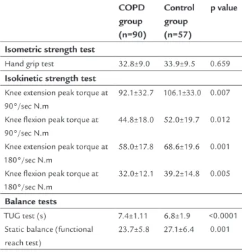
The scores of gait velocity test and TUG test in COPD patients were higher than those of control group (p=0.040 and p=0.000, respectively). The scores of STST, isokinetic strength tests and functional reach test were significantly lower in patients with COPD compared with those of the control group (p≤0.001, Table 2). There was no significant
difference between the patients with COPD and healthy control group regarding hand grip test (p>0.05, Table 2).
TABLE 1 Demographic characteristics, smoking history, lung function and serum 25(OH)D levels of study subjects.
COPD group (n=90)
Control group (n=57)
p value
Age (y)* 60.2±7.8 58.9±6.4 0.116
Gender**
Female 4 (4.5%) 6 (10.5%)
0.154
Male 86 (95.5%) 51 (89.5%)
BMI (kg/m²)* 26.08±4.3 27.24±3.10 0.08
Smoking history**
Never smoker 8 (8.9%) 16 (28.1%)
0.017
Active smoker 40 (44.4%) 18 (31.6%)
Passive smoker 1 (1.1%) 0
Former-smoker 41 (45.6%) 23 (40.4%)
Pack-years***
Duration of smoking 42.7 (0-208) 22.3(0-64) <0.0001
abstinence (years)* 4.7±8.17 4.3±9.0 0.48
Lung function
FVC (L)* 3.2±88 4.2±87 <0.0001
FVC (%) 88.4±19.7 111.9±16 <0.0001
FEV1 (L)* 1.819±681 3.3±66 <0.0001
FEV1 (%) 62.5±20.6 110.2±15.8 <0.0001
FEV1/FVC (%) 55.3±11.1 78.8±5.7 <0.0001
DLCO (mL/mmHg/dk)* 22.7±7.3 31.3±5.6 <0.0001
DLCO (%) 88.0±24.1 118±19.6 <0.0001
PImax (cmH2O)* 56.3±22.9 56.5±24.8 0.95
PImax (%) 52.4±20.6 52.8±22.2 0.89
PEmax (cmH2O)* 71±29 74.3±31 0.52
PEmax (%) 38±24.3 36.8±14.9 0.61
TLC (L)* 5.91±2.6 7.3±1.5 <0.0001
TLC (%) 96.7±50.7 115±19.6 <0.0001
25(OH)D (ng/mL)* 14.5±11.06 16.8±10 0.06
*mean ± SD **n (%)
***median (minimum-maximum)
TABLE 2 Scores of physical performance tests and balance tests of study subjects.
COPD group (n=90)
Control group (n=57)
p value
Physical performance tests
Gait velocity test (s) Sit-to-stand test
27.0±2.6 25.3±7.04
26.3±4.07 31.8±9.7
0.040 <0.0001
TABLE 4 Physical performance tests and balance tests according to serum vitamin D levels in COPD patients.
25(OH)D <15 ng/mL*
25(OH)D 15 ng/mL*
p value
25 (OH)D (ng/mL) 7.8±4 24.5±10.8 <0.0001
Physical performance tests
Gait velocity test (s) Sit-to-stand test (s)
27.1±2.8 24.8±7.7
26.8±2.4 26.1±6
0.07 0.42
Isometric strength test
Hand grip test 30±9.2 37.1±7 <0.0001
Isokinetic strength test
Knee extension peak torque at 90°/sec N.m
Knee flexion peak torque at 90°/sec N.m
Knee extension peak torque at 180°/sec N.m Knee flexion peak torque at 180°/sec N.m
85±29.7
40.7±17.8
55±16.9
30.4±11.5
102.9±34.5
51.0±16.7
62.5±18.5
34.5±12.8 0.01
0.006
0.04
0.15
Balance tests
TUG test (s)
Static balance (functional reach test)
7.6±1 23±6.2
7.3±1 24.7±5.0
0.45 0.09
*mean ± SD
In stage III severe-COPD patients, vitamin D levels were detected to be significantly lower than in the other stag-es (p<0.05).
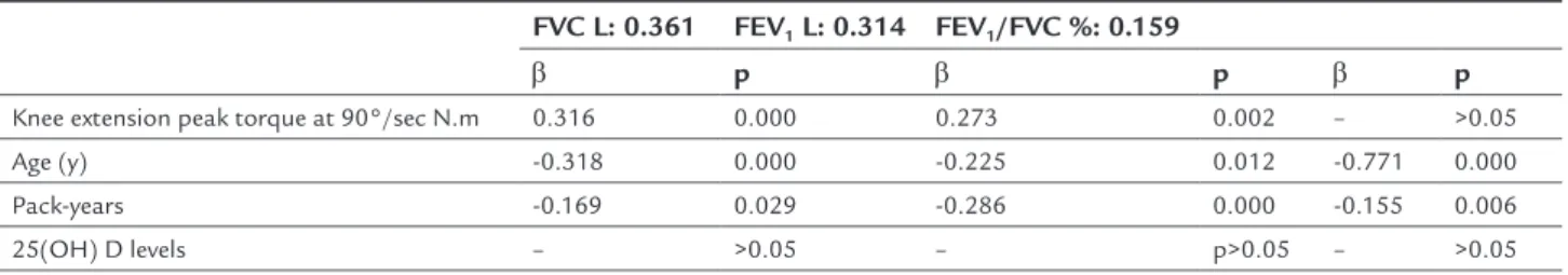
In multiple regression analyses, the independent pre-dictors of FVC in COPD patients were advanced age, pack-years and knee extension peak torque at 90°/sec, whereas 25(OH)D levels had no independent effect. The indepen-dent predictors of FEV1 in COPD patients were age, knee
extension peak torque at 90°/sec and pack-years whereas 25(OH)D levels had no independent effect (Table 5). Age and pack-years were the independent predictors of FEV1/
FVC, whereas knee extension peak torque at 90°/sec and 25(OH)D levels had no independent effect.
25(OH)D levels positively correlated with hand grip test (r= 0.490; p=0.000), knee flexion peak torque at 90°/ sec (r=0.285, p=0.006), FVC (r=0.237, p=0.025), FEV1
(r=0.253, p=0.016), PImax (r=0.214, p=0.049), TLC (r=0.245, p=0.026), and knee extension peak torque at 90° (r=0.254, p=0.016) in COPD patients (Table 6).
D
ISCUSSIONThe present study demonstrated that results of physical performance tests (time up and go, gait velocity test, sit-to-stand test, isokinetic strength), static (functional reach test) and dynamic (time up and go) balance tests in
pa-TABLE 2 (Cont.) Scores of physical performance tests and balance tests of study subjects.
COPD group (n=90)
Control group (n=57)
p value
Isometric strength test
Hand grip test 32.8±9.0 33.9±9.5 0.659
Isokinetic strength test
Knee extension peak torque at 90°/sec N.m
Knee flexion peak torque at 90°/sec N.m
Knee extension peak torque at 180°/sec N.m
Knee flexion peak torque at 180°/sec N.m
92.1±32.7
44.8±18.0
58.0±17.8
32.0±12.1
106.1±33.0
52.0±19.7
68.6±19.6
39.2±14.8 0.007
0.012
0.001
0.005
Balance tests
TUG test (s)
Static balance (functional reach test)
7.4±1.11 23.7±5.8
6.8±1.9 27.1±6.4
<0.0001 0.001
*mean ± SD
Fifty four (60%) of the patients had vitamin D deficiency (mean 25(OH)D: 7.9±3.8 ng/mL) and in 36 (40%) patients’ 25(OH)D levels were >15 ng/mL (mean 25(OH)D: 24.5±10.8 ng/mL).
FVC, FEV1, FEV1/FVC, DLCO levels and TLC were found
to be significantly lower in COPD patients with vitamin D deficiency (p≤0.05), whereas there was no difference be-tween the COPD patients with and without vitamin D de-ficiency, according to PImax and PEmax (p>0.05) (Table 3).
TABLE 3 Lung function according to serum vitamin D levels in COPD patients.
25(OH)D <15 ng/mL*
25(OH)D 15 ng/mL*
p value
FVC (L) 3.08±86 3.47±87 0.02
FEV1 (L) 1.67±67 2.04±63 0.008
FEV1/FVC (%) 53.2±11.4 58.5±9.9 0.04
DLCO mL/mmHg/dk 21.5±7.7 25.1±6.1 0.05
PImax (cmH2O) 51.8±21.9 63.5±22.9 0.20
PEmax (cmH2O) 70.3±29.7 72.1±28.4 0.6
TLC (L) 5.39±1.75 6.74±3.43 0.01
*mean ± SD
tients with COPD were worse than those of controls. Lung functions were significantly lower in COPD patients with vitamin D deficiency than in COPD patients without vi-tamin D deficiency.
It is generally accepted that the combination of sev-eral factors, such as respiratory limitation, elevated oxi-dative stress, systemic inflammation, hypoxemia, frequent steroid intake, age, inactivity, malnutrition and smoking are the main causes for deterioration in muscle function in COPD patients.1,2 Patients with respiratory
dysfunc-tion are at risk of worsening gas exchange and of deteri-oration in functional status if they develop skeletal mus-cle weakness. Kim et al.1 reported that both strength and
endurance of skeletal muscle, limb muscles in particular, are reduced in patients with COPD and that these abnor-malities are associated with attenuated exercise capaci-ty.1 Gosselink et al.16 reported that lung function and
pe-ripheral muscle force are important determinants of exercise capacity in COPD.16
At present, there is no consensus on the optimal 25(OH)D concentration for health. Clinicians variably consider the presence of vitamin D deficiency at levels of 25(OH)D below 20 ng/mL or 30 ng/mL. In contrast, most clinicians agree that vitamin D deficiency is present at 25(OH)D levels less than 15 ng/mL.9,17 In our study, 54
(60%) COPD patients had vitamin D deficiency (less than
TABLE 5 Effective independent variables on airflow rates in COPD patients.
FVC L: 0.361 FEV1 L: 0.314 FEV1/FVC %: 0.159
β p β p β p
Knee extension peak torque at 90°/sec N.m 0.316 0.000 0.273 0.002 – >0.05
Age (y) -0.318 0.000 -0.225 0.012 -0.771 0.000
Pack-years -0.169 0.029 -0.286 0.000 -0.155 0.006
25(OH) D levels – >0.05 – p>0.05 – >0.05
TABLE 6 Relationship between serum vitamin D levels and physical performance tests, balance tests and lung function in COPD patients.
25 (OH)D
r p
Physical performance tests
Gait velocity test (s) Sit-to-stand test (s)
0.011 0.168
0.915 0.113
Isometric strength test
Hand grip test 0.490** <0.0001
Isokinetic strength test
Knee extension peak torque at 90°/sec N.m Knee flexion peak torque at 90°/sec N.m Knee extension peak torque at 180°/sec N.m Knee flexion peak torque at 180°/sec N.m
0.254* 0.285** 0.183 0.149
0.016 0.006 0.084 0.160
Balance tests
TUG test (s)
Static balance (functional reach test)
-0.158 0.198
0.138 0.061
Lung function
FVC (L) 0.237* 0.025
FEV1 (L) 0.253* 0.016
FEV1/FVC (%) 0.169 0.110
DLCO mL/mmHg/dk 0.136 0.323
PImax (cmH2O) 0.214* 0.049
PEmax (cmH2O) 0.074 0.498
TLC (L) 0.245* 0.026
15 ng/mL, mean 25(OH)D: 7.9±3.8 ng/mL). Lung func-tion (FVC, FEV1, FEV1/FVC, DLCO, TLC) was
significant-ly lower in COPD patients with vitamin D deficiency than in COPD patients without vitamin D deficiency, and 25(OH)D levels positively correlated with FVC, FEV1,
PI-max and TLC. Vitamin D deficiency was more common in patients with stage III-severe COPD. Riancho et al.18
reported a significant correlation between vitamin D and the time spent outdoors, suggesting that low exposure to sunlight was the reason for reduced concentrations of vitamin D.18 However, one can speculate that higher
lev-els of vitamin D are associated with better lung functions. The lungs do not grow in size after early adult life.
How-ever, it is likely that tissue remodeling and repair occur in the lungs throughout life. While the number of elas-tic fibers in the alveolar walls decrease with age, type III collagen levels increase with time.19 It was speculated that
vitamin D could influence lung function by influencing tissue remodeling and the synthesis of collagen.20 Lehouck
et al.21 reported that in COPD patients with severe
vita-min D deficiency at baseline vitavita-min D supplementation may reduce incidence of COPD exacerbations.21
In our study, the scores of hand grip test and isokinet-ic strength tests were found to be signifisokinet-icantly lower in COPD patients with vitamin D deficiency. Vitamin D lev-els positively correlated with hand grip test and knee flex-ion and extensflex-ion peak torque at 90°/sec in patients with COPD. Forli et al.22 reported that vitamin D influences
muscle function in patients with advanced pulmonary disease.22 These authors showed that both calcitriol and
PaO2 were significantly associated with handgrip strength.
Our results are in agreement with the results of that study. Taskapan et al.23 showed that vitamin D
supplementa-tion improves muscle strength, funcsupplementa-tional ability and bal-ance in chronic kidney disease (CKD) patients with vita-min D deficiency.23 Whether prevention of vitamin D
deficiency or adequate supplementation in COPD may reverse muscle strength is a burning question.
Vitamin D deficiency has been reported to affect the weight-bearing antigravity lower limbs muscles, which are necessary for postural balance and walking.3 We
demon-strated that balance (TUG and functional reach test) and physical performance (gait velocity test and STST) were worse in COPD patients, however, we could not demon-strate a relationship between serum 25(OH)D levels, bal-ance and physical performbal-ance tests in patients with COPD. Other disease-related factors, such as hypoxemia, dyspnea and fatigue, can affect balance in patients with COPD.24
In our study, in multiple regression analyses, the inde-pendent predictors of FVC were age, knee extension peak
torque at 90°/sec, and pack-years. The independent pre-dictors of FEV1 were age, knee extension peak torque at
90°/sec and pack-years. Age and pack-years were the inde-pendent predictor of FEV1/FVC in COPD patients. COPD
usually develops with advanced age and in the male gen-der. It has been established that limb muscles of older in-dividuals are significantly smaller.25 Alterations in muscle
strength in COPD patients primarily involve the lower limb muscles, with quadriceps muscle strength being 20-30% lower in COPD patients.16,26 The degree of reduced limb
muscle strength correlates with the severity of COPD. Ca-pacity of the muscle to maintain a certain force over time (endurance) of limb muscles is attenuated by about 30% in patients with moderate COPD, and poor muscle endur-ance in COPD correlates positively with FEV1 and resting
partial pressure of oxygen in arterial blood.27,28 Our
find-ings are consistent with the results of these studies. The present study has several limitations. First, the sample size was relatively small. Second, we evaluated the association of serum 25(OH)D levels with lung and mus-cle functions in patients with COPD, however, we did not assess whether vitamin D supplementation can improve lung muscle and muscle functions in patients with COPD.
C
ONCLUSIONThere is peripheral muscle dysfunction in COPD patients. Reduced lung and peripheral muscle functions are more pronounced in COPD patients with vitamin D deficiency.
A
CKNOWLEDGMENTThis study was supported by Inonu University Medical Research Center and Ethics Committee (Approval num-ber and date: 2009-162, 12/22/2009).
R
ESUMORelação entre vitamina D e função pulmonar, desempe-nho físico e equilíbrio em pacientes em estágio I a III de doença pulmonar obstrutiva crônica.
Objetivos: a vitamina D é importante para a função
mus-cular e afeta diferentes aspectos do metabolismo muscu-lar. O objetivo é determinar se os níveis séricos de 25 (OH) D estão relacionados com as funções pulmonares, desem-penho físico e equilíbrio em pacientes com doença pul-monar obstrutiva crônica (DPOC).
Métodos: em 90 pacientes com DPOC e 57 controles
estática (teste de alcance funcional) e dinâmica (tempo de levantar e ir) de equilíbrio foram realizados; e foram ava-liados a associação de níveis de 25 (OH) D com as funções pulmonares, desempenho físico e equilíbrio.
Resultados: os pacientes com DPOC apresentaram
sig-nificativamente mais déficit nos parâmetros de função e equilíbrio físico, e no teste de equilíbrio dinâmico (p<0,005). Força muscular isocinética do joelho (flexores e extenso-res) em pacientes com DPOC foi significativamente me-nor do que nos controles (p<0,05); VEF1 (p=0,008), CVF (p=0,02), VEF1/CVF (p=0,04), CPT (p=0,01) foram mais baixos em pacientes com DPOC e com deficiência de vi-tamina D [25 (OH) D menor do que 15 ng/ml] do que em pacientes com DPOC sem deficiência de vitamina D. Os resultados do teste da força de preensão manual (p=0,000) e força muscular isocinética do joelho (flexor e extensor) (p<0,05) também foram menores nos pacien-tes com DPOC e com deficiência de vitamina D. A defi-ciência de vitamina D foi mais pronunciada em pacien-tes em estágio III da DPOC (p<0,05).
Conclusão: pacientes com DPOC tiveram pior
desempe-nho físico, falta de equilíbrio e menor força muscular. Perturbações graves das funções pulmonares e muscula-res periféricas são mais pronunciadas em pacientes com DPOC e com deficiência de vitamina D.
Palavras-chave: doença pulmonar obstrutiva crônica,
de-ficiência de vitamina D, função muscular.
R
EFERENCES1. Kim HC, Mofarrahi M, Hussain SNA. Skeletal muscle dysfunction in patients with chronic obstructive pulmonary disease. Int J Chron Obstruct Pulmon Dis. 2008;3:637-58.
2. Janssens W, Lehouck A, Carremans C, Bouillon R, Mathieu C, Decramer M. Vitamin D beyond bones in chronic obstructive pulmonary disease. Am J Respir Crit Care Med. 2009;179:630-6.
3. Russell JA. Osteomalacic myopaty. Muscle Nerve. 1994;17:578-80. 4. Koechlin C, Maltais F, Saey D, Michaud A LeBlanc P, Hayot M, Prefaut C.
Hypoxemia enhances peripheral muscle oxidative stres in chronic obstructive pulmonary disease. Thorax. 2005;60:834-41.
5. Black PN, Scragg R: Relationship between serum 25-hydroxyvitamin D and pulmonary function in third national health and nutrition examination survey. Chest. 2005;128:3792-8.
6. Wright RJ, Cohen RT, Cohen S. The impact of stress on the development and expression of atopy. Curr Opin Allergy Clin Immunol. 2005;5:23-9. 7. Holick MF. Vitamin D deficiency. N Engl J Med. 2007;357:266-81.
8. Global Initiative for Chronic Obstructive Lung Disease. Global strategy for the diagnosis, management and prevention of chronic obstructive pulmonary disease 2001, 2006, 2009(update). Availale from://www. goldcopd.com.
9. National Kidney Foundation. K/DOQI clinical practice guidelines for bone metabolism and disease in chronic kidney disease. Am J Kidney Dis. 2003;42(4 Suppl 3):S1-201.
10. American Thoracic Society: Standartized lung function testing: 1987 Update. Am Rev Respir Dis. 1987;136:1285-98.
11. Quanjer PH, Tammeling GJ, Cotes JE, Pederson OF, Peslin R, Yernault JC. Lung volumes and forced ventilatory flows. Report Working Party Standardization of Lung Function Tests, European Community for Steel and Coal. Official Statement of the European Respiratory Society. Eur Respir J Suppl 1993; 16: 5: 40.
12. Hayes KW, Johnson ME. Measures of adult general performance Tests. Arthritis Rheum. 2003;49(Suppl 5):S28-S42.
13. Bohannon RW, Smith J, Hull D, Palmeri D, Barnhard R. Deficits in lower extremity muscle and gait performance among renal transplant candidates. Arch Phys Med Rehab. 1995;76:547-51.
14. Mengshoel AM, Forre O, Komnaes HB. Muscle strength and aerobic capacity in primary fibromyalgia. Clin Exp Rheumatol. 1990;8:475-9.
15. Cetin N, Bayramoglu M, Aytar A, Surenkok O, Yemisci OU. Effects of lower-extremity and trunk muscle fatigue on balance. Open Sports Med J. 2008;2:16-22.
16. Gosselink R, Troosters T, Decramer M. Peripheral muscle weakness contributes to exercise limitation in COPD. Am J Respir Crit Care Med. 1996;153:976-80.
17. Andıran N, Çelik N, Akça H, Doğan G. Vitamin D deficiency in children and adolescents. J Clin Res Pediatr Endocrinol. 2012;4:25-9.
18. Riancho JA, Macias JG, Arco CD, Amado JA, Freijanes, Anton MA. Vertebral compression fractures and mineral metabolism in chronic obstructive lung disease. Thorax. 1987;42:962-6.
19. D’Errico A, Scarani P, Colosimo E, Spina M, Grigioni WF, Mancini AM. Changes in the alveolar connective tissue of the ageing lung: an immunohistochemical study. Virchows Arch A Pathol Anat Histopathol. 1989;415:137-44. 20. Dobak J, Grzybowski J, Liu FT, Landon B, Dobke M. 1,25-dihydroxyvitamin
D3 increases collagen production in dermal fibroblasts. J Dermatol Sci. 1994;8:18-24.
21. Lehouck A, Mathieu C, Carremans C, Baeke F, Verhaegen J, Van Eldere J, High doses of vitamin D to reduce exacerbations in chronic obstructive pulmonary disease: a randomized trial. Ann Intern Med. 2012;156:105-14. 22. Forli L, Bjortuft O, Boe J. Vitamin D status in relation to nutritional depletion and musclle function in patients with advanced pulmonary disease. Exp Lung Res. 2009;35:524-38.
23. Taskapan H, Baysal O, Karahan D, Durmus B, Altay Z, Ulutas O. Vitamin D and muscle strength, functional ability and balance in peritoneal dialysis patients with vitamin D deficiency. Clin Nephrol. 2011;76:110-6. 24. Ozalevli S, Ilgın D, Narin S, Akkoçlu A. Association between disease-related
factors and balance and falls among the elderly with COPD: a cross-sectional study. Aging Clin Exp Res. 2011;23:372-7.
25. Lexell J. Human aging, muscle mass, and fiber type composition. J Gerontol A Biol Sci Med Sci. 1995;50:11-6.
26. Franssen FM, Broekhuizen R, Janssen PP, Wouters EF, Schols AM. Limb muscle dysfunction in COPD: effects of muscle wasting and exercise training. Med Sci Sports Exerc. 2005;37:2-9.
27. Serres I, Gautier V, Prefaut C, Varray A. Impaired skeletal muscle-endurace related to physical inactivity and altered lung funtion in COPD patients. Chest. 1998;113:900-5.