ABSTRACT
Objective: To evaluate diaphragmatic mobility in relation to lung function, respiratory muscle strength, dyspnea, and physical activity in daily life (PADL) in patients with COPD. Methods: We included 25 patients with COPD, classiied according to the Global Initiative for Chronic Obstructive Lung Disease criteria, and 25 healthy individuals. For all of the participants, the following were evaluated: anthropometric variables, spirometric parameters, respiratory muscle strength, diaphragmatic mobility (by X-ray), PADL, and the perception of dyspnea. Results: In the COPD group, diaphragmatic mobility was found to correlate with lung function variables, inspiratory muscle strength, and the perception of dyspnea, whereas it did not correlate with expiratory muscle strength or PADL. Conclusions: In patients with COPD, diaphragmatic mobility seems to be associated with airway obstruction and lung hyperinlation, as well as with ventilatory capacity and the perception of dyspnea, although not with PADL.
Keywords: Pulmonary disease, chronic obstructive; Diaphragm; Spirometry; Dyspnea; Maximal respiratory pressures.
Diaphragmatic mobility: relationship with
lung function, respiratory muscle strength,
dyspnea, and physical activity in daily life in
patients with COPD
Flávia Roberta Rocha1, Ana Karla Vieira Brüggemann1, Davi de Souza Francisco1,
Caroline Semprebom de Medeiros1, Danielle Rosal2, Elaine Paulin1
Correspondence to:
Ana Karla Vieira Brüggemann. Laboratório de Fisioterapia Respiratória, Centro de Ciências da Saúde e do Esporte, Universidade do Estado de Santa Catarina – CEFID/ UDESC – Rua Pascoal Simone, 358, Coqueiros, CEP 88080-350, Florianópolis, SC, Brasil.
Tel.: 55 48 3664-8602 or 55 48 9665-8289. E-mail: [email protected] Financial support: None.
INTRODUCTION
Several studies have shown a decrease in diaphragmatic mobility (DM) in patients with COPD.(1-4) However, COPD also has signiicant extrapulmonary effects,(5) which result in systemic inlammation, loss of muscle mass,(6,7)
malnutrition, depression,(8) physical deconditioning,(9)
and, consequently, reduced health status.(10)
Despite signiicant systemic involvement in patients with COPD, few studies have investigated the relationship
between DM and the systemic changes caused by COPD.
However, the relationship of DM with the six-minute walk
distance,(2,11) dyspnea,(2) and mortality(12) has previously
been described.
Several studies have shown a relationship between
DM and lung function changes.(3,11,13) Recently, Davachi
et al.(11) found that DM was greater in patients classiied as having mild COPD than in those classiied as having very severe COPD. They also found that DM was related
to FVC and slow VC (SVC). It has previously been shown that DM is associated with air trapping,(3,14,15) maximal
voluntary ventilation (MVV),(3) and lung hyperinlation. (1)
These results support the hypothesis that decreased DM is related to lung disease severity.
To date, no studies have investigated the relationship
between DM and physical activity in daily life (PADL), and few have related DM to dyspnea and lung function. (2,3) Therefore, the primary objective of the present study was
to evaluate DM in relation to lung function, dyspnea, and PADL in patients with COPD. A secondary objective was to
compare COPD patients and healthy individuals in terms
of lung function, respiratory muscle strength, and DM.
METHODS
This was a quantitative descriptive cross-sectional study. It was approved by the local human research ethics
committee (Protocol no. CAEE 08871312.7.0000.0118). All participants gave written informed consent. The study sample consisted of 25 patients with COPD (14 males and 11 females) and 25 healthy individuals (5 males and 20 females). We included patients diagnosed with COPD in accordance with the 2015 Global Initiative for Chronic Obstructive Lung Disease criteria(9) and meeting the following criteria: 1) having no associated pulmonary, cardiovascular, or musculoskeletal diseases; 2) having
participated in no training programs in the 6 months prior to
the study; 3) requiring no oxygen therapy supplementation; and 4) being a nonsmoker. The criteria for inclusion of healthy individuals in the present study were as follows: 1) having normal pulmonary function test results (FEV1/ FVC ≥ 0.7; FEV1 ≥ 80% of predicted; and FVC ≥ 80% of predicted); 2) being a nonsmoker; and 3) having no
cardiorespiratory, hepatic, neurological, or oncologic
diseases. The exclusion criteria were as follows: 1) being unable to perform any of the required tests (being unable to understand the instructions or being uncooperative); 1. Curso de Fisioterapia, Universidade do
Estado de Santa Catarina – UDESC – Florianópolis (SC) Brasil.
2. Fundação Universidade Regional de Blumenau – FURB – Blumenau (SC) Brasil.
Submitted: 22 March 2016.
Accepted: 31 October 2016.
2) experiencing COPD exacerbation during the study period; 3) having cardiorespiratory or musculoskeletal complications during the tests; and 4) having a body mass index (BMI) > 30 kg/m2 (i.e., being obese).
Spirometry and respiratory muscle strength Spirometry was performed with a previously calibrated portable digital spirometer (EasyOne®; ndd Medical Technologies, Andover, MA, USA), in accordance
with the methods and criteria recommended by
the American Thoracic Society and the European
Respiratory Society. (16) The following parameters were measured: FVC; FEV1; FEV1/FVC before and 15 min after inhalation of a bronchodilator (albuterol, 400 µg); and inspiratory capacity (IC). A minimum of three acceptable maneuvers and two reproducible maneuvers were performed; for IC, however, the average of three maneuvers was used, as reported by
Miller et al.(16) All spirometric variables are expressed as absolute values and as a percentage of reference
values, in accordance with Pereira et al.(17)
A digital manometer (MVD500®; Globalmed, Porto Alegre, Brazil) attached to a mouthpiece with an air outlet of 1 mm in diameter was used in order to measure inspiratory and expiratory muscle strength. MIP and MEP were measured as indicators of inspiratory and expiratory muscle strength, respectively, in accordance with the Brazilian Thoracic Association guidelines. (18) MIP was measured after a maximal expiratory maneuver (near RV), whereas MEP was measured after a maximal inspiratory maneuver (near TLC). A minimum of three acceptable maneuvers and two reproducible maneuvers were performed. The values of MIP and MEP are expressed as absolute values and as a percentage of reference values, in accordance
with Neder et al.(19) The average of the reproducible
maneuvers was used in the present study.
DM
Patients initially underwent familiarization with dia
-phragmatic breathing for dia-phragmatic proprioception and maximal evaluation of diaphragm amplitude during radiographic examination. Patients were asked to perform two series of ten repetitions of diaphragmatic
breathing, proprioceptive stimulation being provided by placing their hands on their chest and abdomen and verbal encouragement being provided in order to enable patients to direct the air toward the lung bases,
in accordance with Leal.(20)
After having become familiar with diaphragmatic breathing, patients performed three SVC maneuvers using a Wright spirometer (Ferraris Medical Ltd., Hertford, England). SVC maneuvers were performed from TLC to RV and from RV to TLC. The highest value was recorded for comparison with the value obtained during the evaluation of DM, in order to determine whether patient respiratory effort was the same before
and during DM evaluation.
After having become familiar with the diaphragm and having performed all SVC maneuvers, patients
underwent DM evaluation by anteroposterior chest
X-rays, which were taken with patients lying supine on a luoroscopy table. A radiopaque ruler was placed longitudinally under the trunk in the craniocaudal direction, near the thoracoabdominal junction, for subsequent correction of the magniication caused by the divergence of the X-rays. The same ilm was used for all examinations, which were performed during a maximal inspiratory maneuver and a maximal expiratory maneuver.
DM was measured by the method of Saltiel et al.(21): a straight line was drawn from the highest point of the hemidiaphragm during exhalation to the hemidiaphragm during inhalation with the use of a 150-mm digital caliper (Messen; Sensor Technology Co., Guangdong, China; Figure 1).
Dyspnea
Dyspnea was measured with the modiied Medical
Research Council dyspnea scale,(22) the degree of dyspnea ranging from 0 (no dyspnea) to 4 (very severe
dyspnea). Patients were instructed to select the number
that best represented their perception of dyspnea.
PADL
PADL was evaluated with a triaxial accelerometer (DynaPort activity monitor; McRoberts, The Hague, the
Netherlands), which is a small, lightweight device worn on a belt around the waist. It can distinguish among
activities such as sitting, reclining, and walking, and it
measures the time spent in each activity.(23) Patients were monitored 12 h per day for two consecutive
days, patient monitoring beginning immediately
after waking. Patients were subsequently classiied as active (on the basis of the time spent walking) or sedentary (on the basis of the time spent sitting or lying down), the average of the two days being used for analysis. Patients were instructed on how to
position the device and received a manual with clear
instructions and explanatory illustrations. In addition, they were asked not to change their daily activities
while wearing the device.
Sample size calculation
The power of the sample, which consisted of 25 patients, was calculated post hoc with the free statistical software program G*Power, version 3.1.9.2.
Statistics
All statistical analyses were performed with the IBM SPSS Statistics software package, version 20.0 (IBM Corporation, Armonk, NY, USA), descriptive (mean and standard deviation) and inferential statistics being used. Data normality was veriied with the Shapiro-Wilk test. Either a parametric test or a nonparametric test was used
depending on the data distribution. The independent
of dyspnea, and PADL, Pearson’s and Spearman’s correlation coeficients were used for parametric and
nonparametric variables, respectively. The magnitude
of the correlations was described in accordance with
Dancey and Reidy,(24) values of r = 0.10-0.39 indicating a weak correlation, value of r = 0.40-0.69 indicating a moderate correlation, and values of r = 0.70-1.00 indicating a strong correlation. The level of signiicance was set at 5% (p < 0.05) for all tests.
RESULTS
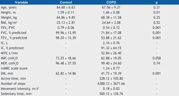
A total of 25 COPD patients and 25 healthy individuals who did not differ in terms of age, weight, or respiratory
muscle strength participated in the present study.
Although the individuals in the control group were classiied as being overweight and those in the COPD group were classiied as being normal weight,(25) there was no statistically signiicant difference between the two groups regarding the BMI. There were differences between the two groups regarding height, FEV1/FVC,
FEV1, FVC, and DM. The results for the two groups are presented in Table 1.
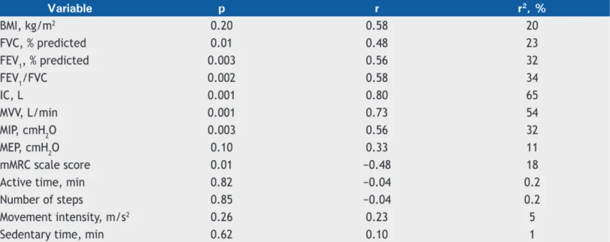
The coeficients of determination for DM and the
study variables in the COPD group are presented in
Table 2. For a signiicance level of 5%, the following powers were found: 0.96 for FEV1/FVC; 0.95 for FEV1; 0.84 for FVC; 0.99 for IC; 0.99 for MVV; 0.95 for MIP; and 0.73 for the perception of dyspnea. Given that neither MEP nor PADL correlated with DM, neither
variable was used.
In the COPD group, DM correlated moderately with
lung function, inspiratory muscle strength, and the perception of dyspnea. In addition, it correlated strongly
with MVV and IC. In the control group, DM did not
correlate with any of the lung function or respiratory
muscle strength variables (Table 2).
DISCUSSION
In the present study, DM was found to correlate moderately with FEV1 and strongly with IC in COPD
Figure 1. Anteroposterior chest X-rays. In A, chest X-ray taken during a maximal expiratory maneuver; in B, chest X-ray taken during a maximal inspiratory maneuver; and in C, superimposition of the two aforementioned images, the image of the radiographic ruler being used as reference to assess diaphragmatic mobility.
A B C
Table 1. Anthropometric, spirometric, and functional characteristics of the groups studied.
Variable Control COPD p
Age, years 64.68 ± 6.63 67.56 ± 9.21 0.21
Height, m 1.59 ± 0.11 1.66 ± 0.08 0.01
Weight, kg 64.86 ± 9.85 68.38 ± 11.58 0.25
BMI, kg/m2 25.13 ± 2.81 24.64 ± 3.08 0.52
FEV1/FVC 0.79 ± 0.06 0.54 ± 0.12 0.001
FVC, % predicted 99.96 ± 13.95 71.84 ± 17.08 0.001
FEV1, % predicted 98.20 ± 12.39 53.88 ± 21.62 0.001
IC, L - 2.16 ± 0.76
-IC, % predicted - 91.32 ± 64.15
-MVV, L/min - 52.84 ± 26.40
-MIP, cmH2O 73.25 ± 18.66 62.88 ± 19.05 0.058
MEP, cmH2O 96.48 ± 37.55 99.40 ± 24.60 0.74
mMRC scale score 1.24 ± 0.77
-DM, mm 62.82 ± 14.86 41.73 ± 19.39 0.001
Active time, min - 228.12 ± 105.80
-Number of steps - 6388.12 ± 3671.66
-Movement intensity, m/s2 - 0.18 ± 0.03
-Sedentary time, min - 500.12 ± 128.76
patients, which is possibly due to the fact that increased airlow obstruction, as assessed by FEV1, and static lung hyperinlation, as assessed by IC, increase the workloads that affect the chest wall, placing the diaphragm at
a geometric and mechanical disadvantage.(26,27) In addition, lung hyperinlation reduces the ability of the diaphragm to generate low and pressure,(28) resulting in decreased diaphragmatic excursion.(29)
Lung hyperinlation is one of the primary changes in patients with COPD; however, air trapping is the principal factor limiting DM in such patients.(3)
Structural changes result in diaphragm remodeling,
which results in lattening of the diaphragm and, consequently, decreased diaphragmatic excursion.(30) The aforementioned changes explain the differences in DM and lung function between COPD patients and healthy individuals; they were expected and have
previously been reported.(1-3,29)
In the present study, a strong correlation was found
between DM and MVV in patients with COPD, showing that a greater DM translates to a better ventilatory
capacity. This inding is in agreement with those of
Kang et al.,(31) who found a signiicant correlation
between DM and MVV and posited that there might be a relationship between decreased DM and hypercapnia in patients with COPD.
In patients with COPD, airlow limitation during exercise is due to reduced ventilatory capacity
associated with increased pulmonary obstruction and,
consequently, lung hyperinlation, as evidenced by
reduced IC and ventilatory reserve.(32) In the present
study, in which patients with moderate to severe obstruction participated, DM correlated moderately
with IC, which also accounted for 65% of the variation in DM, reinforcing the inluence of lung hyperinlation on diaphragmatic mechanics. However, it is known that the inluence of air trapping on DM can be greater than that of lung hyperinlation itself.(3)
Although DM has been shown to correlate with
parameters such as pulmonary obstruction, lung
hyperinlation,(33) and air trapping,(3) Davachi et
al.(11) found no relationship between DM and lung hyperinlation, which is possibly due to the fact that
they selected patients with less severe COPD and,
consequently, reduced airlow obstruction, resulting
in less damage to the diaphragm.
In the present study, a moderate negative correlation
was found between DM and the perception of dyspnea
in patients with COPD, indicating that changes in the
position of the diaphragm make ventilation dificult,
reducing respiratory capacity and increasing the
sensation of dyspnea.(34) These indings corroborate those of Paulin et al.,(2) who found that patients with decreased DM had a greater sensation of dyspnea after submaximal exercise.
Although no correlation was found between PADL
and DM in the COPD patients in the present study, it
is known that exercise capacity decreases with the progression of the disease.(35) This creates a vicious cycle of increasing dyspnea during physical activity,
leading to physical inactivity, decreased physical
conditioning, and an increased number of comorbidities and hospitalizations.(36) It has been shown that, in
comparison with healthy, sedentary elderly individuals, most COPD patients spend more time sitting or lying
down than walking or standing(23,37); however, to date,
no studies have established a relationship between DM
and PADL in COPD patients.
It is of note that assessment of PADL by means of a triaxial accelerometer reveals how much individuals are physically active or inactive in their daily life.(38) However, assessment of PADL with a triaxial accel
-erometer probably depends on several factors other than DM evaluation, and this might explain the lack of correlation between these variables. In addition, it is possible that the number of patients in the study sample and the evaluation period were insuficient to
observe this relationship.
In the present study, no relationship was found between DM and the BMI. Kantarci et al.(39) performed a multiple regression analysis and found that waist circumference apparently plays a more signiicant role
Table 2. Relationship between diaphragmatic mobility and the study variables in the COPD group.
Variable p r r2, %
BMI, kg/m2 0.20 0.58 20
FVC, % predicted 0.01 0.48 23
FEV1, % predicted 0.003 0.56 32
FEV1/FVC 0.002 0.58 34
IC, L 0.001 0.80 65
MVV, L/min 0.001 0.73 54
MIP, cmH2O 0.003 0.56 32
MEP, cmH2O 0.10 0.33 11
mMRC scale score 0.01 −0.48 18
Active time, min 0.82 −0.04 0.2
Number of steps 0.85 −0.04 0.2
Movement intensity, m/s2 0.26 0.23 5
Sedentary time, min 0.62 0.10 1
in the evaluation of DM than does the BMI, a inding that suggests that, although the BMI is a good indicator of nutritional status, it does not relect individual differences in body composition, such as abdominal fat distribution.
We found a relationship between MIP and DM that can be explained by the mechanical disadvantage in which the diaphragm is as a result of air trapping, which leads the inspiratory muscles to work in a shortened position, thus affecting their potential for contraction. (30)
Kodric et al.(40) showed that inspiratory muscle training improved DM in patients with diaphragmatic dysfunction following cardiac surgery, a inding that suggests a
relationship between improved MIP and DM.
Our results suggest that DM is a parameter that can
provide information on respiratory mechanics in patients
with COPD and that is related to certain pulmonary
parameters (FEV1, FEV1/FVC, FVC, IC, and MVV) and
functional parameters. However, studies involving a higher number of patients are needed in order to examine the relationship between DM and PADL.
One potential limitation of the present study is that
no stage I COPD patients were evaluated. However, this is a common problem in the literature, given that stage I COPD patients are usually asymptomatic
and, consequently, do not seek medical attention.
Nevertheless, the results obtained in the present study
cannot be extrapolated to all stages of COPD severity.
In addition, the posture adopted during DM evaluation
might inluence the result obtained; therefore, we
suggest that DM be evaluated in the orthostatic and
supine positions in future studies.
In summary, in patients with COPD, DM is related
to airway obstruction, lung hyperinlation, ventilatory capacity, and the perception of dyspnea. However, it appears to have no relationship with PADL.
REFERENCES
1. Iwasawa T, Kagei S, Gotoh T, Yoshiike Y, Matsushita K, Kurihara H, et
al. Magnetic resonance analysis of abnormal diaphragmatic motion in patients with emphysema. Eur Respir J. 2002;19(2):225-31. https:// doi.org/10.1183/09031936.02.00044602
2. Paulin E, Yamaguti WP, Chammas MC, Shibao S, Stelmach R, Cukier
A, et al. Inluence of diaphragmatic mobility on exercise tolerance
and dyspnea in patients with COPD. Respir Med. 2007;101(10): 2113-8. https://doi.org/10.1016/j.rmed.2007.05.024
3. Dos Santos Yamaguti WP, Paulin E, Shibao S, Chammas MC, Salge
JM, Ribeiro M, et al. Air trapping: The major factor limiting diaphragm mobility in chronic obstructive pulmonary disease patients. Respirology. 2008;13(1):138-44. https://doi.org/10.1111/j.1440-1843.2007.01194.x
4. Yamaguti WP, Claudino RC, Neto AP, Chammas MC, Gomes AC,
Salge JM, et al. Diaphragmatic breathing training program improves abdominal motion during natural breathing in patients with chronic obstructive pulmonary disease: a randomized controlled trial. Arch Physic Med Rehab. 2012;93(4):571-7. https://doi.org/10.1016/j. apmr.2011.11.026
5. Sociedade Brasileira de Pneumologia e Tisiologia. II Consenso Brasileiro sobre Doença Pulmonar Obstrutiva Crônica - DPOC - 2004. J Bras Pneumol. 2004;30(Suppl 5):S1-S42.
6. Watz H, Waschki B, Boehme C, Claussen M, Meyer T, Magnussen H. Extrapulmonary effects of chronic obstructive pulmonary disease
on physical activity: a cross-sectional study. Am J Respir Crit Care Med. 2008;177(7):743-51. https://doi.org/10.1164/rccm.200707-1011OC
7. Bossenbroek L, de Greef MH, Wempe JB, Krijnen WP, Ten Hacken
NH. Daily physical activity in patients with chronic obstructive
pulmonary disease: a systematic review. COPD. 2011;8(4):306-19. https://doi.org/10.3109/15412555.2011.578601
8. Rabe KF, Hurd S, Anzueto A, Barnes PJ, Buist SA, Calverley P, et al.
Global strategy for the diagnosis, management, and prevention of chronic obstructive pulmonary disease: GOLD executive summary.
Am J Respir Crit Care Med. 2007;176(6):532-55 https://doi. org/10.1164/rccm.200703-456SO
9. Global Initiative for Chronic Obstructive Lung Disease [homepage on the Internet]. Bethesda: Global Initiative for Chronic Obstructive Lung Disease. [cited 2016 Jan 10]. Global strategy for the diagnosis,
management, and prevention of COPD – 2015. Available from: http:// goldcopd.org/global-strategy-diagnosis-management-prevention-copd-2015/
10. Skumlien S, Hagelund T, Bjørtuft O, Ryg MS. A ield test of
functional status as performance of activities of daily living in COPD patients. Respir Med. 2006;100(2):316-23. https://doi.org/10.1016/j. rmed.2005.04.022
11. Davachi B, Lari SM, Attaran D, Tohidi M, Ghofraniha L, Amini M,
et al. The relationship between diaphragmatic movements in sonographic assessment and disease severity in patients with stable
chronic obstructive pulmonary disease (COPD). J Cardiothorac Med. 2014;2(3):187-92.
12. Yamaguti WP, Paulin E, Salge JM, Chammas MC, Cukier A, Carvalho
CR. Diaphragmatic dysfunction and mortality in patients with COPD. J Bras Pneumol. 2009;35(12):1174-81. https://doi.org/10.1590/ S1806-37132009001200003
13. Scott S, Fuld JP, Carter R, McEntegart M, MacFarlane NG. Diaphragm ultrasonography as an alternative to whole-body plethysmography in pulmonary function testing. J Ultrasound Med. 2006;25(2):225-32. 14. Decramer M, Jiang TX, Demedts M. Effects of acute hyperinlation
on chest wall mechanics in dogs. J Appl Physiol (1985). 1987;63(4):1493-8.
15. Sinderby C, Spahija J, Beck J, Kaminski D, Yan S, Comtois N, et
al. Diaphragm activation during exercise in chronic obstructive pulmonary disease. Am J Respir Crit Care Med. 2001;163(7):1637-41. https://doi.org/10.1164/ajrccm.163.7.2007033
16. Miller MR, Hankinson J, Brusasco V, Burgos F, Casaburi R, Coates A, et al. Standardisation of spirometry. Eur Respir J. 2005;26(2):319-38. https://doi.org/10.1183/09031936.05.00034805
17. Pereira CA, Sato T, Rodrigues SC. New reference values for forced spirometry in white adults in Brazil. J Bras Pneumol. 2007;33(4):397-406. https://doi.org/10.1590/S1806-37132007000400008 18. Souza RB. Pressões respiratórias estáticas máximas. In: Sociedade
Brasileira de Pneumologia e Tisiologia. Diretrizes para testes de função pulmonar. J Pneumol. 2002;28(Suppl 3):S155-S165. 19. Neder J, Andreoni S, Lerario M, Nery L. Reference values for lung
function tests. II. Maximal respiratory pressures and voluntary ventilation. Braz J Med Biol Res. 1999;32(6):719-27. https://doi. org/10.1590/S0100-879X1999000600007
20. Leal BCE. Validade e coniabilidade da luoroscopia por radiograia digital: uma nova forma de avaliar a mobilidade diafragmática
[dissertation]. Florianópolis: Universidade do Estado de Santa
Catarina; 2014.
21. Saltiel RV, Grams ST, Pedrini A, Paulin E. High reliability of measure of
diaphragmatic mobility by radiographic method in healthy individuals. Braz J Phys Ther. 2013;17(2):128-36. https://doi.org/10.1590/S1413-35552012005000076
22. Hajiro T, Nishimura K, Tsukino M, Ikeda A, Koyama H, Izumi T. Analysis of clinical methods used to evaluate dyspnea in patients with chronic obstructive pulmonary disease. Am J Respir Crit Care Med. 1998;158(4):1185-9. https://doi.org/10.1164/ajrccm.158.4.9802091 23. Pitta F, Troosters T, Spruit MA, Probst VS, Decramer M, Gosselink R.
Characteristics of physical activities in daily life in chronic obstructive pulmonary disease. Am J Respir Crit Care Med. 2005;171(9):972-7. https://doi.org/10.1164/rccm.200407-855OC
global epidemic. Report of a World Health Organization Consultation. Geneva: World Health Organization; 2000. p. 284-56.
26. De Troyer A. Effect of hyperinlation on the diaphragm. Eur Respir J.
1997;10(3):708-13.
27. Poole DC, Sexton WL, Farkas GA, Powers SK, Reid MB. Diaphragm
structure and function in health and disease. Med Sci Sports Exerc. 1997;29(6):738-54. https://doi.org/10.1097/00005768-199706000-00003
28. McKenzie DK, Butler JE, Gandevia SC. Respiratory muscle
function and activation in chronic obstructive pulmonary disease. J Appl Physiol (1985). 2009;107(2):621-9. https://doi.org/10.1152/ japplphysiol.00163.2009
29. Unal O, Arslan H, Uzun K, Ozbay B, Sakarya ME. Evaluation of diaphragmatic movement with MR luoroscopy in chronic obstructive
pulmonary disease. Clin Imaging. 2000;24(6):347-50. https://doi. org/10.1016/S0899-7071(00)00245-X
30. Reid WD, Samrai B. Respiratory muscle training for patients with chronic obstructive pulmonary disease. Phys Ther. 1995;75(11):996-1005.
31. Kang HW, Kim TO, Lee BR, Yu JY, Chi SY, Ban HJ, et al. Inluence of diaphragmatic mobility on hypercapnia in patients with chronic obstructive pulmonary disease. J Korean Med Sci. 2011;26(9):1209-13. https://doi.org/10.3346/jkms.2011.26.9.1209
32. Freitas CG, Pereira CA, Viegas CA. Inspiratory capacity, exercise limitation, markers of severity, and prognostic factors in chronic obstructive pulmonary disease. J Bras Pneumol. 2007;33(4):389-96. https://doi.org/10.1590/S1806-37132007000400007
33. Iwasawa T, Takahashi H, Ogura T, Asakura A, Gotoh T, Shibata H,
et al. Inluence of the distribution of emphysema on diaphragmatic
motion in patients with chronic obstructive pulmonary disease. Jpn J Radiol. 2011;29(4):256-64. https://doi.org/10.1007/s11604-010-0552-8
34. McConnell AK, Romer LM. Dyspnoea in health and obstructive pulmonary disease: the role of respiratory muscle function and training. Sports Med. 2004;34(2):117-32. https://doi. org/10.2165/00007256-200434020-00005
35. Park SK, Meldrum CA, Larson JL. Subgroup analysis of symptoms and their effect on functioning, exercise capacity, and physical activity in patients with severe chronic obstructive pulmonary
disease. Heart Lung. 2013;42(6):465-72. https://doi.org/10.1016/j.
hrtlng.2013.08.008
36. Esteban C, Quintana JM, Aburto M, Moraza J, Egurrola M, Pérez-Izquierdo J, et al. Impact of changes in physical activity on health-related quality of life among patients with COPD. Eur Respir J. 2010;36(2):292-300. https://doi.org/10.1183/09031936.00021409 37. Hernandes NA, Teixeira Dde C, Probst VS, Brunetto AF, Ramos EM,
Pitta F. Proile of the level of physical activity in the daily lives of
patients with COPD in Brazil. J Bras Pneumol. 2009;35(10):949-56. 38. Brandes M, Rosenbaum D. Correlations between the step activity
monitor and the DynaPort ADL-monitor. Clin Biomech (Bristol, Avon). 2004;19(1):91-4. https://doi.org/10.1016/j.clinbiomech.2003.08.001 39. Kantarci F, Mihmanli I, Demirel MK, Harmanci K, Akman C, Aydogan
F, et al. Normal diaphragmatic motion and the effects of body composition: determination with M-mode sonography. J Ultrasound Med. 2004;23(2):255-60.