171
Luiz Guilherme Azevedo de Freitas
1, Natália Silva de Mesquita
2, Juliana Aquino Teixeira Zorrilla
2, Marcos Pereira de Ávila
31 Retina and Vitreous Department, Santa Luzia Eye Hospital, Recife/PE, Brazil; Post-Graduation Programme in Health Science, Federal University of Goiás, Goiânia/GO, Brazil.
2 Residence Programme in Ophthalmology, Santa Luzia Eye Hospital, Recife/PE, Brazil.
3 Federal University of Goiás, Goiânia/GO, Brazil; Reference Centre in Ophthalmology, Goiânia/GO, Brazil.
Institution: Santa Luzia Eye Hospital, Recife/PE, Brazil.
The authors declare no conflict of interest.
Received for publication 19/9/2013 - Acceped for publication 23/2/2014
C
ASER
EPORTRev Bras Oftalmol. 2014; 73 (3): 171-3
Macular edema post-LASIK treated
with ranibizumab
Edema cistoide de mácula pós-LASIK
tratado com ranibizumabe
R
ESUMOOs autores relatam o caso de uma paciente que desenvolveu edema cistóide de mácula pós-cirurgia para correção de miopia pelo método LASIK. Foi submetida a tratamento com injeções intravítrea de ranibizumabe e apresentou resultado visual satisfatório. Descritores: Edema macular/etiologia; Ceratomileuse assistida por excimer laser in situ; Inibidores da angiogênese; Relatos de casos
A
BSTRACTThe authors report the case of a patient who developed cystoid macular edema after surgery for myopia by LASIK method. She was treated with intravitreal injections of ranibizumab and presented satisfactory visual result.
172
I
NTRODUCTIONL
aser in situ keratomileusis (LASIK) is a surgical procedure used to correct refractive errors with high levels of safety and efficacy.1,2 Most post-LASIK complications arerelated to the refractive outcome or to damage to the cornea and anterior segment. The posterior segment is rarely damaged.3
Excessive mechanical stress has been suggested as a cause of vitreoretinal complications during or after LASIK. Changes occur during application of the suction ring to hold the eyeball while creating the corneal flap.4 This exerts an anterior vector
force on the vitreous body where the anterior hyaloid membrane attaches to the posterior lens capsule. The induced traction can act on areas of high vitreoretinal adhesion (vitreous base and macula) causing retinal rupture and macular hole. In areas of low vitreoretinal adhesion, the vector force can cause vitreous detachment.5-7
Another factor is the time required to create the flap. Prolonged suction can be associated with a high incidence of complications, including posterior segment ischemia due to increased intraocular pressure (IOP).
In LASIK, the microkeratome is the main element related to posterior segment complications, and not excimer laser itself. Posterior vitreous detachment, rhegmatogenous retinal detachment and choroidal neovascularisation with macular haemorrhage are all associated with LASIK, with high myopia as the main risk factor.6,8,9 Macular hole and cystoid macular
oedema (CMO) occur within the first six postoperative months and are more common in female patients.10,11
Diseases of the optic nerve, visual field defects and vascular events are associated with high IOP while using the microkeratome.12 Other complications associated with LASIK
include uveal effusion syndrome, serous detachment of the macula, central serous chorioretinopathy, bilateral choroidal infarction, and reactivation of ocular toxoplasmosis.13-16
Despite the large number of refractive surgeries performed each year and the presence of mechanisms responsible for pos-terior segment complications after LASIK, the incidence of such events is low. Candidates for the procedure should be aware that such complications exist and should undergo fundus examination before and after surgery.
C
ASER
EPORTA 52-year-old female patient underwent LASIK in both eyes (BE) in February 2011. Prior to the surgical procedure she had myopia in both eyes. Two months after surgery she presented low visual acuity (LVA) in the right eye (RE).
She sought our Vitreous and Retina Department one year after surgery. She reported having received treatment soon after the onset of symptoms in the RE. She was treated with ketorolac tromethamine (Acular LS, Allergan) 4 times daily for 3 months, without visual improvement.
Ophthalmic examination showed a best corrected visual acuity of 20/40 in the RE 20/20 in the LE. Biomicroscopy found no changes that could explain the LVA.
Retinal mapping of the RE showed macular discolouration and a thinned retinal pigment epithelium in the fovea. The LE had no macular changes.
Fluorescein angiography of the RE showed macular hyperfluorescence. Optical coherence tomography (OCT) of the RE showed a fluid-filled cystoid space in the macular region characteristic of cystoid macular oedema (ECM) (Figure 1).
Treatment with intravitreal injections of ranibizumab was indicated.
The first injection was applied in February 2012. An OCT performed 58 days after the injection already showed a flat foveal depression and the presence of small cystic spaces (Figure 2).
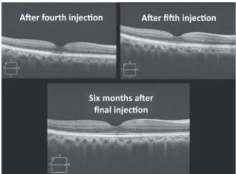
We opted for a new intravitreal injection of ranibizumab in the RE. One month after the second injection, the patient reported a decrease in visual acuity. A new OCT was done, finding a relapse of CMO. Another 3 ranibizumab injections were prescribed, one per month. After the second injection (the fourth in total) a significant reduction of the cyst was noted, without interruption of the internal limiting membrane (Figure 3).
After the fifth and final injection the patient’s visual acuity improved to 20/25 in the RE, and OCT showed a normal foveal depression. The patient then underwent follow up with visits every 6 months (Figure 3).
On examination 6 months after the last ranibizumab injection the patient’s visual acuity remained 20/25 in the RE and 20/20 in the LE. OCT found no anatomical changes in the macula (Figure 3).
Figure 1: Retinography, fluorescein
angiography, and OCT showing cystoid macular oedema before treatment.
Figure 2: Partial improvement of cystoid macular oedema after the first injection, and image showing a relapse of oedema.
Figure 3: Progress of disease throughout treatment and final result.
Freitas LGA, Mesquita NS, Zorrilla JAT, Ávila MP
173
D
ISCUSSIONSurgery to correct refractive errors is becoming increasingly common. LASIK procedures are considered to be safe, but are not exempt from postoperative complications3.
Lin and collaborators17 reported in 2005 a case of bilateral
cystoid macular oedema treated with oral corticosteroids. In our case, we opted not to use corticosteroids because ranibizumab has been showing positive results, and corticosteroids could cau-se undesirable effects such as increacau-sed intraocular pressure and cataract.
Initially it was thought that the patient had a macular microhole, which could explain the relapse of macular fluid. OCT with a high number of slices and in 3D was performed to assess this possibility. This differential diagnosis had to be discarded because the oedema’s pathophysiology and treatment would be entirely different, and ranibizumab would not be effective. The presence of a microhole was discarded, therefore anti-VEGF therapy was resumed.
After a series of injections the patient responded with com-plete resolution of the oedema, and 6 months after the last injection her macular anatomy remained normal.
The result obtained here suggests that treatment with intravitreal injections of ranibizumab can be effective in patients with CMO after LASIK.
R
EFERENCES1. Kohnen T, Steinkamp GW, Schnitzler EM, Baumeister M, Wellermann G, Bühren J, et al. [LASIK with a superior hinge and scanning spot excimer laser ablation for correction of myopia and myopic astigmatism. Results of a prospective study on 100 eyes with a 1-year follow-up]. Ophthalmologe. 2001;98(11):1044-54. German.
2. Sobha S, Rajan MS, Jackson H. Choroidal neovascularization fol-lowing hyperopic LASIK surgery. Clin Experiment Ophthalmol. 2004;32(4):443-5.
3. Loewenstein A, Goldstein M, Lazar M. Retinal pathology occur-ring after excimer laser surgery or phakic intraocular lens im-plantation: evaluation of possible relationship. Surv Ophthalmol. 2002;47(2):125-35. Review.
4. Arevalo JF, Freeman WR, Gomez L. Retina and vitreous pathol-ogy after laser-assisted in situ keratomileusis: is there a cause-effect relationship? Ophthalmology. 2001;108(5):839-40. 5. Luna JD, Artal MN, Reviglio VE, Pelizzari M, Diaz H, Juarez CP.
Vitreoretinal alterations following laser in situ keratomileusis: clinical and experimental studies. Graefes Arch Clin Exp Ophthalmol. 2001;239(6):416-23.
6. Mirshahi A, Schöpfer D, Gerhardt D, Terzi E, Kasper T, Kohnen T. Incidence of posterior vitreous detachment after laser in situ keratomileusis. Graefes Arch Clin Exp Ophthalmol. 2006;244(2):149-53.
7. Smith RJ, Yadarola MB, Pelizzari MF, Luna JD, Juárez CP, Reviglio VE. Complete bilateral vitreous detachment after LASIK retreatment. J Cataract Refract Surg. 2004;30(6):1382-4. 8. Kim HM, Jung HR. Laser assisted in situ keratomileusis for high
myopia. Ophthalmic Surg Lasers. 1996;27(5 Suppl):S508-11. 9. Ozdamar A, Aras C, Sener B, Oncel M, Karacorlu M. Bilateral
retinal detachment associated with giant retinal tear after laser-assisted in situ keratomileusis. Retina. 1998;18(2):176-7 10. Arevalo JF, Mendoza AJ, Velez-Vazquez W, Rodriguez FJ,
Rodriguez A, Rosales-Meneses JL, et al. Full-thickness macular hole after LASIK for the correction of myopia. Ophthalmology. 2005;112(7):1207-12.
11. Arevalo JF, Rodriguez FJ, Rosales-Meneses JL, Dessouki A, Chan CK, Mittra RA, et al. Vitreoretinal surgery for macular hole after laser assisted in situ keratomileusis for the correction of myopia. Br J Ophthalmol. 2005;89(11):1423-6.
12. Bushley DM, Parmley VC, Paglen P. Visual field defect associated with laser in situ keratomileusis. Am J Ophthalmol. 2000;129(5):668-71.
13. Barbara A, Shehadeh-Masha’our R, Sartani G, Garzozi HJ. Reac-tivation of ocular toxoplasmosis after LASIK. J Refract Surg. 2005;21(6):759-61.
14. Butler TK, Sutton G, Moshegov C, McKay DL. Uveal effusion following laser in situ keratomileusis (LASIK) for hypermetro-pia. Am J Ophthalmol. 2004;137(4):763-5.
15. Fontaine F, Fourmaux E, Colin J. [Reactivation of ocular toxo-plasmosis after laser in situ keratomileusis]. J Fr Ophtalmol. 2006;29(5):e11. French.
16. Singhvi A, Dutta M, Sharma N, Pal N, Vajpayee RB. Bilateral serous macular detachment following laser in situ keratomileusis. Am J Ophthalmol. 2004;138(6):1069-71.
17. Lin JM, Tsai YY. Retinal phlebitis after LASIK. J Refract Surg. 2005;21(5):501-4.
Corresponding author:
Luiz Guilherme Freitas Estrada do Encanamento, 909. Casa Forte.Recife/PE, Brazil CEP: 52070-010
Macular edema post-LASIK treated with ranibizumab