·Original article·
Corneal biomechanical properties after femtosecond
laser assisted LASIK with the corneal visualization
Scheimpflug technology and ocular response analyzer
Jing Li, Sheng-Sheng Wei, Ya-Qun Wan, Yong Li, Juan Li, Jing Du, Jian-Guo Liu
Foundation items: Supported by Xi蒺an Social DevelopmentResearch Project Grant [ No. SF1513 (2 )]; Shaanxi Social Development Research Project Grant ( No. 2016SF - 274 ); Shaanxi Provincial Health Research Project (No. 2016E002) Co-first authors: Jing Li and Sheng-Sheng Wei
Refractive surgery Center, Department of Ophthalmology, Xi蒺an No. 4 Hospital, Xi蒺an 710004, Shaanxi Province, China.
Correspondence to: Jian - Guo Liu. Refractive surgery Center, Department of Ophthalmology, Xi蒺an No. 4 Hospital, No. 21, Jiefang Rd, Xi蒺an 710004, Shaanxi Province, China. drljg@ 163. com
Received:2015-11-17摇 摇 Accepted:2016-12-08
飞秒激光制瓣
LASIK
手术后角膜生物力学变
化研究
李摇 晶*,魏升升*, 万雅群, 李摇 勇, 李摇 娟, 杜摇 婧, 刘建国
基金项目:西安市社会发展引导项目[No. SF1513(2)];陕西省 社会发展 项 目 ( No. 2016SF - 274);陕 西 卫 生 科 研 项 目 ( No. 2016E002)
(作者单位:710004 陕西省西安市第四医院眼科激光视力矫正 中心)
*:李晶和魏升升对本文的贡献一致。
作者简介:李晶,西安市第四医院,硕士,主治医师,研究方向:屈 光手术;魏升升,西安市第四医院,硕士,主治医师,研究方向:角
膜屈光手术。
通讯作者:刘建国,西安市第四医院,主任医师,研究方向:屈光 手术、眼科疾病. drljg@ 163. com
摘要
目的:应用可视化角膜生物力学分析仪(Corvis ST) 及眼
反应分析仪(ORA)评估飞秒激光制瓣准分子激光原位角
膜磨镶术(laser in situ keratomileusis,LASIK)术后角膜生
物力学变化,并分析与其他参数的相关性。
方法:收集63例飞秒制瓣LASIK手术患者,所有收集对象
采用右眼数据进行分析。 角膜生物力学测量仪器应用可
视化 角 膜 生 物 力 学 分 析 仪 Corvis ST 及 眼 反 应 分 析 仪
ORA,手术前及手术后1mo进行生物力学数据采集分析。
配对t检验或Mann-Whitney U检验进行手术前后生物力
学对比分析。 Pearson或Spearman统计学方法进行相关性
分析。
结果:与FS-LASIK手术前相比,手术后1stA-time,Vin,2nd
A length,Vout以及 Radius下降,有统计学差异(P= 0. 00, P=0. 00, P=0. 00, P= 0. 00, P= 0. 00)。 2ndA-time,DA以 及PD手术后增加,有统计学差异(P= 0. 00, P= 0. 00, P=
0. 00)。 1stA length以及HC time 手术后改变不明显,无
统计学差异(P= 0. 96,P = 0. 08)。 与FS-LASIK手术前相 比,手术后CH,CRF下降,有统计学差异( P= 0. 00, P= 0郾 00)。 手术后1st A-time,2ndA-time, DA and Radius变
化量与手术前角膜中央厚度有明显相关性(P= 0. 01, P=
0郾 04, P= 0. 03, P= 0. 01)。
结论:飞秒激光LASIK手术后角膜生物力学参数有较明
显改变,可通过Corvis ST及ORA生物力学参数计算得出
相应变化,手术后生物力学参数变化与角膜厚度相关性明显。
关键词:近视;角膜生物力学;飞秒激光;准分子激光原位
角膜磨镶术;可视化角膜生物力学分析仪
引用:李晶,魏升升,万雅群,李勇,李娟,杜婧,刘建国. 飞
秒激光制瓣LASIK手术后角膜生物力学变化研究. 国际眼科杂
志2017;17(2):195-199
Abstract
誗AIM:To investigate the changes of corneal biomechanical properties before and after femtosecond laser assisted LASIK
(FS - LASIK) using Corneal Visualisation Scheimpflug Technology (Corvis ST) and Ocular Response Analyzer
(ORA),and the correlation with other myopic parameters. 誗METHODS:Sixty three patients (63 eyes) who had myopic femtosecond laser assisted LASIK (FS-LASIK)
were enrolled in the study. The right eye of each patient was analyzed in this study. The corneal biomechanical parameters pre-operative and 1mo post-operative was measured with the Corvis ST (Oculus,Wetzlar,Germany)
and ORA(Reichert,Buffalo,New York,USA). Comparison of the biomechanical property values before and after surgery was peformed using Paired t-test or Mann -WhitneyU. Pearson or Spearman correlations were used to evaluate the relationship between parameters.
誗RESULTS: The postoperative 1st A-time, Vin, 2nd A length,Vout,HC time and Radius demonstrate significant decreases comparing with preoperative values(P= 0.00,
coefficient was found between preoperative central corneal thickness(CCT) with postoperative-preoperative changes of 1st A-time, 2nd A-time, DA and Radius respectively(P= 0.01,P= 0.04,P= 0.03,P= 0.01).
誗CONCLUSION: There were significantly changes of corneal biomechanical properties after FS-LASIK surgery. The changes of corneal biomechanical properties after FS -LASIK can be reflected by some parameters of Corvis ST and ORA. The mainly influence of corneal biomechanical alteration was possibly correlation with corneal thickness. 誗KEYWORDS:myopia;corneal biomechanical property;
femtosecond laser;laser in situ keratomileusis;corneal visualization Scheimpflug technology
DOI:10. 3980 / j. issn. 1672-5123. 2017. 2. 01
Citation:Li J, Wei SS, Wan YQ, Li Y, Li J, Du J, Liu JG. Corneal biomechanical properties after femtosecond laser assisted LASIK with the corneal visualization Scheimpflug technology and ocular response analyzer. Guoji Yanke Zazhi(Int Eye Sci) 2017;17 (2):195-199
INTRODUCTION
M
ore and more studies of corneal biomechanical properties are investigated because of the influence of these properties on the predictability and stability of refractive surgery procedures outcomes[1-3]. Previous studies demonstrated that corneal refractive surgery significantly alters the biomechanical properties of the cornea[4-6], which plays an important role in the development of some serious post -operation complications. In some cases, the reduce of corneal biomechanical may contribute to refractive instability and loss of visual acuity, especially keratectasia[7]. Rad et al[8] reported that corneal ectasia represents one of the rarest but also one of the most feared complications with an incidence of 0. 2% to 0. 66% in the literature. Therefore, an effective method of quantifying the biomechanical state of a cornea before and after refractive surgery would be help to reduce the incidence of keratectasia by improving refractive surgery screening. Generally, corneal biomechanics studied by in vitro techniques that assess factors such as viscoelasticity, hydration[9]etc. Until to 2005, the Ocular Response Analyzer (ORA;Reichert Ophthalmic Instruments, Depew, New York, USA), which is based on a dynamic bidirectional applanation process, has enabled us to measure the biomechanical properties of the cornea in clinical[10], such as the corneal hysteresis (CH) and corneal resistance factor ( CRF). This device provides metrics of corneal and / or ocular biomechanics. However, outputs of ORA do not give direct description about the mechanical behavior of cornea. Recently, The Corvis ST ( Corneal Visualisation Scheimpflug Technology, Oculus, Wetzlar, Germany) is a new clinical instrument that allows investigation of the dynamic reaction of the cornea to an air impulse. This novel instrument operates with a Scheimpflug camera taking more than 4 330 images per second along an 8 - mm horizontal corneal coverage during corneal deformation under an air puff indentation. Thecomplete visualization of the deformation process can be shown in a video output. Numerous parameters to describe the corneal viscoelastic properties and stiffness are displayed after internal calculation from the captured video image.
It is common knowledge that there have been some studies on the alteration of corneal biomechanical properties after refractive surgery measured by the ORA[11-13], as well as the repeatability and accuracy in measurement of intraocular pressure (IOP) and central corneal thickness (CCT) by the Corvis ST in healthy subjects, patients with ocular hypertension and glaucoma[14-15]. However, as far as we known, there are few published articles on the alteration of corneal biomechanical properties before and after refractive surgery measured concurrently by the Corvis ST and ORA. Therefore, the purpose of this prospective study was to investigate the biomechanical properties of cornea measured with Corvis ST and ORA before and after laser in situ keratomileusis using femtosecond laser flap creation, and examines the relationship between the corneal biomechanical parameters after femtosecond laser assisted LASIK measured using the Corvis ST and ORA device.
SUBJECTS AND METHODS
Patients摇 Sixty three patients (63 eyes) who had myopic femtosecond laser assisted LASIK (FS-LASIK) were enrolled in the study. Only their right eyes were analyzed. All enrolled patients were confirmed to be having a stable refraction and free of ocular and systemic disease. Contact lens wear was discontinued 2wk before the LASIK operation for soft lenses or 4wk before the LASIK operation for hard lenses. Exclusion criteria were corneal disease, glaucoma, retinal disease, previous intraocular surgery, and any other ocular disease, systemic disease could influence the eye. This study was performed in accordance with the tenets of the Declaration of Helsinki and approved by an Institutional Review Board. The written informed consent was obtained from each patient after receiving an explanation of the benefits and the known risks of the procedure.
Measurements摇 Before the surgical procedure, patients had a complete ophthalmologic examination, including manifest and cycloplegic refraction, uncorrected visual acuity (UCVA) and best spectacle - corrected visual acuity ( BSCVA), and slit - lamp biomicroscopy, ophthalmoscopy through dilated pupils. Best spectacle-corrected visual acuity was 20 / 20 or better in all preoperative eyes. The preoperative central corneal thickness ( CCT) measured by ultrasound and the corneal biomechanical properties measured by Corvis ST and ORA before and 1mo after surgery were recorded.
摇 摇 摇 摇 摇 Table1摇 Preoperative and postoperative findings in corneal biomechanical parameters
Parameter Mean依SDPreoperativeRange Mean依SDPostoperativeRange t(Z),P
1stA-time 7. 37依0. 26 6. 87 to 8. 14 7. 03依0. 29 6. 57 to 7. 98 9. 03,0. 00a
1stA length 1. 79依0. 08 1. 38 to 1. 89 1. 75依0. 15 1. 26 to 1. 92 0. 01,0. 96
Vin 0. 15依0. 02 0. 07 to 0. 18 0. 14依0. 02 0. 03 to 0. 18 13. 70,0. 00a
2ndA-time 21. 72依0. 30 20. 98 to 22. 30 21. 90依0. 44 20. 76 to 22. 65 -3. 07,0. 00a
2ndA length 1. 56依0. 36 0. 78 to 2. 19 1. 36依0. 47 0. 74 to 2. 10 3. 12,0. 00a
Vout -0. 42依0. 07 -0. 66 to -0. 30 -0. 49依0. 09 -0. 72 to -0. 26 6. 84,0. 00a
HC time 16. 72依0. 45 15. 48 to 17. 79 16. 59依0. 38 15. 48 to 17. 56 1. 77,0. 08 DA 1. 06依0. 09 0. 89 to 1. 28 1. 13依0. 13 0. 80 to 1. 43 -4. 78,0. 00a
PD 4. 56依1. 04 2. 37 to 5. 45 4. 69依1. 18 2. 23 to 5. 94 9. 63,0. 00a
Radius 7. 11依0. 83 5. 62 to 10. 37 5. 89依0. 71 2. 21 to 7. 94 10. 74,0. 00a
1stA-time: Time from start until the first applanation;1stA length: Cord length of the cornea in the first applanation;
Vin: Corneal velocity during the first applanation moment;2ndA-time: Time from start until the second applanation;
2ndA length: Cord length of the cornea in the second applanation; Vout: Corneal velocity during the second
applanation moment;HC time: Time from start until the highest concavity of cornea is reached; DA: Maximum deformation amplitude at the corneal apex;PD: Peak distance. aP<0. 05, corneal biomechanical values post-op
were significantly different from pre-op values.
highest concavity[16]. During one measurement process,1st A-time: time from start until the first applanation;1st A length: length of the flattened cornea in the first applanation ;Vin: corneal velocity during the first applanation moment;2ndA -time: time from start until the second applanation; 2nd A length: length of the flattened cornea in the second applanation; Vout: corneal velocity during the second applanation moment;DA: maximum deformation amplitude at the corneal apex;PD: peak distance;Radius can be obtained from the images generated. Furthermore, IOP and corneal pachymetry data are provided in addition to some biomechanical response values[16]. The ORA was used to measure CH and CRF. An experienced ophthalmologist obtained the ORA waveforms, all of which showed symmetric peak heights and similar widths.
Surgical Technique 摇 All flaps were created using a femtosecond laser ( IntraLase Corporation, Irvine, CA). All LASIK procedures were performed by wavefront - guided ablation. The attempted flap thickness was 90 滋m and the flap diameter was 8. 4 mm. After a flap was created, excimer laser ablation was performed with the VISX S4 laser system (Visx USA, Inc. , Santa Clara, CA). After ablation, the corneal flap and stroma surface were cleaned with a balanced salt solution, and the flap was repositioned. The remaining procedures were the same as for the laser ablation.
Statistical Analysis摇 The statistical software used was SPSS 13. 0. All data sets were tested for normality using the Kolmogorov-Smirnov test. Data was expressed as the mean依 standard deviation. Comparison of the preoperative and postoperative biomechanical properties values were performed using Paired t - test or Mann - Whitney U. Pearson or Spearman correlations were used to evaluate the relationship between variables. A P value < 0. 05 was considered to be statistically significant.
RESULTS
The mean age of the 63 patients in this study was 23. 63依4郾 62y (range, 18 to 36 y). Thirty patients (30 eyes) were male and 33 (33 eyes) were female. The mean preoperative spherical equivalent ( SE) of refraction was - 6. 17 依 1. 75 diopters (range, -10. 88 to -2. 50 diopters), with mean astigmatism of -0. 66 依0. 50 diopters ( range, -2郾 00 to 0 diopters). The preoperative central corneal thickness ( CCT) was 531. 05 依 29. 03滋m (range, 492 to 608滋m). The mean ablation depth (AD) was 91. 06依12. 95滋m (range, 52. 20 to 109. 10滋m). The CH was decreased significantly, from 9. 67依1. 34 to 7. 29依 1. 28 (t= 23. 42, P= 0. 00). The CRF was also decreased significantly, from 9. 91依1. 52 to 6. 46依1. 32(t= 20. 18,P= 0. 00 ). Table 1 shows the mean values of all measured preoperative and postoperative corneal biomechanical parameters of Corvis ST. Comparing with preoperative values, the postoperative 1st A - time, Vin, 2nd A length and Vout demonstrate significant decreases after treatment respectively (P = 0. 00, P = 0. 00, P = 0. 00, P = 0. 00 ). The postoperative 2ndA-time significantly increase, however, the 1st A length had no significant difference after surgery. The HC time and Radius had a significant decrease postoperatively (P= 0. 00, P= 0. 00), at the same time, the DA and PD increased significantly (P= 0. 00,P= 0. 00).
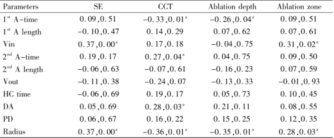
Table2 摇 The correlation of postoperative-preoperative corneal biomechanical parameters with SE,CCT,ablation depth,diameter of ablation zone
Parameters SE CCT Ablation depth Ablation zone 1stA-time 0. 09,0. 51 -0. 33,0. 01a -0. 26,0. 04a 0. 09,0. 51
1stA length -0. 10,0. 47 0. 14,0. 29 0. 07,0. 62 0. 07,0. 61
Vin 0. 37,0. 00a 0. 17,0. 18 -0. 04,0. 75 0. 31,0. 02a
2ndA-time 0. 19,0. 17 0. 27,0. 04a 0. 04,0. 75 0. 09,0. 50
2ndA length -0. 06,0. 63 -0. 07,0. 61 -0. 16,0. 23 0. 07,0. 59
Vout -0. 11,0. 38 -0. 24,0. 07 -0. 13,0. 33 -0. 01,0. 93 HC time -0. 06,0. 69 0. 19,0. 17 0. 05,0. 73 0. 10,0. 45 DA 0. 05,0. 69 0. 28,0. 03a 0. 21,0. 11 0. 08,0. 55
PD 0. 06,0. 67 0. 16,0. 22 0. 15,0. 25 0. 12,0. 35 Radius 0. 37,0. 00a -0. 36,0. 01a -0. 35,0. 01a 0. 28,0. 03a
1stA-time: Time from start until the first applanation;1stA length: Cord length of the cornea in the
first applanation;Vin: Corneal velocity during the first applanation moment;2ndA-time: Time from
start until the second applanation;2ndA length: Cord length of the cornea in the second applanation;
Vout: Corneal velocity during the second applanation moment;HC time: Time from start until the highest concavity of cornea is reached;DA: Maximum deformation amplitude at the corneal apex;PD: Peak distance.aP<0. 05, corneal biomechanical values post-op were significantly different from
pre-op values;SE: Spherical equivalent;CCT: Central corneal thickness. aP<0. 05, changes of corneal
biomechanical values post-op were significantly correlated with procedure parameters.
zone and diameter of ablation zone (Table 2). DISCUSSION
The cornea is a viscoelastic material composited with collagen, proteoglycans, water, and other elements, which can be modeled with quantifiable biomechanical properties[17-18]. Previous studies show that biomechanical properties of the cornea might be changed after myopic femtosecond laser assisted LASIK ( FS - LASIK)[19-20]. Dupps and Roberts[21] thought the reason was predominantly due to an immediate near-circumferential severing of corneal lamellae results in a redistribution of stress and unprogrammed biomechanical shape changes during the surgery. In our study, We also found significant reductions in CH and CRF measurement with ORA at 1mo after LASIK using femtosecond laser for flap creation compared with preoperative. These findings are broadly in line with previous findings from Hamilton et al[1]. Corvis ST is a noncontact tonometer incorporating Scheimpflug technology to measure corneal deformation from air - puff indentation. During the measurement, the cornea reach the maximum deformation amplitude at the corneal apex under the force of air - puff, and rebound to original stations. 1st A -time, 1stA length, Vin, 2ndA-time, 2ndA length, Vout, HC time, DA, PD and Radius were obtained during this course. In previous study[16], the intraexaminer repeatability and intersession reproducibility of Corvis ST were investigated. Only DA and 1st A - time were found repeatable and reproducible, and the DA, as a reliable corneal parameter, could distinguish abnormal corneas from normal corneas. Hon and Lam reported that the CCT, DA, first applanation time (1st A - time ), and IOP were repeatable. In the current study, excepting that the DA was significantly increased after surgical procedures, this was conformed to the result of Hon and Lam[16]. We also demonstrated here that 2nd A - time,
Vout and Peak Distance were increased. In addition, we demonstrated that corneal 1st A -time;Vin;2nd A length and Radius after the surgery captured by Corvis ST were decreased significantly compared with preoperative. Nevertheless, 1st A length and HC time after the surgery were not significant decrease compared with preoperative. For the large variations of some parameters in the measurement might be explained as follows. During the FS-LASIK surgery, the flap creation with femtosecond laser and surgical tissue removal may produce a biochemical parameter related to reduction of corneal stiffness[22-23]. For the DA, the thin cornea after flap creation and tissue ablation could result in decreasing of corneal antagonism to air impulse. The time would be faster from start until the first applanation, at the same time, the deformation amplitude could increase due to the softening of cornea after surgery. Radius could indirectly show the depth of being pressed of cornea. Therefore, the increasing of deformation amplitude could result in reducing of radius. Moreover, the collagen fiber relaxation after a corneal cut may reduce the capacity of the sonic wave to propagate in the corneal surface. The good correlation was demonstrated between biomechanical parameters and CCT in our study. Correlation analysis showed that 1st A - time and radius was negatively correlated with CCT;2ndA-time and DA was positively correlated with CCT. Many previous studies have shown that corneal thickness could influence on deformation amplitude[24-25]. The changes of 1stA -time, radius and 2nd A-time were primarily result from the changing of deformation amplitude ( DA ). Therefore, the CCT plays an important part in the alteration of corneal biomechanical.
and we did not contrasted with 6mo postoperative. We will contrast the forward changing of corneal biomechanical more rigidly in further studies.
In conclusion, there was a significant change in corneal biomechanical following femtosecond laser assisted LASIK (FS-LASIK) procedure. Parameters of Corvis ST and ORA could obviously reflect the changing of corneal biomechanical. CH,CRF, DA and radius had an apparent change in 1mo after surgery. Most of alterations of parameters were correlation with preoperative CCT.
REFERENCES
1 Hamilton DR, Johnson RD, Lee N, Bourla N. Differences in the corneal biomechanical effects of surface ablation compared with laser in situ keratomileusis using a microkeratome or femtosecond laser. J Cataract Refract Surg2008;34(12):2049-2056
2 Kamiya K, Miyata K, Tokunaga T, Kiuchi T, Hiraoka T, Oshika T. Structural analysis of the cornea using scanning-slit corneal topography in eyes undergoing excimer laser refractive surgery. Cornea 2004; 23 (8Suppl):S59-S64
3 Jaycock PD, Lobo L, Ibrahim J, Tyrer J, Marshall J. Interferometric technique to measure biomechanical changes in the cornea induced by refractive surgery. J Cataract Refract Surg2005;31(1):175-184 4 Pepose JS, Feigenbaum SK, Qazi MA, Sanderson JP, Roberts CJ. Changes in corneal biomechanics and intraocular pressure following LASIK using static, dynamic, and noncontact tonometry. Am J Ophthalmol2007;143(1):39-47
5 Dupps WJ Jr, Wilson SE. Biomechanics and wound healing in the cornea. Exp Eye Res2006, 83(4): 709-720
6 Schmack I, Dawson DG, McCarey BE, Waring GO 3rd, Grossniklaus HE, Edelhauser HF. Cohesive tensile strength of human LASIK wounds with histologic, ultrastructural, and clinical correlations. J Refract Surg
2005;21(5):433-445
7 de Medeiros FW, Sinha-Roy A, Alves MR, Wilson SE, Dupps WJ Jr. Differences in the early biomechanical effects of hyperopic and myopic laser in situ keratomileusis. J Cataract Refract Surg2010;36 ( 6 ) : 947 -953
8 Rad AS, Jabbarvand M, Saifi N. Progressive keratectasia after laser in situ keratomileusis. J Refract Surg2004;20(5Suppl):S718-S722 9 Elsheikh A, Wang D, Pye D. Determination of the modulus of elasticity of the human cornea. J Refract Surg2007;23(8):808-818 10 Luce DA. Determining in vivo biomechanical properties of the cornea with an ocular response analyzer. J Cataract Refract Surg2005;31(1): 156-162
11 Chang PY, Chang SW, Wang JY. Assessment of corneal biomechanical properties and intraocular pressure with the Ocular Response Analyzer in childhood myopia. Br J Ophthalmol2010;94(7): 877-881
12 Franco S, Lira M. Biomechanical properties of the cornea measured by the Ocular Response Analyzer and their association with intraocular
pressure and the central corneal curvature. Clin Exp Optom 2009;92 (6):469-475
13 Kotecha A, Elsheikh A, Roberts CR, Zhu H, Garway-Heath DF. Corneal thickness - and age - related biomechanical properties of the cornea measured with the ocular response analyzer. Invest Ophthalmol Vis Sci2006;47(12):5337-5347
14 Hong J, Xu J, Wei A, Deng SX, Cui X, Yu X, Sun X. A new tonometer--the Corvis ST tonometer: clinical comparison with noncontact and Goldmann applanation tonometers. Invest Ophthalmol Vis Sci2013; 54(1):659-665
15 Garcia - Porta N, Fernandes P, Queiros A, Salgado - Borges J, Parafita - Mato M, Gonz佗lez - M佴ijome JM. Cornealbiomechanical properties in different ocular conditions and new measurement techniques. ISRN Ophthalmol2014;2014:724546
16 Hon Y, Lam AK. Corneal deformation measurement using Scheimpflug noncontact tonometry. Optom Vis Sci2013;90(1):e1-e8. 17 Comaish IF, Lawless MA. Progressive post - LASIK keratectasia: biomechanical instability or chronic disease process? J Cataract Refract Surg2002;28(12):2206-2213
18 Guirao A. Theoretical elastic response of the cornea to refractive surgery: risk factors for keratectasia. J Refract Surg 2005;21 ( 2 ) : 176-185
19 Medeiros FW, Sinha - Roy A, Alves MR, Dupps WJ Jr. Biomechanical corneal changes induced by different flap thickness created by femtosecond laser. Clinics (Sao Paulo) 2011;66 ( 6 ) : 1067-1071
20 Uzbek AK, Kamburo姚glu G, Mahmoud AM, Roberts CJ. Change in biomechanical parameters after flap creation using the Intralase femtosecond laser and subsequent excimer laser ablation. Curr Eye Res
2011;36(7):614-619
21 Dupps WJ Jr, Roberts C. Effect of acute biomechanical changes on corneal curvature after photokeratectomy. J Refract Surg2001;17 (6): 658-669
22 Ortiz D, Pi觡ero D, Shabayek MH, Arnalich - Montiel F, Ali佼 JL. Corneal biomechanical properties in normal, post - laser in situ
keratomileusis, and keratoconic eyes. J Cataract Refract Surg2007;33 (8):1371-1375
23 Gatinel D, Chaabouni S, Adam PA, Munck J, Puech M, Hoang -Xuan T. Corneal hysteresis, resistance factor, topography, and pachymetry after corneal lamellar flap. J Refract Surg2007;23(1) : 76-84
24 Dorronsoro C, Pascual D, P佴rez - Merino P, Kling S, Marcos S. Dynamic OCT measurement of corneal deformation by an air puff in normal and cross-linked corneas. Biomed Opt Express2012;3 (3) : 473-487
25 Alonso - Caneiro D, Karnowski K, Kaluzny BJ, Kowalczyk A, Wojtkowski M. Assessment of corneal dynamics with high-speed swept source optical coherence tomography combined with an air puff system.
Opt Express2011;19(15):14188-14199