12 artigo 319
ORIGINAL ARTICLE
IS THERE ANY DIFFERENCE IN THE PROGNOSIS FOR PATIENTS WITH
PRIMARY OSTEOSARCOMA WITH A POOR RESPONSE
TO NEOADJUVANT CHEMOTHERAPY BETWEEN
HUVOS GRADES I AND II?
Rosalvo Zósimo Bispo Júnior1, Olavo Pires de Camargo2
1. MSc and PhD in Orthopedics and Traumatology from the School of Medicine, University of São Paulo (FMUSP). Adjunct Professor, School of Medicine, Federal University of Campina Grande (UFCG), Campina Grande, Brazil.
2. Titular Professor, Department of Orthopedics and Traumatology, School of Medicine, University of São Paulo (DOT/FMUSP), São Paulo, Brazil.
Work performed at the Medical Investigation Laboratory for the Musculoskeletal System (LIM 41), Department of Orthopedics and Traumatology, School of Medicine, University of São Paulo
Correspondence: Campus Universitário - BR-230, km 22 - Caixa Postal 318 – 58053-000 – João Pessoa, PB, Brasil. E-mail: zosimo_jr@yahoo.com.br
Work received for publication: March 30, 2010; accepted for publication: July 17,2010.
ABSTRACT
Objective: Would there be any difference in the prognosis for patients who presented, for example, 8% or 88% tumor necrosis induced by chemotherapy, even though both individuals were considered to be poor responders? The aim of this study was to compare the prognoses for different histological grades (Huvos grade I versus grade II), consequent to chemotherapy, among patients with primary osteosarcoma that was not metastatic at diagnosis. Methods: Twenty-four patients admitted to a referral center between 2000 and 2004 were selected for the study. The accumulated chances of survival were calculated using the Kaplan-Meier technique. Huvos
Rev Bras Ortop. 2011;46(4):420-23
INTRODUCTION
Osteosarcoma is an aggressive malignant type of neoplasia of mesenchymal origin that is characterized by irregular formation of immature bone and production of osteoid matrix and malignant stromal fusiform cells(1). It is one of the
commonest types of primary non-hematopoietic bone sarcoma(2-4), albeit uncommon if compared
with neoplasia in general(2).
In the United States, the annual incidence of osteosarcoma is 6.5 patients per million children, or 900 new cases per year(5). In Brazil, the real incidence
is difficult to determine because of the lack of adequate
grades I and II for necrosis consequent to chemotherapy were evaluated as variables in order to determine their prognostic value, in relation to local recurrence-free survival, metastasis-free survival and overall survival, using the log-rank test. Results: Comparing Huvos grades I and II, the following P values for survival were attained: local recurrence-free survival (P = 0.731), metastasis-free survival (P = 0.596) and overall survival (P = 0.669). Conclusion: In this series, Huvos grades I and II did not have any comparative prognostic value and had similar behavior.
Keywords – Bone Neoplasms/pathology; Drug Therapy;
Prognosis; Survivors
records. However, it has been estimated that there are 350 new cases per year(6).
The appropriate treatment for high-grade malignant osteosarcoma consists of a multidisciplinary approach based on preoperative chemotherapy (neoadjuvant), surgery and postoperative chemotherapy (adjuvant). A recent multi-institutional retrospective study(7)
sug-gested that a combination of surgery and chemothera-py seems to be the standard choice of treatment and, likewise, chemotherapy may function better if used both preoperatively and postoperatively.
The prognosis for patients with osteosarcoma has improved considerably over recent decades. This has
The authors declare that there was no conflict of interest in conducting this work
421
underwent treatment for osteosarcoma. The following inclusion criteria were used: (a) Anatomopathological confirmation of primary osteosarcoma: all the slides were reviewed by a pathologist who was a specialist on musculoskeletal tissues; (b) Patients who were all treated (during the study period) using the same proto-col for neoadjuvant chemotherapy (Study V, GBTO); (c) Patients who underwent operations at our clinic for local control of the primary tumor after the chemo-therapy; (d) Patients who were considered to be poor responders to the preoperative chemotherapy that was instituted (grades I and II according to the classifica-tion of Huvos et al(22). The following exclusion
cri-teria were used: (a) Presence of pulmonary or bone metastases at the initial assessment; (b) Presence of tumors with low-grade malignity; (c) Patients who did not undergo neoadjuvant chemotherapy or who died during this therapy; (d) Patients who had undergone some form of treatment for the tumor prior to regis-tration at our clinic. The anatomopathological review was conducted using paraffin blocks containing tumor fragments from the surgical specimen, in all the cases.
This protocol was approved by our institution’s
research ethics committee.
STATISTICAL ANALYSIS
The study database was compiled in a spreadsheet within the Excel 2007 software for Windows, after applying a protocol to the medical files. The statistical analyses were performed using the SPSS statistical software, version 12.0 (SPSS Inc., Chicago, IL, USA).
The accumulated survival probabilities were calcu-lated by means of the Kaplan-Meier technique. Huvos grades I and II, for the degree of necrosis after the effect of the chemotherapy, were evaluated as varia-bles to determine their prognostic value in relation to survival free from local recurrence, survival free from metastasis and overall survival, using the Log-Rank test. Statistical significance was defined as p < 0.05.
RESULTS
In the sample of 24 cases, the histological respon-ses to preoperative chemotherapy according to the criteria of Huvos et al. (1977) comprised 18 cases (75.0%) with grade I and six cases (25.0%) with grade II (Figure 1). The mean length of follow-up among
IS THERE ANY DIFFERENCE IN THE PROGNOSIS FOR PATIENTS WITH PRIMARY OSTEOSARCOMA WITH A POOR RESPONSE TO NEOADJUVANT CHEMOTHERAPY BETWEEN HUVOS GRADES I AND II?
Rev Bras Ortop. 2011;46(4):420-23
occurred through adequate staging of these tumors, improved imaging methods and, especially, polyche-motherapy. Today, the five-year disease-free survival rate has reached 60-70%, using the most recent che-motherapy protocols(8-12).
Poor histological response to preoperative chemo-therapy, as assessed by the degree of necrosis in the surgical specimen, is also considered to be one of the most important unfavorable prognostic factors rela-ting to the survival of osteosarcoma patients(6,7,8,13-21).
In the literature, the grading method that is still most used is the one described by Huvos et al(22), in 1977.
In this, these authors divided the degree of necrosis into four types: grade I, no effect from chemotherapy; grade II, partial response with more than 50% necro-sis; grade III, more than 90% necrosis, with a viable tumor present; or grade IV, without a viable tumor. According to Rosen et al(23), patients are considered to
be, respectively, good or poor responders if they pre-sent tumor necrosis in the resected specimen greater than or less than 90%. Based on these classifications, the question that we pose is the following: Would there be a difference in prognosis between patients who presented, for example, 8 or 88% tumor necrosis induced by chemotherapy, even though both of these cases would be considered to be poor responders?
The aim of the present study was to compare the prognosis for the histological grading (Huvos grade I versus grade II) after the effect of chemotherapy, among patients with primary osteosarcoma that was not metastatic at the time of diagnosis, with treatment in accordance with Study V of the Brazilian Coope-rative Group for Osteosarcoma Treatment (GBTO).
SAMPLE AND METHODS
The medical files of patients with an anatomopa-thological diagnosis of primary osteosarcoma made by the orthopedic oncology group of a referral clinic between 2000 and 2004 were analyzed. These patients were treated in accordance with Study V of the Brazi-lian Cooperative Group for Osteosarcoma Treatment (GBTO), with cisplatin (120 mg/m2/cycle),
ifosfami-de (13.5 mg/m2/cycle) and Adriamycin® (80 mg/m2/
422
the patients with non-metastatic osteosarcoma in our sample was 39.08 months (with a standard deviation of 28.16 months and median of 47 months), ranging from two to 83 months.
The prognostic values of the degrees of tumor ne-crosis after chemotherapy were evaluated based on the local recurrence-free survival (LRFS), metastasis--free survival (MFS) and overall survival (OS) curves.
The degrees of tissue necrosis after chemotherapy (Huvos I versus Huvos II) did not reach statistically significant levels and were not considered to be prog-nostic factors for LRFS (Figure2).
Figure 1 – Macroscopic preparation of a surgical specimen
together with its radiograph, ready for microscopic analysis (histopathology) on tumor after chemotherapy.
Figure 2 – Local recurrence-free survival curve in months for
the 24 patients with primary non-metastatic osteosarcoma at diagnosis, according to the degree of tumor necrosis after the effect of chemotherapy (Huvos I and II).
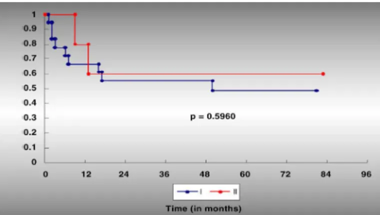
Figure 3 - Metastasis-free survival curve in months for the 24
patients with primary non-metastatic osteosarcoma at diagnosis, according to the degree of tumor necrosis after the effect of chemotherapy (Huvos I and II).
Figure 4 - Overall survival curve in months for the 24 patients with
primary non-metastatic osteosarcoma at diagnosis, according to the degree of tumor necrosis after the effect of chemotherapy (Huvos I and II).
DISCUSSION
Despite the relative rarity of osteosarcoma, several studies(7,12,15-17,20,21,24-27), including some within our
setting, have sought to identify risk factors that might influence the natural history of this tumor and the prognosis for patients.
The efficacy of chemotherapy and its impact on the survival of patients with osteosarcoma are well documented(6,7). There seems to be almost a consensus
in the literature(7,8,12,13,17-19) that the degree of tumor
necrosis induced by preoperative chemotherapy is an important prognostic factor in relation to the survi-val of patients with primary osteosarcoma. This has also been seen in Brazilian studies(6,15,21,24,25). Patients
whose resected surgical specimens present tumor ne-crosis greater than 90% (Huvos types III and IV) have a better prognosis than shown by those whose speci-mens present tumor necrosis of less than 90% (Huvos
Rev Bras Ortop. 2011;46(4):420-23
Comparing the degrees of tumor necrosis (Huvos I versus Huvos II), they did not reach statistically signi-ficant levels and were not considered to be prognostic factors for MFS (Figure 3).
423
types I and II). Thus, we compared the poor respon-ders (< 90% tumor necrosis): Huvos type I (< 50%) versus type II (50 to 90%), in relation to the survival parameters (LRFS, MFS and OS), and observed that these are not adverse factors.
In this study, stratification into groups I and II, following the Huvos criteria, was not shown to be necessary, given the similar behavior presented by the two groups, in the statistical analysis. These sta-tistical results suggest that groups I and II presented homogenous behavior, including in relation to the therapeutic responses made, thus confirming a trend in the worldwide literature, in which the cutoff point is 90% in the histological analysis.
A preliminary evaluation of this study seems to
demonstrate that patients with tumor necrosis of Hu-vos grades I and II after neoadjuvant chemotherapy have similar prognostic behavior, despite the wide difference in percentage range in these two groups when summed (0 to 90%). However, further studies are needed in order to establish a comparative analysis between Huvos groups I and II and thereby corrobo-rate the above affirmation.
CONCLUSION
In this series, comparison between Huvos grades I and II showed that they did not have any prognostic va-lue in relation to any of the survival parameters studied (LRFS, MFS and OS), since they behaved similarly.
Rev Bras Ortop. 2011;46(4):420-23
REFERENCES
1. Schajowicz F, Sissons HA, Sobin LH. The World Health Organization´s histolo-gic classification of bone tumors. A commentary on the second edition. Cancer. 1995;75(5):1208-14.
2. Dorfman HD, Czerniak B. Bone cancers. Cancer. 1995;75(Suppl 1):203-10.
3. Dorfman HD, Czerniak B. Bone tumors. St Louis: Mosby; 1998.
4. Raymond AK, Ayala AG, Knuutila S. Secondary osteosarcoma. In: Fletcher CDM, Unni KK, Mertens F, editors. Pathology and genetics of tumours of the soft tissues and bones. Lyon: IARC Press; 2002. p. 264-70.
5. American Cancer Society. Cancer Reference Inforamtion. Detailed Guide: Os-teosarcoma. What Are The Key Statistics About Osteosarcoma? Dispinível em: http://www.cancer.org/docroot/CRI/content/CRI_2_4_1X_What_are_the_key_sta-tistics_for_osteosarcoma_52.asp?sitearea. Acesso em 08 de outubro de 2009.
6. Petrilli AS, de Camargo B, Filho VO, Bruniera P, Brunetto AL, Jesus-Garcia R, et al. Results of the Brazilian Osteosarcoma Treatment Group Studies III and IV: prognostic factors and impact on survival. J Clin Oncol. 2006;24(7):1161-8.
7. Pakos EE, Nearchou AD, Grimer RJ, Koumoullis HD, Abudu A, Bramer JA, et al. Prognostic factors and outcomes for osteosarcoma: an international colla-boration. Eur J Cancer. 2009;45(13):2367-75.
8. Meyers PA, Heller G, Healey J, Huvos A, Lane J, Marcove R, et al. Chemo-therapy for nonmetastatic osteogenic sarcoma: the Memorial Sloan-Kettering experience. J Clin Oncol. 1992;10(1):5-15.
9. Bacci G, Picci P, Ferrari S, Ruggieri P, Casadei R, Tienghi A, et al. Primary chemotherapy and delayed surgery for nonmetastatic osteosarcoma of the ex-tremities. Results in 164 patients preoperatively treated with high doses of me-thotrexate followed by cisplatin and doxorubicin. Cancer. 1993;72(11):3227-38.
10. Provisor AJ, Ettinger LJ, Nachman JB, Krailo MD, Makley JT, Yunis EJ, et al. Treatment of nonmetastatic osteosarcoma of the extremity with preoperative and postoperative chemotherapy: a report from the Children’s Cancer Group. J Clin Oncol. 1997;15(1):76-84.
11. Souhami RL, Craft AW, Van der Eijken JW, Nooij M, Spooner D, Bramwell VH, et al. Randomised trial of two regimens of chemotherapy in operable osteosarcoma: a study of the European Osteosarcoma Intergroup. Lancet. 1997;350(9082):911-7.
12. Ferrari S, Bertoni F, Mercuri M, Picci P, Giacomini S, Longhi A, Bacci G. Pre-dictive factors of disease-free survival for non-metastatic osteosarcoma of the extremity: an analysis of 300 patients treated at the Rizzoli Institute. Ann Oncol. 2001;12(8):1145-50.
13. Hudson M, Jaffe MR, Jaffe N, Ayala A, Raymond AK, Carrasco H, et al. Pediatric osteosarcoma: therapeutic strategies, results, and prognostic factors derived from a 10-year experience. J Clin Oncol. 1990;8(12):1988-97.
14. Link MP, Goorin AM, Horowitz M, Meyer WH, Belasco J, Baker A, et al.
Ad-juvant chemotherapy of high-grade osteosarcoma of the extremity. Updated results of the Multi-Institutional Osteosarcoma Study. Clin Orthop Relat Res. 1991;(270):8-14.
15. Petrilli AS, Gentil FC, Epelman S, Lopes LF, Bianchi A, Lopes A, et al. Increased survival, limb preservation, and prognostic factors for osteosarcoma. Cancer. 1991;68(4):733-7.
16. Glasser DB, Lane JM, Huvos AG, Marcove RC, Rosen G. Survival, prognosis, and therapeutic response in osteogenic sarcoma. The Memorial Hospital ex-perience. Cancer. 1992;69(3):698-708.
17. Davis AM, Bell RS, Goodwin PJ. Prognostic factors in osteosarcoma: a critical review. J Clin Oncol. 1994;12(2):423-31.
18. Saeter G, Elomaa I, Wahlqvist Y, Alvegård TA, Wiebe T, Monge O, et al. Prog-nostic factors in bone sarcomas. Acta Orthop Scand Suppl. 1997;273:156-60.
19. Bacci G, Ferrari S, Longhi A, Forni C, Zavatta M, Versari M, Smith K. High-grade osteosarcoma of the extremity: differences between localized and metastatic tumors at presentation. J Pediatr Hematol Oncol. 2002;24(1):27-30.
20. Bielack SS, Kempf-Bielack B, Delling G, Exner GU, Flege S, Helmke K, et al. Prognostic factors in high-grade osteosarcoma of the extremities or trunk: an analysis of 1,702 patients treated on neoadjuvant cooperative osteosarcoma study group protocols. J Clin Oncol. 2002;20(3):776-90.
21. Rech A, Castro CG Jr, Mattei J, Gregianin L, Di Leone L, David A, et al. [Cli-nical features in osteosarcoma and prognostic implications]. J Pediatr (Rio J). 2004;80(1):65-70.
22. Huvos AG, Rosen G, Marcove RC. Primary osteogenic sarcoma: pathologic aspects in 20 patients after treatment with chemotherapy en bloc resection, and prosthetic bone replacement. Arch Pathol Lab Med. 1977;101(1):14-8.
23. Rosen G, Caparros B, Huvos AG, Kosloff C, Nirenberg A, Cacavio A, et al. Preoperative chemotherapy for osteogenic sarcoma: selection of postoperative adjuvant chemotherapy based on the response of the primary tumor to preo-perative chemotherapy. Cancer. 1982;49(6):1221-30.
24. Jesus-Garcia R, Consentino E, Camargo OP, Baptista PPR, Croci AT, Korukian M et al. Tratamento ortopédico do osteossarcoma. Grupo cooperativo brasileiro de tratamento do osteossarcoma. Rev Bras Ortop. 1996;31(11):871-8.
25. Cassone AE, Gonçalves JCB, Silva AAM, Epelman S, Amstalden EMI.Trata-mento multidisciplinar do osteossarcoma. Rev Bras Ortop. 1998;33(11):835-40.
26. Bispo Júnior RZ, Camargo OP. Prognostic factors for the survival of patients diagnosed with primary non-metastatic osteosarcoma with a poor response to neoadjuvant chemotherapy. Clinics. 2009;64(12):1177-86.
27. Bispo Júnior RZ. Fatores prognósticos da sobrevida no osteossarcoma primá-rio: grau I versus II de Huvos [tese]. São Paulo: Universidade de São Paulo. Faculdade de Medicina; 2009.