A continuum of executive function
deficits in early subcortical vascular
cognitive impairment
A systematic review and meta-analysis
Felipe Kenji Sudo1,2, Patricia Amado3, Gilberto Sousa Alves4,5, Jerson Laks3,6, Eliasz Engelhardt7
ABSTRACT. Background. Subcortical Vascular Cognitive Impairment (SVCI) is a clinical continuum of vascular-related cognitive impairment, including Vascular Mild Cognitive Impairment (VaMCI) and Vascular Dementia. Deficits in Executive Function (EF) are hallmarks of the disorder, but the best methods to assess this function have yet to be determined. The insidious and almost predictable course of SVCI and the multidimensional concept of EF suggest that a temporal dissociation of impairments in EF domains exists early in the disorder. Objective: This study aims to review and analyze data from the literature about performance of VaMCI patients on the most used EF tests through a meta-analytic approach. Methods: Medline, Web of Knowledge and PsycINFO were searched, using the terms: “vascular mild cognitive impairment” OR “vascular cognitive impairment no dementia” OR “vascular mild neurocognitive disorder” AND “dysexecutive” OR “executive function”. Meta-analyses were conducted for each of the selected tests, using random-effect models. Results: Systematic review showed major discrepancies among the results of the studies included. Meta-analyses evidenced poorer performance on the Trail-Making Test part B and the Stroop color test by VaMCI patients compared to controls. Conclusion: A continuum of EF impairments has been proposed in SVCI. Early deficits appear to occur in cognitive flexibility and inhibitory control.
Key words: mild cognitive impairment, cerebrovascular disorders, neuropsychology, vascular dementia, metabolic syndrome.
UM CONTÍNUO DE DÉFICITS EM FUNÇÃO EXECUTIVA NO COMPROMETIMENTO COGNITIVO SUBCORTICAL INICIAL: REVISÃO SISTEMÁTICA E METANÁLISE
RESUMO. Introdução: O Comprometimento Cognitivo Vascular Subcortical (CCVS) é um contínuo clínico de comprometimento cognitivo de causa vascular, que inclui o Comprometimento Cognitivo Leve Vascular (CCLV) e a Demência Vascular. Déficits em Função Executiva (FE) são marcantes no quadro, mas a melhor metodologia para avaliar essa função ainda necessita ser determinada. A evolução insidiosa e quase previsível do CCVS e o conceito atual multidimensional de FE sugerem que uma dissociação temporal de comprometimentos dos domínios da FE exista nos estágios iniciais do transtorno. Objetivo: Visou-se a revisar e analisar dados da literatura sobre o desempenho de sujeitos com CCLV nos testes mais usados de FE através de uma abordagem metanalítica. Métodos: Foram realizadas buscas no Medline, Web of Knowledge e PsycINFO, usando os seguintes termos: “comprometimento cognitivo vascular” OU “comprometimento cognitivo não demência vascular” OU “transtorno neurocognitivo leve vascular” E “função executiva” OU “disexecutiva”. Metanálises foram conduzidas para cada um dos testes, usando-se modelos de efeitos aleatórios.
Resultados: A revisão sistemática demonstrou grande discrepância entre os resultados dos estudos incluídos. A metanálise evidenciou desempenhos piores no Teste de Trilhas parte B e no teste de cores do Stroop em sujeitos com CCLV em comparação com controles. Conclusão: Um contínuo de comprometimentos em FE no CCVS foi proposto. Déficits iniciais parecem ocorrer em flexibilidade cognitive e controle inibitório.
Palavras-chave: comprometimento cognitivo leve, transtornos cerebrovasculares, neuropsicologia, demência vascular, síndrome metabólica.
This study was conducted at the Departamento de Psicologia, Pontifícia Universidade Católica do Rio de Janeiro, RJ, Brazil.
1Departamento de Psicologia, Pontifícia Universidade Católica do Rio de Janeiro, RJ, Brazil. 2Instituto D’Or de Ensino e Pesquisa, Rio de Janeiro, RJ, Brazil. 3Instituto de Psiquiatria, Universidade Federal do Rio de Janeiro, RJ, Brazil. 4Departamento de Medicina Interna, Universidade Federal do Ceará, CE, Brazil. 5Goethe Universitat Frankfurt Am Main, Germany. 6Programa de Pós-Graduação em Biomedicina Translacional (BIOTRANS), Unigranrio, Duque de Caxias, RJ, Brazil. 7Setor de Neurologia Cognitiva e do Comportamento, INDC/CDA/ IPUB, Universidade Federal do Rio de Janeiro, RJ, Brazil.
Felipe Kenji Sudo. Departamento de Psicologia, Pontifícia Universidade Católica do Rio de Janeiro – Rua Marquês de São Vicente, 225 / Edifício Cardeal Leme – 22451-900 Rio de Janeiro RJ – Brazil. E-mail: [email protected]
Disclosure: The authors report no conflicts of interest.
Received October 01, 2017. Accepted in final form November 13, 2017.
Dement Neuropsychol 2017 December;11(4):371-380
372 Executive dysfunction in subcortical vascular cognitive Impairment Sudo et al.
INTRODUCTION
S
ubcortical Vascular Cognitive Impairment (SVCI), a clinical continuum of cognitive impairments due to small-vessel disease, has been recognized as a com-mon cause of cognitive dysfunction and, ultimately, of dementia in the elderly population.1,2 Metabolic risk factors, such as diabetes mellitus, smoking and dyslip-idemia, as well as systemic arterial hypertension, are assumed to promote continuous insults to the small perforating arteries, which may sufer occlusion or sub-occlusion due to progressive arteriolosclerosis, lipohya-linosis and ibrinoid necrosis.3 hese vascular changes result in subcortical lesions: lacunar infarcts and white matter lesions, which can be detectable as punctuate periventricular white-matter hyperintensities (WMH) to extensive areas of leukoaraiosis on neuroimaging.4 hese white matter lesions may impair deep (punctu-ate or conluent WMH on T2 and FLAIR images) and periventricular regions (WMH bordering the lateral ventricles).5Small-vessel disease consistently disrupts intercon-nections among prefrontal, sensory, motor and limbic cortices, causing disturbances to complex cognitive functions dependent on the tight integration of high order decisional neurons in the prefrontal cortex with primary and association cortical areas.6 Among the afected abilities, impairment in executive function (EF) has been widely described in SVCI and resulted in its inclusion among the early cognitive changes associated with vascular-related neurocognitive disorders in the 5th Edition of the Diagnostic and Statistical Manual of Mental Disorders (DSM-5).7
Although the relationship between SVCI and execu-tive dysfunction is clear, some shortcomings regarding the best methods to assess EF still exist. he lack of harmonization on the choice of cognitive tests adopted in research has resulted in diiculties integrating and interpreting indings from diferent studies.8 To address these issues, the National Institute of Neurological Dis-orders and Stroke-Canadian Stroke Network Vascu-lar Cognitive Impairment Harmonization Standards (NINDS-CSN) work group proposed, in 2006, speciic neuropsychological protocols to evaluate cerebrovascu-lar cognitive disorders.8 However, the long time required to apply these batteries has made their use unfeasible in most clinical settings.9 he same problem may afect the VADAS-cog, another proposed neuropsychological instrument for vascular-related cognitive impairment.10 In addition, EF has been increasingly recognized as a multidimensional rather than a unitary construct; the diverse range of abilities grouped under the
umbrella-term of EF, such as cognitive lexibility, inhibitory control, working memory and “complex” unspeciic EF, precludes direct comparison of results from difer-ent EF tests.11,12 Moreover, since small-vessel disease commonly exhibits an insidious course, which follows a largely predictable temporal pattern of subcortical lesions - from periventricular to juxtacortical regions - it follows that impairments in EF domains may also be subject to a temporal dissociation according to site of damage.13,14
Characterizing the EF changes in early SVCI could contribute toward a better understanding of the natural history of the disorder, thereby enabling early interven-tion to control vascular risk factors. his measure could help prevent the onset of Vascular Dementia.15 Further-more, brief and widely accessible tests would be more suitable for clinical use than extensive sophisticated neuropsychological batteries. A recent systematic review has listed the most frequently used EF tests in studies, including the Trail-Making test part B (TMTB), the semantic and phonemic Verbal Fluency (sVF and pVF), the Clock Drawing Test (CDT), the Digit Span backwards (DSb), the Stroop test and the Wisconsin Card Sorting Test.16 he present study aims to review data on the per-formance of patients with early SVCI, namely Vascular Mild Cognitive Impairment (VaMCI), on these tests.
METHODS
Literature search. Database searches were performed on Medline, Web of Knowledge and PsycINFO using the following combination of terms: “vascular mild cognitive impairment” OR “vascular cognitive impair-ment no deimpair-mentia” OR “vascular mild neurocognitive disorder” AND “dysexecutive” OR “executive func-tion”. No restriction on date of publication or ield was placed. he reference lists of the selected articles were hand searched for potentially relevant papers.
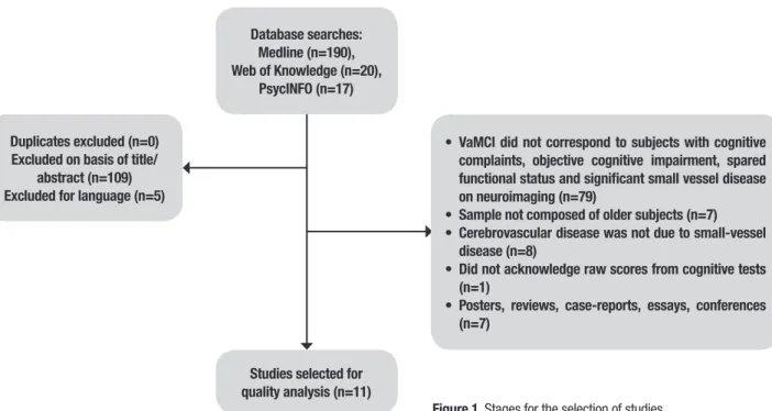
Database searches: Medline (n=190), Web of Knowledge (n=20),
PsycINFO (n=17)
Duplicates excluded (n=0) Excluded on basis of title/
abstract (n=109) Excluded for language (n=5)
• VaMCI did not correspond to subjects with cognitive complaints, objective cognitive impairment, spared functional status and significant small vessel disease on neuroimaging (n=79)
• Sample not composed of older subjects (n=7)
• Cerebrovascular disease was not due to small-vessel disease (n=8)
• Did not acknowledge raw scores from cognitive tests (n=1)
• Posters, reviews, case-reports, essays, conferences (n=7)
Studies selected for quality analysis (n=11)
Figure 1. Stages for the selection of studies.
Procedures. Screening of the retrieved articles was carried out by two independent researchers (F.K.S. and E.E.). Data extraction was performed independently and discordant results were resolved through discussion with the whole team of authors.
Quality assessment strategy. he risk of biases in the selected studies was appraised through the following criteria derived from a published checklist.17
1. A representative sample of general elderly pop-ulation was recruited with a minimum baseline partici-pation rate of 70%;
2. Participation rate at follow-up was 70% of the baseline sample or greater (when applicable);
3. Follow-up duration was at least 1 year (when applicable);
4. Diagnosis of VaMCI was based on recognized international criteria by a consensus committee.4,7,18,19
Items 2 and 3 were not applied for cross-sectional studies. Samples were deemed representative of the overall elderly population if no limits for recruitment of participants regarding age (subjects were aged 60-65 years or older and no additional restrictions for age were adopted), gender, race, education and cognitive and clinical status (exclusion of subjects with dementia at baseline was accepted) were used. Studies which fulilled these criteria were assigned as higher quality studies. Data derived from at least two higher quality studies was classiied as “Grade 1 level of evidence”. “Grade 2 level of evidence” was attributed to single higher
qual-ity studies, whereas “inconsistent evidence” was that obtained from lower quality studies.
Data synthesis. Mean scores, standard deviations and sample sizes were extracted for each of the EF tests. hese values were subjected to meta-analyses stratiied by EF test. Data were assessed using R Statistics version 3.3.3. he Random efect approach was preferred over the ixed efect, so that weights of studies of diferent sizes could be balanced.20 Presence of heterogeneity across studies was identiied through the determination of I.2
RESULTS
Eleven studies were selected from a total of 227 retrieved from the database searches. he stages for the selection of the studies are depicted in Figure 1.
Dement Neuropsychol 2017 December;11(4):371-380
374 Executive dysfunction in subcortical vascular cognitive Impairment Sudo et al. Diagnosis of VaMCI. Some variations in the diagnostic criteria used to identify subjects with VaMCI were detected among studies. Modiied versions of Petersen`s criteria for Mild Cognitive Impairment (MCI)24 were adopted in ive of the studies.22,25-28 In ive of the articles, the detection of memory impairment was mandatory for the diagnosis of MCI.21,25,26,29,30 he criteria proposed by Frisoni (2002) for VaMCI31 was adopted by one of the studies, which included the presence of dysexecutive syndrome, memory deicits and functional preservation as necessary features for the diagnosis.32
Speciic cutof values in tests were applied to deter-mine objective cognitive impairment: cognitive scores lower than 1.5 standard-deviations (16th percentile) from normative data22,23,27,28 or at least two scores below established cutof points in episodic memory tasks were employed in studies.26 Absence of functional impair-ment was determined through scores on structured questionnaires, such as the Pfefer Functional Activities Questionnaire,28 the Functional Autonomy Measure-ment System27 and the Seoul Instrumental Activities of Daily Living scale.23
Subjects were classiied as MCI if they had Clinical Dementia Rating scores of 0.5 in two studies.21,29 For the present review, in Ishii et al. (2007), normal controls were deined as subjects with CDR = 0 and without cerebro-vascular disease, whereas VaMCI patients were deined as those with CDR = 0.5 and non-strategic infarcts.21
he presence of cerebrovascular disease was deter-mined by the identiication of focal neurological symp-toms or signs and of severe white-matter abnormali-ties on neuroimaging.22,23,25,29,32 Some studies deined the presence of small-vessel disease in VaMCI accord-ing to the extension of WMH: infarcts less than 2 cm in size,26 lesions of at least 4 mm21 or the presence of WMH afecting over 19.375% of the total white-matter volume.25 Neuroimaging criteria deined by Erkinjuntti et al. (2000), comprising the presence of severe WMH (periventricular WMH > 10 mm and deep WMH ≥ 25 mm in maximum diameter) or moderate WMH with at least 5 lacunes,33 was adopted by two studies.22,34 he presence of moderate WMH, as indicated by scores > 1 on the modiied-Fazekas Scale, and the absence of hip-pocampal atrophy, deined as scores on De Leon’s scale of ≤ 1, were considered indicative of pure cerebrovascu-lar disease in one study.28 Presence of conluent WMH deined the presence of vascular burden in one study.27
Performance on EF tests. Table 2 summarizes the scores on EF assessments of normal controls and VaMCI. he choice of EF tests varied greatly among studies, ranging from single screening tasks25 to long batteries.26,30 Since the Clock-Drawing Test was only included in one of the selected studies,28 it has not been shown in the table. he Wisconsin Card Sorting Test was not used in any of the included articles.
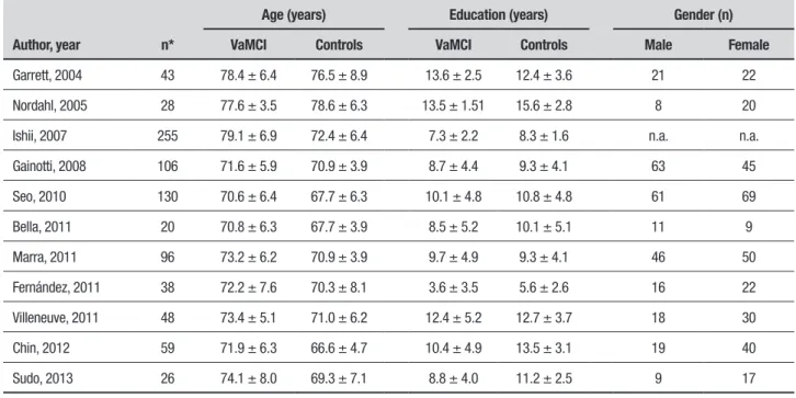
Table 1. Sociodemographic characteristics of the samples.
Author, year n*
Age (years) Education (years) Gender (n) VaMCI Controls VaMCI Controls Male Female
Garrett, 2004 43 78.4 ± 6.4 76.5 ± 8.9 13.6 ± 2.5 12.4 ± 3.6 21 22
Nordahl, 2005 28 77.6 ± 3.5 78.6 ± 6.3 13.5 ± 1.51 15.6 ± 2.8 8 20
Ishii, 2007 255 79.1 ± 6.9 72.4 ± 6.4 7.3 ± 2.2 8.3 ± 1.6 n.a. n.a.
Gainotti, 2008 106 71.6 ± 5.9 70.9 ± 3.9 8.7 ± 4.4 9.3 ± 4.1 63 45
Seo, 2010 130 70.6 ± 6.4 67.7 ± 6.3 10.1 ± 4.8 10.8 ± 4.8 61 69
Bella, 2011 20 70.8 ± 6.3 67.7 ± 3.9 8.5 ± 5.2 10.1 ± 5.1 11 9
Marra, 2011 96 73.2 ± 6.2 70.9 ± 3.9 9.7 ± 4.9 9.3 ± 4.1 46 50
Fernández, 2011 38 72.2 ± 7.6 70.3 ± 8.1 3.6 ± 3.5 5.6 ± 2.6 16 22
Villeneuve, 2011 48 73.4 ± 5.1 71.0 ± 6.2 12.4 ± 5.2 12.7 ± 3.7 18 30
Chin, 2012 59 71.9 ± 6.3 66.6 ± 4.7 10.4 ± 4.9 13.5 ± 3.1 19 40
Sudo, 2013 26 74.1 ± 8.0 69.3 ± 7.1 8.8 ± 4.0 11.2 ± 2.5 9 17
Dement Neuropsychol 2017 December;11(4):371-380
375
Sudo et al.
Executive Dysfunction in Subcortical
Vascular Cognitive Impairment
Study n VaMCI NC p–value VaMCI NC p–value VaMCI NC p–value VaMCI NC p–value VaMCI NC p–value
Garrett, 2004
VaMCI = 18; NC = 25
190.5 ± 76.3 90.8 ± 33.5 <.0001 31.8 ± 9.6 29.4 ± 8.4 n.s. 14.3 ± 4.1 17.8 ± 6.2
n.s. – – – – – –
Nordahl, 2005
VaMCI = 10; NC = 17
– – – – – – 12.6
± 4.2
16.6 ± 3.9
.03 – – – – – –
Ishii, 2007
VaMCI = 21; NC = 234
346.9 ± 122.1 221.4 ± 103.6
n.a. – – – 6.2
± 1.5
7.7 ± 2.4
n.a. – – – – – –
Gainotti, 2008
VaMCI = 41; NC = 65
– – – 23.2
± 8.43 24.5 ± 9.6 .63 14.6 ± 3.8 14.9 ± 3.7 .84 3.3 ± 0.81 3.9 ± 1.0 .01 70.3 ± 26.5 53.7 ± 16.5 .001 Seo, 2010
VaMCI = 34; NC = 96
– – – 14.0
± 7.6 26.4 ± 11.2 <.05 11.2 ± 4.5 16.5 ± 4.2 <.05 3.4 ± 1.0 3.7 ± 1.1
n.s. – – –
Marra, 2011
VaMCI = 36; NC = 60
– – – 25.2
± 10.3 24.4 ± 9.6 .91 14.9 ± 4.3 14.9 ± 3.8 .86 3.6 ± 1.2 3.9 ± 1.35 .096 66.8 ± 29.5 53.7 ± 16.5 .017 Bella, 2011
VaMCI = 10; NC = 10
– – – – – – – – – – – – 41.1
± 15.9 26.3 ± 11.8 <.05 Fernández, 2011
VaMCI = 19; NC = 19
– – – – – – 12.1
± 2.8 16.1 ± 2.2 <.05 3.1 ± 0.7 3.1 ± 0.5
n.s. – – –
Villeneuve, 2011
VaMCI = 21; NC = 27
– – – – – – – – – – – – 38.3
± 11.4 27.5 ± 7.9 <.05 Chin, 2012
VaMCI = 31; NC = 28
– – – 17.0
± 8.7 33.8 ± 8.4 <.05 23.2 ± 7.4 39.6 ± 6.9 <.05 3.5 ± 0.9 4.3 ± 1.0
<.05 – – –
Sudo, 2013
VaMCI = 15; NC = 11
265.8 ± 136.4 127.4 ± 46.7
.004 – – – 15.7
± 4.5
16.6 ± 3.4
Dement Neuropsychol 2017 December;11(4):371-380
376 Executive dysfunction in subcortical vascular cognitive Impairment Sudo et al. Trail-Making Test B. VaMCI subjects performed signii-cantly worse than controls in two studies.28,29 A signii-cant number of participants could not complete the TMTB in Fernandez et al. (2011) due to low educa-tion and results on the test were not analyzed by the authors.32 he presence of signiicant diferences in time required to complete the TMTB between controls and VaMCI was not acknowledged in Ishii et al. (2007).21
Verbal Fluency. Controls performed signiicantly better than VaMCI patients on the sVF in some studies,22,23,25,32 whereas these diferences were not found in other arti-cles.26,28-30 Most of the studies used the sum of words beginning with F, A and S as the method for calcu-lating performance on the pVF task. VaMCI patients performed poorer than controls on the pVF,22,23 but these indings were not replicated in other studies.26,29,30 Diferent application methods for the VF were adopted in some studies, such as sVF using categories of objects found in a supermarket22 and pVF tests involving words beginning with the letter P (Fernández et al., 2011). he presence of signiicant diferences in sVF scores between controls and VaMCI patients were not acknowledged in Ishii et al. (2007).21
Stroop test. he number of correct items during the reading (Stroop word test) and the inhibiting (Stroop color test) tasks were measured in two of the studies,22,23 while time for completion of these tasks was computed in other studies.26,27,30,34 Some authors included the number of errors during the color task as an additional measurement of inhibitory control.26,30,34 No signiicant diference was identiied between controls and VaMCI subjects in the reading test, but time to complete the color reading was signiicantly higher in VaMCI subjects than controls in three of the studies.26,27,34 he number of errors was signiicantly higher in VaMCI patients compared to controls.30,34
Digit Span backwards. Controls performed signiicantly better than VaMCI participants in one of the studies.23 On the other hand, no signiicant diferences were iden-tiied between VaMCI subjects and controls in the other articles which used this test.22,26,30,32
Clock Drawing Test. No diference was identiied between controls and VaMCI patients on the clock drawing test, measured according to the CLOX method.28
Risk of bias. None of the selected studies had higher quality according to the criteria used in this review. Most of the studies recruited unrepresentative samples drawn from tertiary facilities. In all cases, diagnoses of VaMCI were based on diferent criteria from those determined by teams of specialists, or were highly dependent on performances on screening tests (e.g., MMSE) or global assessment scales (e.g., CDR). In some of the studies, only amnestic MCI subjects were included.21,25,29,30
Meta-analysis. Mean scores on EF tests were combined in a meta-analytic approach. Analyses were performed for each EF test.
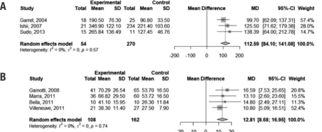
For the completion of the TMTB, pooled-anal-ysis indicated that VaMCI subjects performed 112.59 seconds slower than normal controls (95% CI 84.10,141.08). Time to complete the Stroop color test was 12.81 seconds higher in VaMCI patients than controls (95% CI 8.68, 16.95). he I2 of 0% indicated absence of heterogeneity among studies for both tests (Figure 2).
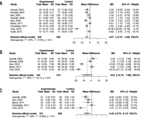
Performance on the sVF (animals) was slightly worse in VaMCI subjects than in controls, with a diference of 3.67 points favoring the latter group (95% CI –5.70, –1.65). However, the presence of heterogeneity was marked among studies (I2 = 91%). Pooled analysis of scores on the pVF (letters FAS) showed inconclusive results (95% CI -12.74, 1.69). A very small mean
difer-A
B
ence was found in performances on the DSb between controls and VaMCI subjects (mean diference = –0.45 points; 95% CI –0.79, –0.11), but heterogeneity of studies was signiicant (I2 = 68%). Figure 6 depicts this result. Figure 3 illustrates these indings.
DISCUSSION
Based on the present meta-analysis, in which we assessed performance of VaMCI subjects and normal controls on the most used EF tests in research, we suggest that a temporal dissociation of cognitive impair-ments may exist in SVCI. Results for at least two of the cognitive instruments have shown diferences between early SVCI and healthy older subjects, with no signii-cant heterogeneity among the studies. Most remark-ably, data suggested a marked increase in the time required to complete the TMTB among VaMCI patients compared to normal healthy subjects. A slightly shorter time to perform the Stroop color test was also identiied in controls relative to VaMCI individuals. On the other hand, results on the DSb, pVF and sVF tasks were either not signiicant or the studies were too heterogeneous to allow conclusions to be drawn.
Executive dysfunction has been regarded as the dis-tinctive cognitive marker of SVCI by many authors.35-37 he EF tests analyzed in this study were those con-sidered the most commonly used in research.16 Factor
analyses and regression analyses studies indicated that they measure diferent aspects of EF. According to one of these studies, TMTB was identiied as an index of cogni-tive lexibility.38 Diiculties on the Stroop color test have been associated with inhibitory control impairment.39 Moreover, multiple regression analyses indicated that sVF assesses semantic memory and working memory, whereas pVF is dependent on episodic verbal memory and cognitive speed.40
When individual EF domains were analyzed in sub-jects with Vascular Dementia, data in the literature sug-gested that all of them exhibited impairment when com-pared to controls.35,36 he present study suggests that speciic EF domains, namely cognitive lexibility and inhibitory control, might be impaired in early SVCI (in the VaMCI stage), while other domains, such as work-ing memory, might be initially preserved. Evidence from functional neuroimaging and pathology studies might support this idea. For instance, periventricular white matter receives blood supply from terminal vessels of long perforating branches of a watershed circulation (areas in which branches of the diferent cerebral large vessels meet). Most long perforating arteries are very tortuous and these anatomical characteristics make those locations especially vulnerable to hypoperfusion due to arteriosclerosis.41,42 herefore, it could be pre-dicted that periventricular WMH occur early in SVCI,
A
B
C
Dement Neuropsychol 2017 December;11(4):371-380
378 Executive dysfunction in subcortical vascular cognitive Impairment Sudo et al.
because of chronic insults to the small vasculature associated with metabolic risk factors. Juxtacortical white matter, on the other hand, could be considered less susceptible to vascular damage and expected to be impaired later in the disorder.13,41 Because of this tem-poral-anatomical dissociation in white-matter lesions, neuronal pathways with periventricular trajectories, which include long interlobar ibers, could be disrupted in initial SVCI, whereas short corticocortical juxtacorti-cal U-ibers might still be preserved in this stage.13,14,41 his mechanism might account for the continuum of EF impairments in SVCI, with earlier diiculties observed in abilities dependent on long ibers with periventricular trajectories, while other functions associated with short juxtacortical U-ibers might be subject to deicits later in the disorder.14
One study which evaluated neural correlates of TMTB scores in a post-stroke sample suggested that poor performance could be predicted by lesions situ-ated in the lateral cholinergic pathways and in the left superior longitudinal fasciculus.43 In accordance, one fMRI study demonstrated that during the TMTB, left frontal and parietal areas, associated with motor, atten-tional, decisional, linguistic and sensory functions, are activated.44 Interlobar integration is also required dur-ing the Stroop color test, which may demand increased activity in frontal, parietal and occipital areas.45 On the other hand, a meta-analysis indicated that tasks which measure verbal working memory are mostly dependent on activation of the left dorsolateral prefrontal cortex.46 Also, another study showed that deep, but not periven-tricular WMH, correlated with working memory impair-ment.47 hese data might explain the indings of early impairments in cognitive lexibility and inhibitory con-trol in VaMCI, which we theorize might occur due to increased vulnerability to interlobar disconnection due to periventricular WMH in these cases. In addition, according to this hypothesis, working memory, which relies on the integrity of speciic prefrontal areas, might be initially spared in SVCI.
he strength of the present study was allowing the identiication of the EF domains afected in early SVCI, namely, cognitive lexibility and inhibitory control, through statistical combination of results from studies. his was not the case for the systematic review, given that major disparities exist among the selected articles, as revealed in this study. Moreover, assessment of the heterogeneity of the studies, performed by a meta-ana-lytic method, raises questions over whether data derived from diferent sources are comparable or not. For this
reason, conclusions drawn in a previous review on the cognitive correlates of white matter lesions in non-demented subjects should be considered with caution due to the selection of studies with potentially hetero-geneous samples and methods.48 In addition, the current concept that EF comprises multiple distinct abilities and not a unitary entity may preclude direct comparisons between diferent EF tasks, which might have been a problem in a previous meta-analysis on the theme.49
Limitations of this study should be acknowledged. he quality assessment of the selected articles showed that risk of selection or diagnostic biases were signii-cant in all. Since diagnostic criteria for VaMCI have been evolving over the years, diferences in the charac-terization of this disorder varied greatly. For instance, some studies included only subjects presenting memory deicits, while others identiied the cases based on the Clinical Dementia Rating of 0.5. Moreover, detection of cerebrovascular disease adopted diferent criteria, which might have led to variations in the severity of brain lesions among samples from diferent studies. Also, results on performances of VaMCI subjects in sVF, pVF and DSb were inconclusive due to high levels of heterogeneity among studies, despite the random efect models applied. Only one of the selected studies used the CDT and none employed the Wisconsin Card Sorting Test, hence, data on these tests remain unavail-able. Finally, a low number of studies was selected and included small sample sizes, indicating that further research in this ield is still needed.
Early identiication of SVCI is crucial to allow inter-vention to control vascular risk factors before the onset of dementia. he hypothesis of a temporal continuum of dysexecutive syndrome, based on a multidimensional concept of EF and on pathophysiological aspects of lesion progression in SVCI, might be of great value for this purpose. However, further studies are needed to validate these theories.
Author contribution. Felipe Kenji Sudo: search in data-bases, articles selection, methodology, discussion of results, article writing. Patricia Amado and Gilberto Sousa Alves: statistical analysis, discussion of results, methodology, article writing. Jerson Laks: discussion of results, methodology, inal revision of the text. Eliasz Engelhardt: search in databases, article selection, meth-odology, discussion of results, inal revision of the text.
REFERENCES
1. Kramer JH, Reed BR, Mungas D, Weiner MW, Chui HC. Executive
dysfunction in subcortical ischaemic vascular disease. J Neurol Neuro-surg Psychiatr. 2002;72:217–20.
2. Meyer JS, Xu G, Thornby J, Chowdhury MH, Quach M. Is mild cognitive
impairment prodromal for vascular dementia like Alzheimer’s disease? Stroke. 2002;33(8):1981-5.
3. Jellinger KA. Pathogenesis and treatment of vascular cognitive
impair-ment. Neurodegener Dis Manag. 2014;4(6):471-90.
4. Gorelick PB, Scuteri A, Black, SE, DeCarli C, Greenberg SM, Iadecola
C et al. Vascular Contributions to Cognitive Impairment and Dementia: A Statement for Healthcare Professionals From the American Heart Association/American Stroke Association. Stroke. 2011;42(9):2672-713.
5. Smith CD, Johnson ES, Van Eldik LJ, Jicha GA, Schmitt FA, Nelson
PT, et al. Peripheral (deep) but not periventricular MRI white matter hyperintensities are increased in clinical vascular dementia compared to Alzheimer’s disease. Brain Behav 2016;6(3):e00438.
6. Kim KW, MacFall JR, Payne ME. Classification of white matter
lesions on magnetic resonance imaging in the elderly. Biol Psychiatr. 2008;64(4):273-80.
7. American Psychiatric Association. Diagnostic and statistical manual of
mental disorders (5th ed.). Washington, DC: 2013.
8. Hachinski V, Iadecola C, Petersen RC, Breteler MM, Nyenhuis DL,
Black SE, et al. National Institute of Neurological Disorders and Stroke-Canadian Stroke Network vascular cognitive impairment harmonization standards. Stroke. 2006;37:2220-41.
9. Brookes RL, Hollocks MJ, Khan U, Morris RG, Markus HS. The Brief
Memory and Executive Test (BMET) for detecting vascular cognitive impair-ment in small vessel disease: a validation study. BMC Med. 2015;13:51. 10. Ferris SH. General measures of cognition. Int Psychogeriatr. 2003;15
Suppl 1:215-7.
11. Miyake CA, Friedman NP, Emerson MJ, Witzki AH, Howerter A, Wager TD. The unity and diversity of executive functions and their contribu-tions to complex “Frontal Lobe” tasks: a latent variable analysis. CogN Psychol. 2000;41:49-100.
12. Sudo FK, Alves GS, Tiel C, Ericeira-Valente L, Moreira DM, Laks J, Engelhardt E. Neuroimaging criteria and cognitive performance in vascular mild cognitive impairment: a systematic review. Dement. neuro-psychol. 2015;9(4):394-404.
13. de Groot JC, de Leeuw FE, Oudkerk M, van Gijn J, Hofman A, Jolles J, Breteler MM. Cerebral white matter lesions and cognitive function: the Rotterdam Scan Study. Ann Neurol. 2000;47(2):145-151.
14. Sudo FK. Aspectos clinicos, cognitivos e de neuroimagem do compro-metimento cognitivo vascular. PhD [thesis]. Rio de Janeiro: Universi-dade Federal do Rio de Janeiro;2016. Available from: https://minerva. ufrj.br/F?RN = 723486506.
15. Douiri A, McKevitt C, Emmett ES, Rudd AG, Wolfe CD. Long-term effects of secondary prevention on cognitive function in stroke patients. Circulation. 2013;128(12):1341-8.
16. Faria CA, Alves HVD, Charchat-Fichman H. The most frequently used tests for assessing executive functions in aging. Dement Neuropsychol. 2015;9(2):149-55.
17. Biessels GJ, Staekenborgm S, Brunner E, Brayne C, Scheltens P. Risk of dementia in diabetes mellitus: a systematic review. Lancet Neurol. 2006;5:64-74.
18. Zhao QL, Zhou Y, Wang YL, Dong KH, Wang YJ. A new diagnostic algo-rithm for vascular cognitive impairment: the proposed criteria and evalu-ation of its reliability and validity. Chin Med J (Engl). 2010;123(3):311-9. 19. Sachdev P, Kalaria R, O’Brien J, Skoog I, Alladi S, Black SE, et al. Diag-nostic criteria for vascular cognitive disorders: a VASCOG statement. Alzheimer Dis Assoc Disord, 2014;28(3):206-28.
20. Borenstein M, Hedges L, Rothstein H. Meta-Analysis. Fixed effect vs. random effects. 2007. Available on: https://www.meta-analysis.com/
downloads/M-a_f_e_v_r_e_sv.pdf. Assessed in September 28th 2017.
21. Ishii H, Meguro K, Yamaguchi S, Ishikawa H, Yamadori A. Prevalence and cognitive performances of vascular cognitive impairment no dementia in Japan: the Osaki-Tajiri Project. Eur J Neurol. 2007;14(6):609-16.
22. Seo SW, Ahn J, Yoon U, Im K, Lee JM, Tae Kim S, et al. Cortical thin-ning in vascular mild cognitive impairment and vascular dementia of subcortical type. J Neuroimaging. 2010;20(1):37-45.
23. Chin J, Seo SW, Kim SH, Park A, Ahn HJ, Lee BH, et al. Neurobe-havioral dysfunction in patients with subcortical vascular mild cogni-tive impairment and subcortical vascular dementia. Clin Neuropsychol. 2012;26(2):224-38.
24. Petersen RV, Smith GE, Waring SC, Ivnik RJ, Tangalos EG, Kokmen E. Mild cognitive impairment: clinical characterization and outcome. Arch Neurol. 1999;56(3):303-8.
25. Nordahl CW, Ranganath C, Yonelinas AP, DeCarli C, Reed BR, Jagust WJ. Different mechanisms of episodic memory failure in mild cognitive impairment. Neuropsychologia 2005;43(11):1688-97.
26. Gainotti G, Ferraccioli M, Vita MG, Marra C. Patterns of neuropsycho-logical impairment in MCI patients with small subcortical infarcts or hippocampal atrophy. J Int Neuropsychol Soc. 2008;14(4):611-9. 27. Villeneuve S, Massoud F, Bocti C, Gauthier S, Belleville S. The nature
of episodic memory deficits in MCI with and without vascular burden. Neuropsychologia. 2011;49(11):3027-35.
28. Sudo FK, Alves CEO, Alves GS, Ericeira-Valente L, Tiel C, Moreira DM, et al. White matter hyperintensities, executive function and global cognitive performance in vascular mild cognitive impairment. Arq Neuro-Psiquiatr. 2013;71(7):431-6.
29. Garrett KD, Browndyke JN, Whelihan W, Paul RH, DiCarlo M, Moser DJ et al. The neuropsychological profile of vascular cognitive impairment-no dementia: comparisons to patients at risk for cerebrovascular disease and vascular dementia. Arch Clin Neuropsychol. 2004;19(6):745-57. 30. Marra C, Ferraccioli M, Vita MG, Quaranta D, Gainotti G. Patterns of
cogni-tive decline and rates of conversion to dementia in patients with degen-erative and vascular forms of MCI. Curr Alzheimer Res. 2011;8 (1):24-31. 31. Frisoni GB, Galluzzi S, Bresciani L, Zanetti O, Geroldi C. Mild cognitive
impairment with subcortical vascular features: clinical characteristics and outcome. J Neurol. 2002;249(10):1423-32.
32. Fernandez PJ, Campoy G, Garcıa Santos JM, Antequera MM, Garcıa-Sevilla J, Castillo, A et al. Is there a specific pattern of attention deficit in mild cognitive impairment with subcortical vascular features? Evidence from the Attention Network Test. Dem Geriatr Cog Dis. 2011;31(4):268-75.
33. Erkinjuntti T, Inzitari D, Pantoni L, Wallin A, Scheltens P, Rock-wood K, Roman GC, Chui H, Desmond DW. Research criteria for subcortical vascular dementia in clinical trials, J. Neural. Transm. 2000;(Suppl.)59:23-30.
34. Bella R, Ferri R, Pennisi M, Cantone M, Lanza G, Malaguarnera G, et al. Enhanced motor cortex facilitation in patients with vascular cognitive impairment-no dementia. Neurosc Let. 2011;503(3):171-5.
35. Graham N, Emery T, Hodges J. Distinctive cognitive profiles in Alzheim-er’s disease and subcortical vascular dementia. J Neurol Neurosurg Psych. 2004;75(1):61-71.
36. Desmond DW. The neuropsychology of vascular cognitive impairment: is there a specific cognitive deficit? J Neurol Sci. 2004;226(1-2):3-7. 37. Gorelick PB, Counts SE, Nyenhuis D. Vascular cognitive impairment and
dementia. Biochim Biophys Acta. 2016;1862(5):860-8.
38. Kortte KB, Horner MD, Windham WK. The trail making test, part B: cognitive flexibility or ability to maintain set? Appl Neuropsychol. 2002;9(2):106-9.
39. Troyer A, Leach L, Strauss E. Aging and Response Inhibition: Norma-tive Data for the Victoria Stroop Test. Aging Neuropsychol Cogn. 2006;13:20-35.
40. Kraan C, Stolwyk RJ. The Abilities Associated with Verbal Fluency Perfor-mance in a Young, Healthy Population Are Multifactorial and Differ Across Fluency Variants. Appl Neuropsychol. 2013;20(3):159-68.
41. Mangla R, Kolar B, Almast J, Ekholm SE. Border zone infarcts: pathophys-iologic and imaging characteristics. Radiographics. 2011;31: 1201-14. 42. Lin J, Wang D, Lan L, Fan Y. Multiple Factors Involved in the
Dement Neuropsychol 2017 December;11(4):371-380
380 Executive dysfunction in subcortical vascular cognitive Impairment Sudo et al.
43. Muir RT, Lam B, Honjo K, Harry RD, McNeely AA, Gao FQ, et al. The Trail Making Test elucidates neural substrates of specific post-stroke executive dysfunctions. Stroke. 2015;46(10):2755-61.
44. Moll J, Oliveira-Souza R, Moll FT, Bramati IE, Andreiuolo PA. The cere-bral correlates of set-shifting: an fMRI study of the trail making test. Arq Neuropsiquiatr. 2002;60(4):900-5.
45. Banich MT, Milham MP, Atchley R, Cohen NJ, Webb A, Wszalek T, et al. fMRI Studies of Stroop Tasks Reveal Unique Roles of Anterior and Posterior Brain Systems in Attentional Selection. J Cog Neurosc. 2000; 12(6):988-1000.
46. Wager TD, Smith EE. Neuroimaging studies of working memory: A meta-analysis. Cog Affect Beh Neurosc. 2003;3(4):255-74.
47. Oosterman JM, Van Harten B, Weinstein HC, Scheltens P, Sergeant JA, Scherder EJ. White matter hyperintensities and working memory: an explorative study. Neuropsychol Dev Cogn B Aging Neuropsychol Cogn. 2008;15(3):384-99.
48. Gunning-Dixon FM, Raz N. The Cognitive Correlates of White Matter Abnormalities in Normal Aging: A Quantitative Review. Neuropsychologia 2000;14(2):224-32.