Influenza A (H1N1) Pneumonia in an Immunossupressed Patient
after Heart Transplantation
Fernando Bacal, Luis Fernando Seguro, Tania Ogawa, Sandrigo Mangini, Alfredo Fiorelli, Edimar Bocchi
Instituto do Coração (InCor) – Faculdade de Medicina da Universidade de São Paulo, São Paulo, SP – BrazilThe role of the immune response during Influenza H1N1 virus infection is not yet fully established, but it is believed that it decisively participates in the severity of the disease as well as in the development of acute respiratory distress syndrome. The role of immunomodulating therapies in the control of viral infections is not a consensus either, and data from the literature defining the indications for their use are lacking. The present report is, to our knowledge, the first on a heart transplant patient who developed H1N1 virus infection and had a favorable outcome, thus generating discussion on the real role of immunosuppressive therapy as a risk factor for the severe form of the disease.
Key Words
Influenzavirus A (H1N1); Heart Transplantation; Immunosuppression; Pneumonia.
O papel da resposta imunológica durante a infecção pelo vírus Influenza H1N1 não está totalmente estabelecido, mas acredita-se que atue de forma decisiva no agravamento do quadro e no aparecimento da síndrome de desconforto respiratório agudo. O papel de terapias imunomoduladoras no controle de infecções virais também não é consensual e faltam dados de literatura para se definir as indicações de seu uso. Neste relato de caso, apresentamos, segundo nosso conhecimento, pela primeira vez, o relato de um paciente transplantado cardíaco que apresentou infecção pelo vírus H1N1 e evoluiu de forma favorável, trazendo um questionamento sobre o real papel da terapia imunossupressora como fator de risco para a forma grave da doença.
Mailing Address: Fernando Bacal •
Av. Divino Salvador 395 / 201 - 04078-011 - São Paulo, SP – Brasil E-mail: [email protected]
Manuscript received September 04, 2009; revised manuscript received October, 2009; accepted October, 2009
Case Report
e91
Introduction
In the current influenza pandemic with the Influenza H1N1 virus, some factors have been associated with an increased risk for a more severe disease, among which, immunosupressive conditions. In this case report, for the first time, to our knowledge, we present a heart transplant patient who was receiving immunossuppressants and developed Influenza A (H1N1) virus infection; we also discuss the role of these drugs in the treatment and consequent clinical outcome of the disease.
Case Report
A 45-year-old female Caucasian patient underwent heart transplantation one year and seven months earlier due to cardiomyopathy secondary to chemotherapeutic agent toxicity. The patient had a history of hypertension, diabetes mellitus, and multiple sclerosis. She received home oxygen therapy via nasal cannula, at a 2 L/min flow, due to a chronic lung disease resulting from recurrent pneumonia and right paralysis of the diaphragm following previous cardiac surgeries. She was receiving immunosuppressive therapy with corticosteroid, cyclosporine and sodium mycophenolate, in addition to medication to control hypertension and diabetes mellitus (oral hypoglycemic agents).
The patient sought the emergency care service of Instituto do Coração on August 2, 2009 with a complaint of productive cough and progressive worsening of dyspnea for ten days. She reported to have had fever in the three previous days. On baseline physical examination, the patient was in good general state of health, hemodynamically stable, however with worsening of hypoxia, thus requiring a higher oxygen concentration (oxygen mask). A nasopharyngeal swab was collected for Influenza A (H1NI) virus test using the real-time PRC technique (RT-PCR). Empirical treatment was started with oseltamivir, ceftriaxone and clarithromycin.
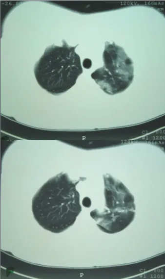
CT scan of the chest was performed at the beginning of treatment (Figure 1), showing: right middle and inferior lung lobes with reduced volume with elevation of the right diaphragm dome (already seen in previous tests); pulmonary consolidation in the right inferior lobe with air bronchograms; and extensive involvement of the left lung with diffuse ground glass opacities. Baseline C-reactive protein level was 160.00 mg/mL (reference value of 5 mg/L) and complete blood count was normal: hemoglobin 13.1g/dL; white blood cells 5,600/ mm3; neutrophils 4,032/mm3 with no band cells; and platelets 201,000/mm3. Blood cultures (two aerobic/anaerobic pairs) were negative and qualitative test for Influenza A (H1N1) virus using RT-PCR was positive.
References
Arq Bras Cardiol 2009; 93(6) : e91-e92
Bacal et al
Influenza A (H1N1) in a Transplant Patient
e92
Case Report
Discussion
In March 2009, there was an outbreak of Influenza A H1N1 virus infection in Mexico1. It rapidly spread worldwide and, in June 11, 2009, the World Health Organization (WHO) moved the alert level to the highest level of a pandemic. The
latest WHO’s data show more than 200 thousand confirmed cases worldwide, almost 60% of which concentrated in the Americas2. The exact number of cases in Brazil is not available, since as from July 16, the Ministry of Health has prioritized notification, investigation and treatment only of the cases with Acute Respiratory Distress Syndrome (ARDS) and of people with risk factors for disease complications: obese individuals; pregnant women; immunocompromised individuals; those with chronic diseases; children under two years of age; and the elderly. Until August 22, there were 5,206 confirmed cases of ARDS due to Influenza A H1N1 in Brazil, with a total of 557 deaths3,4.
In this pandemic, a trend has been observed of a greater number of cases of ARDS and deaths among populations at younger age ranges for Influenza A H1N1 than for seasonal influenza; a significant percentage of those (approximately 45%) had no risk factors for complications3-5.
Most of the deaths result from severe pulmonary involvement with rapid progression to ARDS and multi-organ failure6. Lung injury is caused, in most of the patients, by an effect of the influenza virus infection itself and not by secondary nosocomial infections. Possible mechanisms include direct injury to the respiratory epithelium by the virus and lesion secondary to the exuberant inflammatory response generated by a storm of cytokines and other inflammatory mediators7.
The case reported refers to a heart transplant patient, therefore classified in the population at a higher risk of complications due to immunodepression and a small baseline pulmonary function reserve. At baseline, extensive pulmonary involvement by the infection was identified. Thus, the inflammatory response generated was expected to lead to severe ARDS. However, this was not the outcome observed. Despite worsening of the baseline hypoxia and need for non-invasive ventilation, the patient did not develop respiratory failure or other organ failure.
We believe that the fact that the patient was receiving immunosuppressive therapy may have modulated the deleterious effect that an exacerbated inflammatory response would have generated, thus sparing her from a more severe form of Influenza A H1N1 virus infection. Should this finding be observed in similar cases, it will possibly influence the management of the disease, since immunosuppressive therapy may modulate the pulmonary and systemic inflammatory response that significantly contributes to the complications related to the disease.
Figure 1 – CT-scan of the chest images showing extensive involvement of the left lung with diffuse ground glass opacities.
1. Outbreak of swine-origin influenza A (H1N1) virus infection - Mexico, March-April 2009. MMWR Morb Mortal Wkly Rep. 2009; 58 (17): 467-70.
2. World Health Organization. Influenza A (H1N1) update [Acessed 2008 Aug 28]. Available from: http://www.who.int/csr/don/2009.
3. Situação epidemiológica da nova influenza A (H1N1) no Brasil. 2009. [Acesso em 2009 ago 28]. Disponível em: http://portal.saude.gov.br.
4. Ministério da Saúde. Secretaria de Vigilância em Saúde. Gabinete Permanente de Emergências de Saúde Pública. Brasília; 2009.
5. Chowell G, Bertozzi SM, Colchero MA, Lopez-Gatell H, Alpuche-Aranda C, Hernandez M, et al. Severe respiratory disease concurrent with the circulation of H1N1 Influenza. N Engl J Med. 2009; 361 (7): 674-9.
6. Peiris JS, Poon LL, Guan Y. Emergence of a novel swine-origin influenza A virus (S-OIV) H1N1 virus in humans. J Clin Virol. 2009; 45: 169-73.