Review Article
Key Words
Echocardiography / techniques; heart failure; pacemaker artificial.
Echocardiography In Cardiac Resynchronization Therapy
Viviane Cordeiro Veiga
1,2, Henry Abensur
1, Salomón Soriano Ordinola Rojas
1,2Real e Benemérita Associação Portuguesa de Beneficência1, São Paulo, SP; Universidade Estadual de Campinas (UNICAMP)2, Campinas, SP - Brazil
Mailing address: Viviane Cordeiro Veiga •
Rua Martiniano de Carvalho, 864 - cj 310 - Bela Vista – 01321-001 São Paulo, SP - Brazil
E-mail: [email protected]
Manuscript received October 13, 2008; revised manuscript received November 27, 2008; accepted December 11, 2008.
Summary
Cardiac resynchronization therapy has been an effective option in patients with advanced heart failure. However, 20 to 30% of the patients do not benefit from this therapy. Clinical, electrocardiographic and echocardiographic criteria have been studied in an attempt to select patients who will benefit from a cardiac resynchronization therapy, and the echocardiogram is important both in the selection and in the evaluation and optimization of the therapy. The objective of this review is to describe the main echocardiographic parameters used in the evaluation of the cardiac resynchronization therapy.
Introduction
Heart failure is a condition with high morbidity and mortality, which affects approximately 23 million people in the world1 approximately two million new cases are diagnosed
each year2, with an annual cost estimated at US$ 198 million,
according to DATASUS3.
Cardiac resynchronization therapy (CRT) has been an effective option in patients with advanced heart failure, providing clinical benefits, such as improved functional class, ability to exercise, and quality of life, as well as reduced hospitalization and mortality4.
The first clinical report of biventricular pacing is credited to Cazeau et al5 in 1994, and the therapy had its approval for clinical
use by the Food and Drug Administration (FDA) in 20016.
The indication for CRT, according to the guidelines of the American Heart Association, is class I for patients in functional class III or IV (New York Heart Association), with optimized heart failure therapy, an ejection fraction of less than 35% on the echocardiogram, and QRS ≥ 120 ms and sinus rhythm on the electrocardiogram7.
However, approximately 20 to 30% of patients do not respond to CRT8,9, and various clinical, electrocardiographic
and echocardiographic parameters have been used for the selection of candidates for this therapy.
The objective of this review is to describe the main echocardiographic parameters used in the evaluation of the cardiac resynchronization therapy.
Echocardiographic parameters for selecting
patients for CRT
The echocardiographic evaluation before the implantation of a biventricular pacemaker consists mainly of the following: confirming the presence of left ventricular function deterioration, assessing the presence of atrioventricular, interventricular and intraventricular dyssynchrony, and evaluating associated structural abnormalities that may hinder the implantation of the pacemaker10.
Assessment of left ventricular function
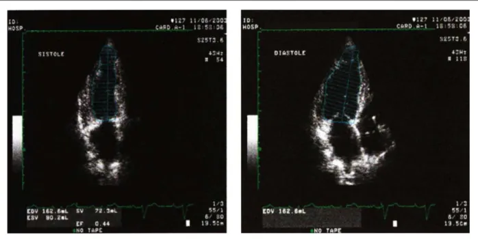
The assessment of systolic function should be performed by the two-dimensional method (Simpson) to assess the left ventricle ejection fraction indication for CRT is a value of less than 35%9 (Figure 1).
Assessment of atrioventricular dyssynchrony
The atrioventricular dyssynchrony may affect ventricular function due to almost simultaneous atrial and ventricular contractions, which cause a reduction in the preload by decreasing the atrial contraction. Atrioventricular dyssynchrony is characterized when the pre-ejection aortic time exceeds 140 ms, or if the diastolic filling time is less than 40% of the cardiac cycle, by the measurement of the time interval between the beginning of the E wave and the end of the A wave in the mitral flow11-13.
Assessment of interventricular dyssynchrony
The interventricular dyssynchrony may be assessed by pulsed Doppler ultrasonography, using the difference between the left and right ventricular electromechanical delays by measuring the interval between the R wave of the electrocardiogram and the beginning of the aortic and pulmonary flow velocity curves. If the difference between the two intervals is greater than 40 ms, this is indicative of interventricular dyssynchrony12,14-17 (Figure 2). The limitation
of this analysis is that the ventricles can not be assessed simultaneously. Furthermore, pathological conditions such as pulmonary hypertension may be associated with a prolongation of the pulmonary pre-ejection time interval, thus limiting its specificity.
Figure 1 -Assessment of left ventricular function by two-dimensional method (A - systole, B - diastole).
Figure 2 -Assessment of interventricular dyssynchrony, measured by the difference between the electromechanical delay of the ventricles (A - left ventricle,
B - right ventricle).
segments. The assessment is done by measuring the difference between the time intervals characterized by the onset of the QRS complex on the electrocardiogram to the peak (or beginning) of the S-wave on the tissue Doppler recording, respectively measured in the right ventricular free wall and the left ventricular lateral wall18-20. However, there is no consensus
about the cut-off for this measurement.
Assessment of intraventricular dyssynchrony
Intraventricular dyssynchrony promotes electromechanical delays between the walls of the left ventricle, and there are several ways to analyze it through an echocardiography.
Pitzalis et al21, in 2002, used the M mode for the assessment
Review Article
Veiga et al
Echocardiography in cardiac resynchronization
Figure 3 -Assessment of intraventricular dyssynchrony by M mode .
the left ventricle. This is done in the parasternal short axis view, by measuring the time interval between the maximum systolic excursion of the two walls. More than 130ms denotes significant dyssynchrony.
This method has limitations in patients with coronary artery disease who present hypokinetic or akinetic areas, where the identification of the peak systolic excursion is often limited. Moreover, only the middle regions of the two walls are evaluated10,12,21-23 (Figure 3).
Tissue Doppler can be used to obtain the myocardial velocities of the basal segments of septal, lateral, anterior and inferior walls in the apical plane. The time interval between the beginning of the QRS complex to the peak systolic myocardial wave (S-wave) is measured in these segments. A delay between any two segments of more than 65 ms is indicative of significant dyssynchrony19,20,24-29.
Yu et al30 evaluated the delay between the beginning of the
QRS complex and the tissue Doppler peak systolic myocardial wave in twelve segments, concluding that there are two parameters which are indicative of intraventricular dyssynchrony: maximum time difference between 2 distinct segments greater than 100 ms, and standard deviation of time to peak systolic velocity in 12 segments exceeding 33ms30.
Another technique for the assessment of intraventricular dyssynchrony derived from tissue Doppler is tissue tracking, which expresses the full speed of myocardial motion in color, from the ventricular apex to the base. The absence of displacement is characterized by the absence of color31,32.
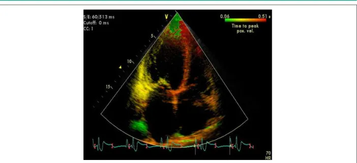
Tissue synchronization is a technique that uses color coding for electromechanical delay in each segment of the myocardium. The equipment measures the time interval between the beginning of the QRS complex and the peak systolic wave (S wave) at a specific point of the myocardium. If the time interval is less than 150ms (normal), the myocardium is represented in green; if it ranges between 150-300ms the myocardium is represented in yellow; and if it is greater than 300ms the myocardium is
represented in red. It should be used with caution in apical segments18 (Figure 4).
More recently, the three-dimensional echocardiography has been used for indicating and assessing patients undergoing cardiac resynchronization therapy with a biventricular pacemaker.
The three-dimensional echocardiography provides the percentage of cardiac dyssynchrony by measuring the rate of cardiac dyssynchrony (sigma). In this method, the left ventricle is studied in models of 16 or 17 parietal segments, and the regional and global contractility is analyzed (Figure 5). The sigma represents the standard deviation of the mean end-systolic contraction time of each segment compared to global end-systolic contraction (a lower index indicates lower dyssynchrony); a value of less than 8% is considered normal. The disadvantage of this method is the low number of frames33-35.
New techniques have been developed to evaluate intraventricular dyssynchrony, such as the two-dimensional strain, in which a computer software analyzes the deformation suffered by the muscle in two dimensions and not only toward the Doppler36-38 beam. Assessment of the ventricular electrode
implant site.
Evaluation of the site of the ventricular electrode implant Ansalone et al36 demonstrated that when the electrode was
implanted in the left ventricle, in the site of greatest activation delay, a better response to CRT was obtained, and in 35% of cases the lateral wall was the most affected. Therefore, the determination by echocardiography of the region with higher electromechanical delay indicates the best place to implant the electrode37.
Echocardiographic parameters in the
evaluation of patients undergoing CRT
Figure 4 -Avaliação da dissincronia ventricular pela técnica de sincronização tecidual.
Figure 5 -Assessment of ventricular dyssynchrony by tissue synchronization technique.
evaluation of patients undergoing CRT: • Increase in ejection fraction; • Reduction in mitral regurgitation;
• Regression of ventricular remodeling, characterized by a reduction by at least 15% in the left ventricle end systolic volume21;
• Presence of atrioventricular, interventricular and intraventricular synchrony;
• Adjustment of the atrioventricular interval (AV synchrony), performed using pulsed Doppler in mitral flow, by measuring the time interval between the onset of the E wave and the end of the A wave. The optimization of the atrioventricular interval is recommended when: the A wave of mitral flow is not identified, when the E and A waves are merged, or when the E wave is truncated by mitral valve closure 14.
Conclusion
Echocardiography is a diagnostic method that has been widely
used both in the indication and in the evaluation and optimization of post-operative patients who undergo cardiac resynchronization therapy. However, this technology is still evolving and there are no definitive echocardiographic parameters that can firmly determine or exclude the presence of significant dyssynchrony.
Potential Conflict of Interest
No potential conflict of interest relevant to this article was reported.
Sources of Funding
There were no external funding sources for this study.
Study Association
Review Article
Veiga et al
Echocardiography in cardiac resynchronization
References
1. McAlister FA, Teo KK, Taher M, Montague TJ, Humen D, Cheung L, et al. Insights into the contemporary epidemiology and outpatient management of congestive heart failure. Am Heart J. 1999; 138 (1 Pt 1): 87-94.
2. Rossi Neto JM. A dimensão do problema da insuficiência cardíaca no Brasil e no mundo. Rev Soc Cardiol Estado de São Paulo. 2004: 1: 1-10.
3. Ministério da Saúde. Sistema de Informações Hospitalares do SUS. Morbidade hospitalar do SUS. [Acesso em 2008 nov 10]. Disponível em: http://www.datasus. gov.br
4. Galvão Filho SS, Vasconcelos JTM, Barcelos CB, Rabello AC. Seleção de pacientes e modos de estimulação cardíaca no tratamento da disfunção ventricular. Rev Soc Cardiol Estado de São Paulo. 2004; 1: 43-54.
5. Cazeau S, Ritter P, Bakdach S, Lazarus A, Limousin M, Henao L, et al. Four chamber pacing in dilated cardiomiopathy. Pacing Clin Electrophysiol. 1994; 17 (11 Pt 2): 1974-9.
6. Aranda JM, Woo GW, Schofield RS, Handberg EM, Hill JA, Curtis AB, et al. Management of heart failure after cardiac resynchronization therapy – integrating advanced heart failure treatment with optimal device function. J Am Coll Cardiol. 2005; 46 (12): 2193-8.
7. Epstein AE, DiMarco JP, Ellenboger KA, Estes NA 3rd, Freedman RA, Gettes LS, et al. ACC/AHA/HRS 2008. Guidelines for device-based therapy of cardiac rhythm abnormalities: executive summary. Heart Rhytm. 2008; 5 (6): 934-55.
8. Leclercq C, Faris O, Tunin R, Johnson J, Kato R, Evans F, et al. Systolic improvement and mechanical resynchronization does not require electrical synchrony in the dilated failing heart with left bundle-branch block. Circulation. 2002; 106 (14): 1760-3.
9. Abraham WT, Fisher WG, Smith AL, Delurgio DB, Leon AR, Loh E, et al. MIRACLE Study Group Multicenter InSync Randomized Clinical Evaluation. Cardiac resynchronization in chronic heart failure. N Engl J Med. 2002; 346 (24): 1845-53.
10. Mullens W, Tang WH, Grimm RA. Using echocardiography in cardiac resynchronization therapy. Am Heart J. 2007; 154 (6): 1011-20.
11. Cazeau S, Gras D, Lazarus A Ritter P, Mugica J. Multisite stimulation for correction of cardiac asynchrony. Heart. 2000; 84 (6): 579-81.
12. Rodrigues ACT, Tsutsui JM, Mathias Jr W. Avaliação de dissincronia cardíaca pelo ecocardiograma: estratégias para um resultado adequado. Rev Bras Ecocardiogr. 2006; 19 (3): 46-51.
13. Auricchio A, Stellbrink C, Block M, Sack S, Vogt J, Bakker P, et al. Effect of pacing chamber and atrioventricular delay on acute systolic function of paced patients with congetive heart failure: the pacing therapies for congestive heart failure study group. The Guidant congestive heart failure research group. Circulation. 1999; 99 (23): 2993-3001.
14. Gorcsan J, Abraham T, Agler DA, Bax JJ, Derumeaux G, Grimm RA, et al. Echocardiography for cardiac resynchronization therapy: recommendations for performance and reporting – a report from the American Society of Echocardiography Dyssynchrony writing Group Endorsed by the Heart Rhytm Society. J Am Soc Echocardiogr. 2008; 21 (3): 191-213.
15. Agler DA, Adams DB, Waggoner AD. Cardiac resynchronization therapy and the emerging role of echocardiography (Part 2): the comprehensive examination. J Am Soc Echocardiogr. 2007; 20 (1): 76-90.
16. Bax JJ, Ansalone G, Breithardt O, Derumeaux G, Leclercq C, Schalij MJ, et al. Echocardiographic evaluation of cardiac resynchronization therapy: ready for routine clinical use? A critical appraisal. J Am Coll Cardiol. 2004; 44 (1): 1-9.
17. Bordachar P, Lafitte S, Reuter S, Sanders P, Jais P, Haissaguerre M, et al. Echocardiographic parameters of ventricular dyssynchrony validation in patients with heart failure using sequential biventricular pacing. J Am Coll Cardiol. 2004; 44 (11): 2157-65.
18. Penicka M, Bartunck J, De Bruync B, Vanderheyden M, Goethals M, De Zutter M, et al. Improvement of left ventricular function after cardiac resynchronization therapy is predicted by tissue Doppler imaging echocardiography. Circulation. 2004; 109 (8): 978-83.
19. Yu CM, Fung JW, Zhang Q, Chan CK, Chan YS, Lin H, et al. Tissue Doppler imaging is superior to strain rate imaging and postsystolic shortening on the prediction of
reverse remodeling in both ischemic and nonischemic heart failure after cardiac resynchronization therapy. Circulation. 2004; 110 (1): 66-73.
20. Bax JJ, Bleeker GB, Marwick TH, Molhoek SG, Boersma E, Steendijk P, et al. Left ventricular dyssynchrony predicts response and prognosis after cardiac resynchronization therapy. J Am Coll Cardiol. 2004; 44 (9): 1834-40.
21. Pitzalis MV, Iacoviello M, Romito R, Massari F, Rizzon B, Luzzi G, et al. Cardiac resynchronization therapy tailored by echocardiographic evaluation of ventricular asynchrony. J Am Coll Cardiol. 2002; 40 (9): 1615-22.
22. Waggoner AD, Agler DA, Adams DB. Cardiac resynchronization therapy and the emerging role of echocardiography (part 1): indications and results from current studies. J Am Soc Echocardiogr. 2007; 20 (1): 70-5.
23. Pitzalis MV, Iacoviello M, Romito R, Guida P, De Tommasi E, Luzzi G, et al. Ventricular asynchrony predicts a better outcome in patients with chronic heart failure receiving cardiac resynchronization therapy. J Am Coll Cardiol. 2005; 45 (1): 65-9.
24. Parro Jr A, Paulitsch FS, Cherubin MC, Miola L, Armstron SF, Sierra MAC, et al. Emprego da ecocardiografia convencional na avaliação de pacientes com miocardiopatia dilatada, candidatos à terapia de ressincronização. Reblampa. 2006; 19 (1): 34-44.
25. Bleeker GB, Schalij MJ, Molhoek SG, Verwey HF, Holman ER, Boersma E, et al. Relationship between QRS duration and left ventricular dyssynchrony in patients with end-stage heart failure. J Cardiovasc Electrophysiol. 2004; 15 (5): 544-9.
26. Yu CM, Fung WH, Lin H, Zhang Q, Sanderson JE, Lau CP. Predictors of left ventricular reverse remodeling after cardiac resynchronization therapy for heart failure secondary to idiopathic dilated a ischemic cardiomyopathy. Am J Cardiol. 2003; 91 (6): 684-8.
27. Yu CM, Zhang Q, Fung JW, Chan HC, Chan YS, Yip GW, et al. A novel tool to assess systolic asynchrony and identify responders of cardiac resynchronization therapy by tissue synchronization imaging. J Am Coll Cardiol. 2005; 45 (1): 677-84.
28. Bax JJ, Molhoek SG, van Erven LVoogd PJ, Somer S, Boersma E, et al. Usefulness of myocardial tissue Doppler echocardiography to evaluate left ventricular dyssynchrony before and after biventricular pacing in patients with idiopathic dilated cardiomyopathy. Am J Cardiol. 2003; 91 (1): 94-7.
29. Gorcsan J, Kanzaki H, Bazaz R, Dohi K, Schwartzman D. Usefulness of echocardiographic tissue synchronization imaging to predict acute response to cardiac resynchronization therapy. Am J Cardiol. 2004; 93 (9): 1178-81.
30. Yu CM, Lin H, Zhang Q, Sanderson JE. High prevalence of left ventricular systolic and diastolic asynchrony in patients with congestive heart failure and normal QRS duration. Heart. 2003; 89 (1): 54-60.
31. Silva CES, Barreto ACP. Avaliação ecocardiográfica da terapia de ressincronização cardíaca. Arq Bras Cardiol. 2005; 84 (6): 50-7.
32. Pan C, Hoffmann R, Kühl H, Severin E, Franke A, Hanrath P. Tissue tracking allows rapid and accurate visual evaluation of left ventricular function. Eur J Echocardiogr. 2001; 2 (3): 197-202.
33. Vieira MLC, Morhy SS, Maddukuri P, Phang R, Fischer C, Lira Fº E, et al. O ecocardiograma na terapia de ressincronização. Einstein. 2005; 3 (1): 41-5.
34. Roelondt JR, Yao J, Kasprzak JD. Three-dimensional echocardiography. Curr Opin Cardiol. 1998; 13 (6): 386-96.
35. Vieira MLC, Cury AF, Naccarato G, Oliveira WA, Monaco CG, Cordovil A, et al. Índice de dissincronia ventricular: comparação com a fração de ejeção bidimensional e tridimensional. Arq Bras Cardiol. 2008; 91 (3): 142-7.
36. Ansalone G, Giannantoni P, Ricci R, Trambaiolo P, Fedele F, Santini M. Doppler myocardial imaging to evaluate the effectiveness of pacing sites in patients receiving biventricular pacing. J Am Coll Cardiol. 2002; 39 (3): 489-99.
37. Kindermann M, Frolhig G, Doerr T, Schieffer H. Optimizing the AV delay in DDD pacemaker patients with high degree AV block: mitral valve Doppler versus impedance cardiography. Pacing Clin Electrophysiol. 1997; 20 (10 Pt 1): 2453-462.