AR
TIGO ORIGINAL / ORIGINAL AR
TICLE
INTRODUCTION
The relation between liver iron stores and al-coholic liver disease (ALD) is well established. In 1964, hemochromatosis was thought to be a peculiar manifestation of alcoholic cirrhosis(10, 11). Despite hemochromatosis being a different disease, up to
33% of ALD patients(8) and more than half of ALD
patients with advanced cirrhosis present with excessive liver iron stores(27). Even so, iron stores never reach the same intensity in ALD as they do in hereditary hemochromatosis(20, 32).
Excessive liver iron content has been characterized as an independent risk factor for ibrosis, while in alcoholic cirrhosis the accumulated liver iron concen-trations correlate inversely with patient survival(13). It is known that serum iron and ferritin increase linearly with daily alcohol consumption(34). Moreover, serum ferritin levels are higher in ALD patients than in non-alcoholic patients with other chronic liver diseas-es, such as autoimmune disorders or chronic hepatitis
HFE
MUTATIONS AND IRON OVERLOAD IN
PATIENTS WITH ALCOHOLIC LIVER DISEASE
Luís
COSTA
-
MATOS
1, 2, Paulo
BATISTA
2, Nuno
MONTEIRO
2,
Pedro
HENRIQUES
2, Fernando
GIRÃO
2and Armando
CARVALHO
1ABSTRACT – Context - Alcoholic liver disease (ALD) is generally associated with iron overload, which may contribute to its patho-genesis, through increased oxidative stress and cellular damage. There are conlicting reports in literature about hemochromatosis (HFE) gene mutations and the severity of liver disease in alcoholic patients. Objectives - To compare the prevalence of mutations in the hemochromatosis (HFE) gene between patients with ALD and healthy controls; to assess the relation of HFE mutations with liver iron stores and liver disease severity. Methods - Liver biopsy specimens were obtained from 63 ALD patients (during routine treatment) and 52 healthy controls (during elective cholecystectomy). All individuals underwent routine liver function tests and
HFE genotyping (to detect wild-type sequences and C282Y, H63D, S65C, E168Q, E168X, V59M, H63H, P160delC, Q127H, Q283P, V53M and W164X mutations). Associations between HFE mutations and risk of excessive liver iron stores, abnormal serum ferritin, liver ibrosis, or necroinlammatory activity were assessed by multivariate logistic regression analysis. Results - ALD patients had signiicantly higher serum ferritin and transferrin saturation than controls (both P<0.05), but the distribution of HFE mutations was similar between the two groups. For ALD patients, the odds ratio for having at least one HFE mutation and excessive liver iron stores was 17.23 (95% conidence interval (CI): 2.09-142.34, P = 0.008). However, the presence of at least one HFE mutation was not associated with an increased risk of liver ibrosis or necroinlammatory activity. Active alcohol ingestion showed the strongest association to increased serum ferritin (OR = 8.87, 95% CI: 2.11-34.78, P = 0.003). Conclusions - ALD patients do not present with a differential proile of HFE mutations from healthy controls. In ALD patients, however, the presence of at least one HFE mutation increases the risk of having excessive liver iron stores but has no detectable effects on liver disease activity or severity.
HEADINGS - Alcoholic liver disease. Membrane proteins. Iron. Hemochromatosis.
Conflicts of interest: The authors declare they have no conflicts of interest related to the publication of this study. Supportive foundations: Tondela-Viseu Hospital Centre E.P.E., Portugal.
1Faculty of Medicine of the University of Coimbra, 3004-504 Coimbra, Portugal; 2Tondela-Viseu Hospital Centre E.P.E., 3504-509 Viseu, Portugal.
Correspondence: Dr. Luís Costa Matos - Serviço de Medicina Interna I, Centro Hospitalar Tondela-Viseu E.P.E. - 3504-509, Portugal. E-mail: [email protected]
C virus (HCV) infection. Interestingly, upon alcohol withdrawal, the increased serum ferritin levels have been shown to rapidly decrease(2, 24).
Alcohol consumption has been implicated as the main cause of increased serum ferritin levels in the general population(17, 24). The apparent sensitivity of this pathological process was further indicated by a study showing that even moderate alcohol ingestion can affect iron metabolism(18).
In 1996, a G→A transition at nucleotide 845 in the HLA-H gene was found to be present as a homozy-gous mutation in 80% of hereditary hemochromatosis patients(7). According to this disease association, the gene and its encoded protein were renamed as
hemochromatosis (HFE) gene and HFE protein.
Several other HFE mutations have been identiied and
characterized, including those with very high allelic frequen-cy (H63D: 14%) and others with very low allelic frequenfrequen-cy (S65C: 0.5%). Interpreting the presence of these mutations, however, requires some caution. Homozygous carriers of H63D have been found to not be at risk for iron overload. The S65C allele appears to exert its inluence only when inherited with homozygous C282Y, wherein it worsens the iron
over-load(1, 16). In contrast, compound heterozygous C282Y/H63D
or H63D homozygosity may produce only slight increases in serum ferritin and transferrin saturation levels. However, in patients presenting with overt iron overload of unknown cause, it is necessary to investigate both the presence of HFE
mutations and other non-genetic causes(4, 22).
Hereditary hemochromatosis is considered a functional paradigm of the synergy between alcohol and iron. Under conditions of excessive alcohol ingestion, the phenotypical expression of iron overload increases(31). Patients with he-mochromatosis who ingest more than 60 g of alcohol daily have been characterized as having a 9-fold increased risk of developing cirrhosis(9). In addition, some studies have
suggested that HFE mutations may play a role in ALD
severity, but other studies have not conirmed these results. The current study was designed to compare the prevalence
of HFE mutations between ALD patients and a healthy
control population and assess their relations with excessive liver iron stores and liver disease severity (ibrosis grade and necroinlammatory activity).
METHODS
Participants
The study design conformed to the ethical guidelines of the 1975 Declaration of Helsinki and was approved by the local Ethics Committees of the University of Coimbra and the Tondela-Viseu Hospital Centre, Portugal.
Patients were recruited from the Liver Diseases Outpatient Consultation Clinic at Tondela-Viseu Hospital Centre. Study enrolment was based on the following inclusion criteria: ALD diagnosis; recent liver biopsy for disease grading and staging, doubts about diagnosis, or the presence of concurrent liver disease. Patients were refused enrolment according to the following exclusion criteria: gastrointestinal bleeding; blood transfusion within the previous 3 months; other relevant liver or systemic diseases; and refusal to provide informed written consent. Of the 69 patients who fulilled the criteria, 2 were subsequently excluded for lack of suficient liver tissue and 4 for other liver or systemic diseases that were identiied during the follow-up exams.
Healthy controls were recruited from the patient popu-lation that was admitted for elective cholecystectomy due to non-complicated gallstones, during which a small liver sample had been obtained during surgery for another study. The inclusion criteria for healthy controls were: alcohol con-sumption <20 g/day in women and <40 g/day in men; level of serum ferritin above the lower limit of normal (LLN); and levels of alanine aminotransferase (ALT), aspartate
aminotransferase (AST), g-glutamyl transpeptidase (GGT), and alkaline phosphatase (ALP) lower than 3 times the ULN. Patients were excluded according to: anemia; other relevant liver or systemic diseases; of evidence of inlammation, de-ined as C reactive protein (CRP) >2.5 mg/dL; and refusal to provide informed written consent. Finally, 52 controls were enrolled in the study.
Histopathologic evaluation
Liver tissue samples were ixed in 10% formalin buffer, embedded in parafin, sectioned, and stained with hematoxy-lin and eosin (H-E) and Perls’ Prussian blue. Since a validated histological score for ALD does not yet exist, the non-alco-holic steatohepatitis (NASH) scoring method was adapted(19). Briely, a semi-quantitative evaluation was performed for the following parameters: steatosis (absent, light, moderate, severe); necroinlammatory activity (absent, light, moderate, severe); ibrosis (absent, periportal or perisinusoidal, peri-portal and perisinusoidal, bridging ibrosis, cirrhosis); and iron stores grade (with Perls coloration and evaluation with Scheuer’s scale). “Signiicant liver iron stores” were deined as Perls grade 2 or higher. “Signiicant ibrosis” was assigned according to the presence of periportal and perisinusoidal, bridging ibrosis, or cirrhosis. Necroinlammatory activity was generally categorized as absent or present, regardless of grade. Any amount of alcohol ingested on a daily basis led to categorization as “active alcoholism”.
Biochemical and imaging evaluations
Cases and controls underwent routine laboratory test-ing for markers of liver disease, inlammation, and kidney function. Speciically, measurements were taken of complete blood count, international normalized ratio (INR), glucose, ALT, AST, GGT, ALP, CRP, ceruloplasmin, a1-antitripsin, serum iron, serum ferritin, and serum transferrin saturation. In addition, serological markers for hepatitis B virus (HBV) and hepatitis C virus (HCV) infections were assayed.
All study participants had undergone a previous liver ultrasound evaluation and the scan images were collected for assessment.
Blood samples were collected from all study participants
and used for HFE genotyping with the Haemochromatosis
StripAssay A (Vienna Lab, Vienna, Austria) according to the manufacturer’s protocol. This test detects some of the
very rare HFE mutations (E168Q, E168X, V59M, H63H,
P160delC, Q127H, Q283P, V53M, and W164X) as well as mutations in the ferroportin 1 gene and the transferrin receptor type 2 gene.
Statistical analysis
variables were analysed for normal distribution by using the Shapiro-Wilk test; then, the difference between two sample means was assessed for statistical signiicance by using the Student’s t-test or the non-parametrical Mann–Whitney U
test for non-normally distributed variables.
Multivariate logistic regression analysis was carried out to calculate odds ratios (OR) for the presence of at least
one HFE mutation using the following covariates: age,
sex, body mass index (BMI), active alcoholism, model for end-stage liver disease (MELD), haemoglobin, CRP, serum ferritin, and transferrin saturation. OR and 95% conidence intervals (CI) were determined for the following outcomes: signiicant liver iron stores vs no signiicant liver iron stores; necroinlammatory activity vs no necroinlammatory activity; and signiicant ibrosis vs no signiicant ibrosis. The same model, without the covariates serum ferritin and transferrin saturation, was used to evaluate abnormal serum ferritin (>322 ng/mL) vs normal serum ferritin.
Correlations between variables were evaluated with Spearman’s correlation coeficient. All P-values were
two-sid-ed and P<0.05 was considered statistically signiicant.
RESULTS
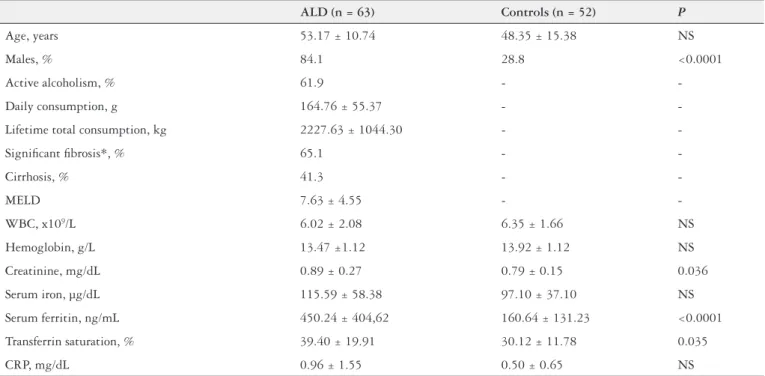
Table 1 presents the clinical and laboratory characteristics of ALD patients and healthy controls. The average age was similar between the two groups; however, the ALD patients had a signiicantly higher proportion of men. The majority of ALD patients had signiicant ibrosis, as evidenced by histological grading of liver samples. In addition, 41.3% of ALD patients were cirrhotic.
The ALD patients showed signiicantly higher levels of creatinine, but for both groups the levels were within normal range (considering standard deviations). Serum ferritin levels and transferrin saturation were also signiicantly higher in ALD patients than in controls. No signiicant differences were observed between the two groups for hemoglobin values, white blood cell count, or CRP.
Genotyping detected only three HFE mutations (C282Y,
H63D and S65C) among all the ALD patients and controls. In addition, neither the ferroportin 1 mutation nor the trans-ferrin receptors type 2 mutation was detected in any of the study samples. None of the ALD patients (or controls) were diagnosed as hereditary hemochromatosis (C282Y/C282Y) upon genotyping. There were no signiicant differences in genotype frequencies observed between the ALD patients and controls (Table 2). Heterozygous C282Y and compound heterozygous C282Y/H63D were detected only in ALD patients, and the prevalence of wild-type (WT) genotypes was slightly lower in the ALD patients; however, neither of
TABLE 1. Clinical and laboratory characteristics of ALD patients and healthy controls
ALD (n = 63) Controls (n = 52) P
Age, years 53.17 ± 10.74 48.35 ± 15.38 NS
Males, % 84.1 28.8 <0.0001
Active alcoholism, % 61.9 -
-Daily consumption, g 164.76 ± 55.37 -
-Lifetime total consumption, kg 2227.63 ± 1044.30 -
-Signiicant ibrosis*, % 65.1 -
-Cirrhosis, % 41.3 -
-MELD 7.63 ± 4.55 -
-WBC, x109/L 6.02 ± 2.08 6.35 ± 1.66 NS
Hemoglobin, g/L 13.47 ±1.12 13.92 ± 1.12 NS
Creatinine, mg/dL 0.89 ± 0.27 0.79 ± 0.15 0.036
Serum iron, µg/dL 115.59 ± 58.38 97.10 ± 37.10 NS
Serum ferritin, ng/mL 450.24 ± 404,62 160.64 ± 131.23 <0.0001
Transferrin saturation, % 39.40 ± 19.91 30.12 ± 11.78 0.035
CRP, mg/dL 0.96 ± 1.55 0.50 ± 0.65 NS
*perisinusoidal and portal/periportal ibrosis, bridging ibrosis, or cirrhosis
TABLE 2. HFE genotype frequencies*
ALD (n = 63) Controls (n = 52) P
WT/WT 61.9 65.4 NS
WT/C282Y 1.6 0 NS
C282Y/H63D 3.2 0 NS
WT/H63D 27.0 26.9 NS
H63D/H63D 3.2 1.9 NS
WT/S65C 1.6 5.8 NS
H63D/S65C 1.6 0 NS
At least one HFE mutation 38.1 32.7 NS
these differences reached statistical signiicance. The most frequently detected non-WT genotype, heterozygous H63D, was identically distributed between the two groups. The S65C mutations were slightly more prevalent in the controls.
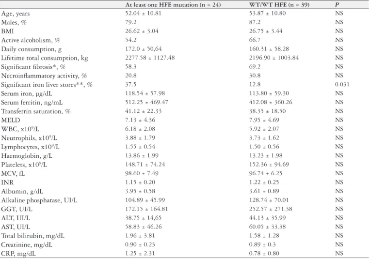
Table 3 presents the distribution of ALD patients with at least one HFE mutation in comparison to those with WT
genotypes. Correlation analysis indicated that the ALD pa-tients with at least one HFE mutation had signiicantly higher
liver iron stores (Perls grade 2 or higher) than those with WT genotypes. There was also a trend toward higher serum ferritin values in the ALD patients with at least one HFE mutation.
All other variables were similar between the two subgroups. Multivariate logistic regression analysis of the ALD pa-tients having at least one HFE mutation with adjustments
for age, sex, BMI, active alcoholism, MELD, haemoglobin, CRP, serum ferritin and transferrin saturation indicated that the OR for signiicant liver iron stores was 17.23 (95% CI: 2.09-142.34, P = 0.008). In contrast, the analysis indicated no
signiicantly increased risk associated with signiicant ibrosis (OR = 0.60, 95% CI: 0.14-2.67) or necroinlammatory activity (OR = 0.42, 95% CI: 0.07-2.68) (Figure 1).
The strongest determinant factor for having a serum ferri-tin higher than normal was the presence of active alcoholism (OR = 8.87, 95% CI: 2.11-34.78, P = 0.003). The presence of
TABLE 3. Comparison between ALD patients with and without any HFE mutation
At least one HFE mutation (n = 24) WT/WT HFE (n = 39) P
Age, years 52.04 ± 10.81 53.87 ± 10.80 NS
Males, % 79.2 87.2 NS
BMI 26.62 ± 3.04 26.75 ± 3.44 NS
Active alcoholism, % 54.2 66.7 NS
Daily consumption, g 172.0 ± 50,64 160.31 ± 58.28 NS
Lifetime total consumption, kg 2277.58 ± 1127.48 2196.90 ± 1003.84 NS
Signiicant ibrosis*, % 58.3 69.2 NS
Necroinlammatory activity, % 20.8 30.8 NS
Signiicant iron liver stores**, % 37.5 12.8 0.031
Serum iron, µg/dL 118.54 ± 57.98 113.80 ± 59.30 NS
Serum ferritin, ng/mL 512.25 ± 469.47 412.08 ± 360.26 NS
Transferrin saturation, % 41.12 ± 22.33 38.35 ± 18.50 NS
MELD 7.13 ± 4.36 7.95 ± 4.69 NS
WBC, x109/L 6.18 ± 2.08 5.92 ± 2.07 NS
Neutrophils, x109/L 3.88 ± 1.79 3.73 ± 1.62 NS
Lymphocytes, x109/L 1.55 ± 0.54 1.50 ± 0.56 NS
Haemoglobin, g/L 13.86 ± 1.99 13.23 ± 1.98 NS
Platelets, x109/L 148.71 ± 74.24 152.36 ± 94.69 NS
MCV, fL 98.60 ± 7.49 96.74 ± 6.25 NS
INR 1.15 ± 0.20 1.22 ± 0.25 NS
Albumin, g/dL 3.95 ± 0.58 3.61 ± 0.89 NS
Alkaline phosphatase, UI/L 104.89 ± 45.99 128.74 ± 70.01 NS
GGT, UI/L 172.15 ± 164.81 252.57 ± 271.38 NS
ALT, UI/L 38.75 ± 14,65 44.13 ± 35.99 NS
AST, UI/L 58.83 ± 46.26 60.05 ± 33.38 NS
Total bilirubin, mg/dL 1.96 ± 3.81 1.58 ± 1.28 NS
Creatinine, mg/dL 0.90 ± 0.23 0.89 ± 0.3 NS
CRP, mg/dL 1.25 ± 2.31 0.78 ± 0.80 NS
*perisinusoidal and portal/periportal ibrosis, bridging ibrosis or cirrhosis **Iron stores Perls grade 2 or higher
at least one HFE mutation (when adjusted for age, sex, BMI,
active alcoholism, MELD, haemoglobin, and CRP) did not signiicantly increase the risk of having an increased serum ferritin level (OR = 1.79, 95% CI: 0.47-6.79) (Figure 2).
Correlation analysis indicated that the serum ferritin level in ALD patients was signiicantly associated with Perls grade (rs = 0.542, P<0.0001), GGT levels (r
s = 0.374, P = 0.003), and ALT (rs = 0.265 P = 0.036), but not with AST, steatosis,
or ibrosis grades.
The variables showing the strongest associations with ibrosis grade were the amount of daily alcohol consumption (rs = 0.308, P = 0.014) and lifetime total alcohol consumption
(rs = 0.302, P = 0.016).
DISCUSSION
Our study population mirrored the known impact of alcoholism in our country, wherein most ALD patients are young/middle aged (mean: 53 years old) males with high daily alcohol intake, in many cases since childhood. In ad-dition, these patients often present with advanced ibrosis grades. Meanwhile, the control group relected the common trend of more women having gallstones than men. Unfor-tunately, these distinguishing features of the two groups may have induced a bias in our comparison of serum iron, ferritin, and transferrin saturation. We attempted to reduce the effects of this bias as much as possible by excluding all controls with hemoglobin and serum ferritin below normal or with biochemical evidence of inlammation. However, it is important to note that the purpose of this study was not to demonstrate that ALD patients have iron overload, as this fact is already suficiently well established in the literature. Since HFE mutations are not sex-linked, it is
unnecessary to match groups of ALD patients and healthy controls for sex when studying the prevalence of these mu-tations between the two groups.
In the current study, we did not ind evidence that HFE
mutations are more frequent in ALD patients; therefore, the iron overload that was present in this group must be explained by some other factor. We recently identiied inappropriately low liver hepcidin mRNA expression in alcoholic patients
as a potential cause of ALD iron overload, and this may be one such factor(5).
Comparison between the ALD patient subgroups
— with and without HFE mutations — provided more
interesting results. The subgroups were similar for age, percentage of males, BMI, percentage of active alcohol-ism, and amounts of alcohol ingested. We found that the presence of at least one HFE mutation increased the risk
of higher liver iron stores, but no evidence to suggest that it may have inluence on the disease severity; speciically, these carriers were not at an increased risk for signiicant ibrosis or necroinlammatory activity in their liver tissues.
The fact that having at least one HFE mutation did not
increase the risk of having ferritin above normal, despite active alcohol intake, is intriguing and future studies should assess the potentially different impact of HFE mutations
on systemic and liver iron stores.
Our study’s indings also conirmed that the most relevant factor for ALD progression to cirrhosis is the lifetime amount of alcohol consumed.
As serum ferritin levels showed a strong association with the liver biopsy Perls grade, we believe that this measurement represents a simple, cheap, effective, non-invasive marker for estimating the severity of liver iron stores in ALD patients.
Two previous studies have demonstrated correlation
between HFE mutations and iron overload/ALD
progres-sion(3, 33). In a Spanish population with advanced ALD,
H63D mutations were more frequent than in the general
population (OR = 1.66, 95% CI: 1.10-2.52, P = 0.01) and
no association was found with C282Y in particular(30).
Another study from Portugal found a correlation with the H63D mutation and liver disease in alcoholics who consumed >80 g/day (OR = 1.57, 95% CI: 1.02-2.40,
P = 0.02)(23). However, all other related studies have found no evidence of correlation for the HFE mutations and the
risk of developing ALD. For example, one British study
from Newcastle(15), involving 257 ALD patients and 117
controls, did not show any association between HFE
mu-tations and iron liver stores or advanced ibrosis. Another, from Shefield(14), also failed to demonstrate any difference
between the frequencies of HFE mutations between two
alcoholic groups, one with established cirrhosis and another without liver disease.
In general, studies from Caucasian populations, mainly
in Europe, have shown that HFE mutations are not
asso-ciated with alcohol-related liver disorders. A study from Spain found no differences in the distribution of C282Y or H63D mutations between alcoholic cirrhosis patients, HCV cirrhosis patients, and healthy controls. However, C282Y heterozygosity was much more frequent (20.9%) in a subgroup of the alcoholic cirrhosis patients with hepato-cellular carcinoma (vs those without tumour)(21). Similarly, a study from Switzerland found no association with C282Y heterozygosity and alcoholic cirrhosis(12). Yet another study from New Zealand(29), using patients in an inpatient alcohol addiction clinic, found no differences the frequencies of HFE
mutations compared to the general population and no
sociation of these mutations with liver function or liver iron stores. Similar reports came from Australia(26) and Poland(28).
Our data agrees with the majority of previously published studies showing no differences in the distributions of HFE
mutations among ALD patients and healthy controls. Like-wise, the evidence that these mutations are not associated with an increased risk of severe ibrosis or impaired liver function are concordant. The novelty of our work, however, lies in our demonstration that the presence of at least one
HFE mutation in ALD patients is signiicantly associated
with higher liver iron stores. This is an interesting inding, as tissue iron overload is thought to be relevant to ibrosis
and inlammation but neither of these features were strongly associated in our ALD patient group.
The collective indings from our study indicate that HFE
mutations may play a minor role in the ALD feature of in-creased liver iron stores while not conferring inin-creased risk for ibrosis grade or disease activity.
ACKNOWLEDGEMENTS
We are grateful to the Centro Hospitalar Tondela-Viseu E.P.E. for funding this research.
Costa-Matos L, Batista P, Monteiro N, Henriques P, Girão F, Carvalho A. Mutações HFE e sobrecarga de ferro em pacientes com doença hepática alcoólica. Arq Gastroenterol. 2013;50(1):35-41.
RESUMO – Contexto - A doença hepática alcoólica (DHA) está geralmente associada à sobrecarga de ferro, que pode contribuir para a sua patogênese, através do aumento do estresse oxidativo e dano celular. As descrições existentes na literatura sobre a associação entre mutações HFE e a gravidade da DHA nem sempre são concordantes. Objetivos - Comparar a prevalência de mutações HFE entre um grupo de pacientes com DHA e uma população de controle. Avaliar a relação entre mutações HFE e os depósitos de ferro hepático. Avaliar se a presença dessas mutações está associada com a gravi-dade da DHA. Métodos – Compararam-se 63 pacientes com DHA que efetuaram biopsia hepática com 52 controles saudáveis. A genotipagem HFE
(wild type, C282Y, H63D, S65C, E168Q, E168X, V59M, H63H, P160delC, Q127H, Q283P, V53M, W164X) e uma avaliação laboratorial de rotina (incluindo cinética do ferro) foram feitos em todos os indivíduos. Realizou-se regressão logística multivariada nos casos para avaliar se a presença de mutações HFE estava relacionada com risco aumentado de depósitos de ferro hepático aumentados, ferritina sérica anormal, ibrose hepática signiicativa ou atividade necroinlamatória. Resultados - Os pacientes apresentaram ferritina sérica e saturação da transferrina mais elevadas que os controles, mas não existiram diferenças signiicativas na distribuição de mutações HFE entre pacientes e controles. Considerando apenas os pacientes, o risco relativo de estes apresentarem pelo menos uma mutação HFE e depósitos de ferro hepático signiicativos foi de 17.23 (CI 95% 2.09-142.34,
P = 0.008). Contudo, a presença de pelo menos uma mutação HFE não esteve associada ao risco signiicativamente aumentado de ibrose ou ativi-dade necroinlamatória signiicativas. O fator mais determinante para apresentar ferritina sérica acima do normal foi a presença de alcoolismo ativo, com risco relativo de 8.87 (CI 95% 2.11-34.78, P = 0.003). Conclusões - Não existiram diferenças na distribuição de mutações HFE entre pacientes com DHA e controles normais. Nos pacientes, o achado de pelo menos uma mutação HFE aumentou o risco de ter depósitos de ferro hepático mais elevados, mas não para ter ibrose ou atividade necroinlamatória signiicativas.
REFERENCES
1. Aranda N, Viteri FE, Montserrat C, Arija V. Effects of C282Y, H63D, and S65C HFE gene mutations, diet, and life-style factors on iron status in a general Medi-terranean population from Tarragona, Spain. Ann Hematol. 2010;89:767-73. 2. Bell H, Skinningsrud A, Raknerud N, Try K. Serum ferritin and transferrin
saturation in patients with chronic alcoholic and non-alcoholic liver diseases. J Intern Med. 1994;236:315-22.
3. Bonkovsky HL, Lambrecht RW, Shan Y. Iron as a co-morbid factor in nonhe-mochromatotic liver disease. Alcohol. 2003;30:137-44.
4. Bulaj ZJ, Griffen LM, Jorde LB, Edwards CQ, Kushner JP. Clinical and bio-chemical abnormalities in people heterozygous for hemochromatosis. N Engl J Med. 1996;335:1799-805.
5. Costa-Matos L, Batista P, Monteiro N, Simões M, Egas C, Pereira J, Pinho H, Santos N, Ribeiro J, Cipriano MA, Henriques P, Girão F, Rodrigues A, Carvalho A. Liver hepcidin mRNA expression is inappropriately low in alcoholic patients compared with healthy controls. Eur J Gastroenterol Hepatol. 2012;24:1158-65. 6. European Association for the Study of the Liver. EASL clinical practice guidelines
for HFE hemochromatosis. J Hepatol. 2010;53:3-22. [PMID: 20471131]. 7. Feder JN, Gnirke A, Thomas W, Tsuchihashi Z, Ruddy DA, Basava A, Dormishian
F, Domingo R Jr., Ellis MC, Fullan A, Hinton LM, Jones NL, Kimmel BE, Kronmal GS, Lauer P, Lee VK, Loeb DB, Mapa FA, McClelland E, Meyer NC, Mintier GA, Moeller N, Moore T, Morikang E, Prass CE, Quintana L, Starnes SM, Schatzman RC, Brunke KJ, Drayna DT, Risch NJ, Bacon BR, Wolff RK. A novel MHC class I-like gene is mutated in patients with hereditary haemochro-matosis. Nat Genet. 1996;13:399-408. [PMID: 8696333].
8. Fletcher LM, Halliday JW, Powell LW. Interrelationships of alcohol and iron in liver disease with particular reference to the iron-binding proteins, ferritin and transferrin. J Gastroenterol Hepatol. 1999;14:202-14.
9. Fletcher LM, Dixon JL, Purdie DM, Powell LW, Crawford DH. Excess alcohol greatly increases the prevalence of cirrhosis in hereditary hemochromatosis. Gastroenterology. 2002;122:281-9.
10. Fletcher LM, Bridle KR, Crawford DH. Effect of alcohol on iron storage diseases of the liver. Best Pract Res Clin Gastroenterol. 2003;17:663-77.
11. Fletcher LM, Powell LW. Hemochromatosis and alcoholic liver disease. Alcohol. 2003;30:131-6.
12. Frenzer A, Rudzki Z, Norton ID, Butler WJ, Roberts-Thomson IC. Heterozygo-sity of the haemochromatosis mutation, C282Y, does not inluence susceptibility to alcoholic cirrhosis. Scand J Gastroenterol. 1998;33:1324.
13. Ganne-Carrié N, Christidis C, Chastang C, Ziol M, Chapel F, Imbert-Bismut F, Trinchet JC, Guettier C, Beaugrand M. Liver iron is predictive of death in alco-holic cirrhosis: a multivariate study of 229 consecutive patients with alcoalco-holic and/ or hepatitis C virus cirrhosis: a prospective follow up study. Gut. 2000;46:277-82. 14. Gleeson D, Evans S, Bradley M, Jones J, Peck RJ, Dube A, Rigby E, Dalton A. HFE genotypes in decompensated alcoholic liver disease: phenotypic expression and comparison with heavy drinking and with normal controls. Am J Gastro-enterol. 2006;101:304-10.
15. Grove J, Daly AK, Burt AD, Guzail M, James OF, Bassendine MF, Day CP. Heterozygotes for HFE mutations have no increased risk of advanced alcoholic liver disease. Gut. 1998;43:262-6.
16. Hanson EH, Imperatore G, Burke W. HFE gene and hereditary hemochromatosis: a HuGE review. Human genome epidemiology. Am J Epidemiol. 2001;154:193-206.
17. Hearnshaw S, Thompson NP, McGill A. The epidemiology of hyperferritinaemia. World J Gastroenterol. 2006;12:5866-9.
18. Ioannou GN, Dominitz JA, Weiss NS, Heagerty PJ, Kowdley KV. The effect of alcohol consumption on the prevalence of iron overload, iron deiciency, and iron deiciency anemia. Gastroenterology. 2004;126:1293-301.
19. Kleiner DE, Brunt EM, Van Natta M, Behling C, Contos MJ, Cummings OW, Ferrell LD, Liu YC, Torbenson MS, Unalp-Arida A, Yeh M, McCullough AJ, Sanyal AJ; Nonalcoholic Steatohepatitis Clinical Research Network. Design and validation of a histologic scoring system for nonalcoholic fatty liver disease. Hepatology 2005;41:1313–21.
20. Kohgo Y, Ohtake T, Ikuta K, Suzuki Y, Hosoki Y, Saito H, Kato J. Iron accu-mulation in alcoholic liver diseases. Alcohol Clin Exp Res. 2005;29:189s-93s. 21. Lauret E, Rodríguez M, González S, Linares A, López-Vázquez A, Martínez-Borra
J, Rodrigo L, López-Larrea C. HFE gene mutations in alcoholic and virus-re-lated cirrhotic patients with hepatocellular carcinoma. Am J Gastroenterol. 2002;97:1016-21.
22. Lim EM, Rossi E, De Boer WB, Reed WD, Jeffrey GP. Hepatic iron loading in patients with compound heterozygous HFE mutations. Liver Int. 2004;24:631-6. 23. Machado MV, Gonçalves MS, Cortez-Pinto H. Metabolismo do ferro em alcoóli-cos crónialcoóli-cos em consumo activo e durante a abstinência. J Port Gastrenterol. 2004;11:186-90.
24. Machado MV, Ravasco P, Martins A, Almeida MR, Camilo ME, Cortez-Pinto H. Iron homeostasis and H63D mutations in alcoholics with and without liver disease. World J Gastroenterol. 2009;15:106-11.
25. Milman N, Ovesen L, Byg K, Graudal N. Iron status in Danes updated 1994. I: prevalence of iron deiciency and iron overload in 1332 men aged 40-70 years. Inluence of blood donation, alcohol intake, and iron supplementation. Ann Hematol. 1999;78:393-400.
26. Olynyk JK, Knuiman MW, Divitini ML, Bartholomew HC, Cullen DJ, Powell LW. Effects of HFE gene mutations and alcohol on iron status, liver biochemistry and morbidity. J Gastroenterol Hepatol. 2005;20:1435-41.
27. Pascoe A, Kerlin P, Steadman C, Clouston A, Jones D, Powell L, Jazwinska E, Lynch S, Strong R. Spur cell anaemia and hepatic iron stores in patients with alcoholic liver disease undergoing orthotopic liver transplantation. Gut. 1999;45:301-5.
28. Raszeja-Wyszomirska J, Kurzawski G, Zawada I, Suchy J, Lubinski J, Milkiewicz P. HFE gene mutations in patients with alcoholic liver disease. A prospective study from northwestern Poland. Pol Arch Med Wewn. 2010;120:127-31. 29. Robinson G, Narasimhan S, Weatherall M, Beasley R. Hemochromatosis gene
mutations, liver function tests and iron status in alcohol-dependent patients admitted for detoxiication. J Gastroenterol Hepatol. 2007;22:852-4.
30. Ropero Gradilla P, Villegas Martínez A, Fernández Arquero M, García-Agúndez JA, González Fernández FA, Benítez Rodríguez J, Díaz-Rubio M, de la Concha EG, Ladero Quesada JM. C282Y and H63D mutations of HFE gene in patients with advanced alcoholic liver disease. Rev Esp Enferm Dig. 2001;93:156-63. 31. Scotet V, Mérour MC, Mercier AY, Chanu B, Le Faou T, Raguénes O, Le Gac G,
Mura C, Nousbaum JB, Férec C. Hereditary hemochromatosis: effect of excessive alcohol consumption on disease expression in patients homozygous for the C282Y mutation. Am J Epidemiol. 2003;158:129-34.
32. Sebastiani G, Walker AP. HFE gene in primary and secondary hepatic iron overload. World J Gastroenterol. 2007;13:4673-89.
33. Wallace DF, Subramaniam VN. Co-factors in liver disease: the role of HFE-related hereditary hemochromatosis and iron. Biochim Biophys Acta. 2009;1790:663-70. 34. Whitield JB, Zhu G, Heath AC, Powell And LW, Martin NG. Effects of alcohol
consumption on indices of iron stores and of iron stores on alcohol intake markers. Alcohol Clin Exp Res. 2001;25:1037-45.