Key words:
Prostatic Neoplasms; Biopsy; Pathology; Sex Hormone-Binding Globulin; Androgens
Int Braz J Urol. 2013; 39: 793-9
__________________
Submitted for publication: May 17, 2013
__________________
Accepted after revision: August 15, 2013 Purpose: To explore the association between serum levels of Sex Hormone Binding
Glo-bulin (SHBG) and the risk of developing prostate cancer (PCa) as well as high grade dise-ase in men undergoing prostate biopsy.
Materials and Methods: Between 2006 and 2012, we prospectively enrolled 740 patients with no history of PCa undergoing prostate biopsy. Before biopsy general data of the patient DRE, PSA and BMI were recorded. The risk of detecting cancer and high grade cancer was assessed as a function of SHBG using crude and adjusted logistic regressions. Results: Serum levels of SHBG were not associated with an increased risk of PCa or high grade disease. Age (OR 1.027 95% CI 1.003-1.052 p = 0.027), DRE (OR 3.391 95% CI 2.258-5.092 p = 0.000) and PSA (OR 1.078 95% CI 1.037-1.120 p = 0.000) were found to be independent predictors of prostate cancer risk. Age (OR 1.051 95% CI 1.009-1.095 p = 0.016), DRE (OR 2.519 95% CI 1.384-4.584 p = 0.000), BMI (OR 1.098 95% CI 1.011-1.193 p = 0.027) and PSA (OR 1.074 95% CI 1.014-1.137 p = 0.015) were found to be indepen-dent predictors of high grade disease.
Conclusions: In our cohort of patients, serum levels of SHBG are not predictive of PCa or high grade disease. According to our experience SHBG should not be considered a biomarker in PCa diagnosis neither a marker for high grade disease.
Serum levels of Sex Hormone Binding Globulin (SHBG)
are not predictive of prostate cancer diagnosis and
aggressiveness: results from an italian biopsy cohort
_______________________________________________
Cosimo De Nunzio, Riccardo Lombardo, Simone Albisinni, Mauro Gacci, Andrea Tubaro
Department of Urology, Ospedale Sant’Andrea, University La Sapienza (CDN, RL, SA, AT), Rome, and Department of Urology, Careggi Hospital (MG), University of Florence, Florence, Italy
ABSTRACT
ARTICLE
INFO
_________________________________________________________ ___________________
INTRODUCTION
Prostate cancer (PCa) is a major health concern worldwide, being the second most com-mon neoplasm and sixth cause of cancer-related death in the entire world (1). PSA is considered the standard marker to identify patients at risk of PCa. However, specially in men with low PSA values, several markers have been investigated in order to identify patients at risk of high grade, poorly differentiated PCa (2,3).
of PCa (3,6-9). The role of these hormones in the pathogenesis of PCa is complex and no real con-sensus has been reached regarding their possible clinical implications.
The use and identification of new PCa bio-markers would help urologists in decision making and developing normograms with the aim of bet-ter identifying patients at risk of PCa and particu-larly high grade disease (3). The aim of our study was to evaluate the relationship between levels of SHBG and PCa in a group of patients undergoing transrectal prostate biopsies in order to identify a possible relationship between SHBG serum levels and PCa diagnosis and aggressiveness.
MATERIALS AND METHODS
After an Internal Review Board approval, between 2006 and 2012, a consecutive series of patients with no known history of PCa were refer-red to our department to undergo initial prostate biopsy because of an abnormal finding on digital rectal examination (DRE) and/or an elevation of serum levels of PSA (> 4ng/mL). Patients with un-controlled diabetes, thyroid disease, hyperprolac-tinemia, hypoalbuminemia or liver disease were excluded from the cohort. DRE was performed by a senior staff urologist and judged positive if su-ggestive of cancer. Body mass index (BMI) was calculated as kg/m2. Every patient underwent 12-core TRUS (Transrectal Ultrasound) biopsy with the same scheme, following our department’s pro-tocol (10). All biopsies were performed and pa-thologically reviewed in the same institute. Low grade disease was defined as Gleason ≤ 6 and high grade disease as Gleason ≥ 7.
On the day of biopsy, after obtaining a de-dicated informed consent, blood specimens were drawn before the procedure at 8:00 AM (all men were fasting from the night before). PSA, SHBG and testosterone were measured. Blood samples were kept at 4° until serum was separated by cen-trifugation. Serum and plasma aliquots were then stored at -80° until assay. The IMMULITE 2000 (Siemens, Germany) chemoluminescent enzyme immunoassay method which is a solid phase, se-quential chemiluminescent immunometric assay was used to determine PSA (ng/mL), SHBG (pg/
mL) and testosterone (ng/mL) concentrations. Ho-wever, testosterone serum levels were not evalu-ated in this study as they have been previously investigated by our group (9).
Statistical analysis
Statistical analysis was performed using SPSS software. Evaluation of data distribution confirmed a not normal distribution of the study dataset. Differences between groups of patients in medians for quantitative variables and differenc-es in distributions for categorical variabldifferenc-es were tested with the Kruskal Wallis one way analysis of variance and Chi-square test, respectively. Using multiple logistic regression with the enter method, the statistical significant variables as assessed in univariate analysis were entered and investigated as predictors of prostate cancer presence vs. ab-sence and in a separate model comparing high grade vs. low-grade among men with cancer at biopsy. The logistic regression analysis was car-ried out using date from patients whose complete data were available. The variables considered for entry into the model included Age, PSA, BMI, DRE and SHBG. Receiver operator characteristic curves (ROC) were produced to evaluate the area under the curve (AUC) and the diagnostic performance of PSA, SHBG and BMI for the detection of PCa and high grade PCa. An alpha value of 5% was considered as the threshold for significance. Data are presented as median with Inter Quartile Range (IQR) and mean ± standard deviation (SD). Odds ratios and 95% Confidence Interval (CI) were cal-culated for the parameters in each group using PCa negative and Gleason 6 as reference group.
RESULTS
7). More precisely 157/275 (57%) had a Gleason 6, 73/275 (27%) a Gleason 7, 23/275 (8%), a Glea-son 8, 18/275 (7%), a GleaGlea-son 9 and 4/270 (1%) a Gleason 10 disease.
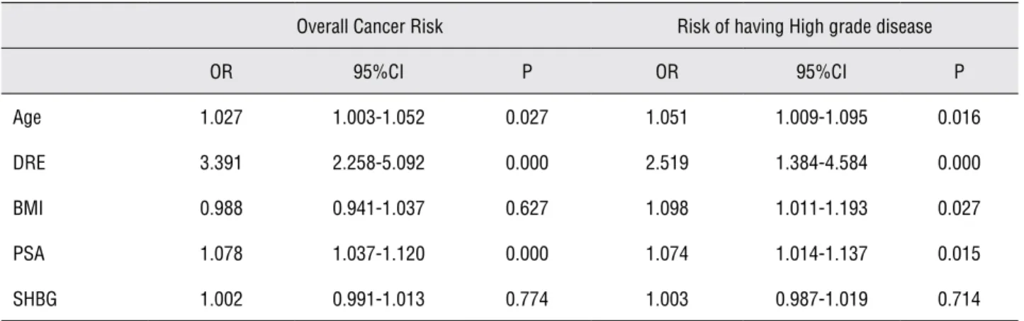
Patients with PCa were significantly older (p = 0.000), presented a higher PSA (p = 0.000) when compared to patients with a negative bi-opsy. No significant differences were observed in terms of BMI and SHBG levels (Table-2). On the multivariate analysis Age, PSA and DRE were found to be independent predictors of prostate cancer risk (Table-3). Risk of having prostate can-cer is increased by 2.7% per unit of age, by 239% if DRE was positive and by 7.8% per unit of PSA.
Patients with high grade disease were significantly older (p = 0.000), presented a
high-er PSA (p = 0.035) and highhigh-er BMI (p = 0.050) while no difference was found in terms of SHBG serum levels (Table-2). On multivariate analysis Age, DRE, BMI and PSA were found to be in-dependent predictors of high grade disease (Ta-ble-3). Risk of high grade disease is increased 5.1% per unit of age, by 152% if DRE, by 9.8% per unit of BMI and by 7.8% per unit of PSA. In addition ROC analyses were applied to evalu-ate SHBG, BMI and the presence of cancer (Fig-ure-1). SHBG produced an area under the curve (AUC = 0.517; CI: 0.474-0.561; p = 0.428) that did not seem to qualify to predict PCa. BMI produced an area under the curve that did not seem to qualify to predict PCa (AUC = 0.503 ;CI: 0.459-0.547; p = 0.882). PSA served as a control (AUC = 0.595; CI: 0.553-0.638; p = 0.000). Fur-thermore, ROC analyses were applied to evalu-ate 17BE levels, BMI and the presence of high grade PCa (Figure-1). SHBG produced an area under the curve (AUC = 0.524; CI: 0.453-0.596; p = 0.596) that did not seem to qualify to predict high grade PCa. BMI produced an area under the curve (AUC = 0.569; CI: 0.500-0.639; p = 0.05) that qualified to predict high grade disease. PSA served as a control (AUC = 0.575; CI: 0.505-0.645; p = 0.035).
Table 1 - Baseline characteristics of the cohort population.
Mean ± SD Median (IQR)
Age (years) 67.0 ± 7.94 68 (62/73)
BMI (kg/m²) 27.45 ± 3.79 27.1(25.0/29.4)
PSA (ng/mL) 7.2 ± 4.8 6.0 (4.3/8.8)
SHBG (pg/mL) 41.4 ± 17.4 38.4(29.7/49.4)
Table 2 - Clinical and pathological characteristics of the cohort.
No cancer Cancer p Gleason Score ≤ 6
Gleason
Score ≥ 7 p
Patients 465/740 (62.8%) 275/740 (37.2%) 155/275 (56%) 120/275 (44%)
DRE + 58/465 (12%) 92/275 (33%) 0.000 35/155 (23%) 57/120 (48%) 0.000
Age (years)
Mean ± SD 66.02 ± 7.9 68.9 ± 7.6
0.000
67.37 ± 7.72 70.91 ± 7.1
0.000 Median (IQR) 67 (60/72) 70 (64/74) 68 (61/73) 72 (66/76)
BMI (Kg/m²)
Mean ± SD 27.45 ± 3.6 27.45 ± 4.02
0.881
27.02 ± 3.62 28.00 ± 4.44
0.050 Median (IQR) 27.0 (25.0/29.4) 27.2 (24.8/29.6) 26.8 (24.8/29.3) 27.7 (25.2/30.5)
PSA (ng/mL)
Mean ± SD 6.53 ± 4.0 8.4 ± 5.6
0.000
7.5 ± 4.61 9.6 ± 6.7
0.035 Median (IQR) 5.6 (4.0/8.0) 6.7 (4.6/10.9) 6.2 (4.5/9.6) 7.2 (4.8/12.3)
SHBG (pg/mL)
Mean ± SD 40.7 ± 16.2 42.7 ± 19.3
0.428
41.1 ± 16.5 24.7 ± 22.5
DISCUSSION
Our experience confirmed how older pa-tients with higher PSA are at increased risk of PCa diagnosis. Moreover higher PSA and higher BMI in patients scheduled for prostate biopsy have an increased risk of developing high grade disease as showed by our previous studies (11). Clearly the strong association between DRE and prostate can-cer risk at biopsy in our series confirms the inter-nal validity of our results and it is in line with the peer review literature (3,12).
In our series obesity is a prevalent condi-tion and it is associated with an increased risk of high grade disease in patients with PCa. This data confirmed our previous experiences where we ob-served how obesity is a significant independent risk factor for high grade Gleason score when PCa is diagnosed by prostate biopsy (3,9,13). Further-more, the same observations were reported in other studies evaluating the possible relationship be-tween obesity and PCa development and progres-sion (14,15). Although further studies are needed to confirm these findings and to evaluate which
Table 3 - Exploring the association of SHBG and prostate cancer risk.
Overall Cancer Risk Risk of having High grade disease
OR 95%CI P OR 95%CI P
Age 1.027 1.003-1.052 0.027 1.051 1.009-1.095 0.016
DRE 3.391 2.258-5.092 0.000 2.519 1.384-4.584 0.000
BMI 0.988 0.941-1.037 0.627 1.098 1.011-1.193 0.027
PSA 1.078 1.037-1.120 0.000 1.074 1.014-1.137 0.015
SHBG 1.002 0.991-1.013 0.774 1.003 0.987-1.019 0.714
a population of patients at high risk of prostate cancer while most of the available evidence con-sidered patients scheduled for RRP.
Case-control studies have also evaluated the relationship between hormonal status and PCa diagnosis. Sawada et al. analysed plasma testoster-one and SHBG and the risk of PCa among Japanese men and found that neither testosterone neither SHBG levels were associated with an increased risk for total PCa (respectively: p = 0.49; p = 0.89) (18). In the same way Heikkila et al. in their case control study found no correlation between SHBG levels and PCa risk (23). On the other hand, Gann et al. made a prospective study of sex hormone levels and risk of PCa and found an inverse trend of risk for PCa with increasing levels of SHBG (p = 0.07) (24). Finally, a metanalysis from Roddam et al., consid-ering 18 prospective studies on sex hormones and PCa found no correlation between serum concen-trations of sex hormones and PCa risk and a mod-est inverse association between SHBG levels and PCa risk with a relative risk reduction of 14% (25). It is important to point out that one of the possible limitations in comparing these studies is the time of venous sampling. SHBG levels are quite stable during the day although variations in SHBG levels in a longer period of time hasn’t been evaluated yet (26). Moreover serum levels of SHBG are influenti-ated by IGF-1 levels and BMI (26). Data analysed by the available studies differ in these terms and so far are difficult to compare. From all these studies it is evident that the exact role of SHBG in PCa gene-sis and progression is still an open debate and more standardised and multicentric studies are needed to clarify these preliminary observations.
It is correct to point out the limitation of our study. Cancer aggressiveness was classified accord-ing to biopsy specimens and TRUS-guided prostate biopsy is associated with a certain degree of under and overestimating of PCa, however with a single centre study and a single uropathologist reviewing all biopsies this possible bias was reduced. Another limitation of our study is that a single assessment of circulating SHBG concentrations may not be re-liable clinically; however, as reported by Salonia et al., a single fasting morning venous blood sample determines the assessment of the hormone status more user friendly in every day clinical practice biological factors are responsible for the observed
results, our experience add further evidence on the relationship between obesity and PCa.
SHBG is a serum globulin that binds tes-tosterone in plasma regulating though its free component. SHBG has been associated with PCa genesis (16-18). Berndt et al. (16) found a strong relationship between SHBG gene polymorphism and risk of developing PCa. Moreover SHBG re-ceptors have been found on prostate cells and their activation leads to an increase in cAMP pos-sibly activating oncological pathways (17,18).
(20). However, at present our study is the largest prospective series evaluating the association be-tween SHBG levels and PCa risk among men with elevated PSA or/and abnormal digital rectal exami-nation scheduled for prostate biopsy and using the same biopsy template for the entire cohort.
We have also considered the possibility of running a separate analysis for patients with Glea-son 8-10 (excluding patients with GleaGlea-son 7) but for the sake of homogeneity with papers published in the peer review literature (7,9,10,11,13,21), we decided to run a dichotomous analysis grouping in the high risk population also men with a Glea-son score 7. Furthermore, our approach to define high grade prostate cancer by Gleason score ≥ 7 is also in line with a number of published papers (27,28). However, we acknowledge that further re-search and large number of patients are needed to explore potential differences in the different Glea-son score subgroups.
CONCLUSIONS
In our cohort of patients at risk of PCa, the serum level of SHBG is not a significant predictor of prostate cancer diagnosis and aggressiveness on initial prostate biopsy. According to our expe-rience, SHBG, should not be considered a reliable marker to predict poorly differentiated cancer in the setting of initial prostate biopsy. Further stud-ies are needed to confirm our findings in other populations and to evaluate which biological fac-tors related to serum SHBG levels are involved to prostate cancer development and progression.
ABBREVIATIONS
PCa = Prostate Cancer
SHBG = Sex Hormone Binding Globulin
DRE = Digital Rectal Examination
BMI = Body Mass index
ROC = Receiver Operator Characteristics
AUC = Area Under the Curve
IQR = Inter Quartile Range
CI = Confidence interval
RRP = Radical retropubic prostatectomy
TRUS = Transrectal Ultrasound
CONFLICT OF INTEREST
None declared.
REFERENCES
1. Center MM, Jemal A, Lortet-Tieulent J, Ward E, Ferlay J, Brawley O, et al.: International variation in prostate cancer incidence and mortality rates. Eur Urol. 2012; 61: 1079-92. 2. Castelli T, Cimino S, Magno C, Morgia G: Molecular mark-ers for prostatic cancer. Front Biosci (Elite Ed). 2010; 2: 641-56.
3. De Nunzio C, Lombardo R, Leonardo C, Franco G, Gacci M, Presicce F, et al.: Serum levels of 17-β-estradiol are not predictive of prostate cancer diagnosis and aggressive-ness: Results from an Italian biopsy cohort. Urol Oncol. 2013. In Press
4. De Nunzio C, Miano R, Trucchi A, Finazzi Agrò E, Tubaro A: Finasteride for prostatic disease: an updated and com-prehensive review. Expert Opin Drug Metab Toxicol. 2008; 4: 1561-8.
5. Huggins G, Hodge GB: Studies on prostatic cancer. I. The effect of castration, of estrogen and of androgen injec-tion on serum phosphatase in metastatic carcinoma of the prostate. Cancer Res. 1941; 1: 293-7.
6. Wirén S, Stocks T, Rinaldi S, Hallmans G, Bergh A, Sten-man UH, et al.: Androgens and prostate cancer risk: a pro-spective study. Prostate. 2007; 67: 1230-7.
7. Schatzl G, Madersbacher S, Thurridl T, Waldmüller J, Kramer G, Haitel A, et al.: High-grade prostate cancer is associated with low serum testosterone levels. Prostate. 2001; 47: 52-8.
8. Hoffman MA, DeWolf WC, Morgentaler A: Is low serum free testosterone a marker for high grade prostate cancer? J Urol. 2000; 163: 824-7.
9. Albisinni S, De Nunzio C, Tubaro A, Barry WT, Banez LL, Freedland SJ: Greater percent-free testosterone is associ-ated with high-grade prostate cancer in men undergoing prostate biopsy. Urology. 2012; 80: 162-7.
10. De Nunzio C, Trucchi A, Miano R, Stoppacciaro A, Fattahi H, Cicione A, et al.: The number of cores positive for high grade prostatic intraepithelial neoplasia on initial biopsy is associated with prostate cancer on second biopsy. J Urol. 2009; 181: 1069-74; discussion 1074-5.
12. Wolf AM, Wender RC, Etzioni RB, Thompson IM, D’Amico AV, Volk RJ, et al.: American Cancer Society guideline for the early detection of prostate cancer: update 2010. CA Cancer J Clin. 2010; 60: 70-98.
13. De Nunzio C, Freedland SJ, Miano L, Finazzi Agrò E, Bañez L, Tubaro A: The uncertain relationship between obesity and prostate cancer: an Italian biopsy cohort analysis. Eur J Surg Oncol. 2011; 37: 1025-9.
14. Allott EH, Masko EM, Freedland SJ: Obesity and prostate cancer: weighing the evidence. Eur Urol. 2013; 63: 800-9. 15. Freedland SJ, Wen J, Wuerstle M, Shah A, Lai D, Moalej B,
et al.: Obesity is a significant risk factor for prostate cancer at the time of biopsy. Urology. 2008; 72: 1102-5.
16. Berndt SI, Chatterjee N, Huang WY, Chanock SJ, Welch R, Crawford ED, et al.: Variant in sex hormone-binding globu-lin gene and the risk of prostate cancer. Cancer Epidemiol Biomarkers Prev. 2007; 16: 165-8.
17. Weigel NL, Zhang Y: Ligand-independent activation of ste-roid hormone receptors. J Mol Med (Berl). 1998; 76: 469-79. 18. Sawada N, Iwasaki M, Inoue M, Sasazuki S, Yamaji T,
Shimazu T, et al.: Plasma testosterone and sex hormone-binding globulin concentrations and the risk of prostate cancer among Japanese men: a nested case-control study. Cancer Sci. 2010; 101: 2652-7.
19. Lee SE, Chung JS, Han BK, Park CS, Moon KH, Byun SS, et al.: Preoperative serum sex hormone-binding globulin as a predictive marker for extraprostatic extension of tumor in patients with clinically localized prostate cancer. Eur Urol. 2008; 54: 1324-32.
20. Salonia A, Gallina A, Briganti A, Zanni G, Suardi N, Capi-tanio U, et al.: Sex hormone-binding globulin is a signifi-cant predictor of extracapsular extension in men undergo-ing radical prostatectomy. BJU Int. 2011; 107: 1243-9. 21. Salonia A, Briganti A, Gallina A, Karakiewicz P, Shariat S,
Freschi M, et al.: Sex hormone-binding globulin: a novel marker for nodal metastases prediction in prostate cancer patients undergoing extended pelvic lymph node dissec-tion. Urology. 2009; 73: 850-5.
22. Salonia A, Gallina A, Briganti A, Suardi N, Capitanio U, Ab-dollah F, et al.: Circulating estradiol, but not testosterone, is a significant predictor of high-grade prostate cancer in patients undergoing radical prostatectomy. Cancer. 2011; 117: 5029-38.
23. Heikkilä R, Aho K, Heliövaara M, Hakama M, Marniemi J, Reunanen A, et al.: Serum testosterone and sex hormone-binding globulin concentrations and the risk of prostate carcinoma: a longitudinal study. Cancer. 1999; 86: 312-5. 24. Gann PH, Hennekens CH, Ma J, Longcope C, Stampfer MJ:
Prospective study of sex hormone levels and risk of pros-tate cancer. J Natl Cancer Inst. 1996 ; 88: 1118-26. 25. Roddam AW, Allen NE, Appleby P, Key TJ, Endogenous
Hormones and Prostate Cancer Collaborative Group: En-dogenous sex hormones and prostate cancer: a collabora-tive analysis of 18 prospeccollabora-tive studies. J Natl Cancer Inst. 2008; 100: 170-83.
26. Pasquali R, Casimirri F, Cantobelli S, Melchionda N, Mor-selli Labate AM, Fabbri R, et al.: Effect of obesity and body fat distribution on sex hormones and insulin in men. Me-tabolism. 1991; 40: 101-4.
27. Thompson IM, Goodman PJ, Tangen CM, Lucia MS, Miller GJ, Ford LG, et al.: The influence of finasteride on the de-velopment of prostate cancer. N Engl J Med. 2003; 349: 215-24.
28. Andriole GL, Bostwick DG, Brawley OW, Gomella LG, Mar-berger M, Montorsi F, et al.: Effect of dutasteride on the risk of prostate cancer. N Engl J Med. 2010; 362: 1192-202.
_______________________ Correspondence address: