AR
TIGO ORIGINAL / ORIGINAL AR
TICLE
ASSOCIATION OF
HELICOBACTER PYLORI
RESTRICTION ENDONUCLEASE-REPLACING
GENE,
hrgA
WITH OVERT
GASTROINTESTINAL DISEASES
Manoj
G
1, Santosh K.
TIWARI
1, Vishwas
SHARMA
1, Mohammed Aejaz
HABEEB
1,2,
Aleem A.
KHAN
1and Habibullah
CM
1ABSTRACT - Background and Aim - Helicobacter pylori has been proven to be responsible for causing various gastrointestinal disorders
including gastric adenocarcinoma. Several genes of pathogen (the genes of the cag-PAI, vacA, iceA, and babA) either in combination or independently have been reported to significantly increase the risk of ulceration/gastric carcinoma, with the cagA gene having the strongest predictive value. Pursuit to identify new genes which could serve as a marker of overt disease progression, lead to the discovery of hrgA gene. Methods - Fifty-six indigenous strains of H. pylori from subjects with various gastric disorder were screened to assess the status of hrgA gene along with the cagA gene using simple polymerase chain reaction using specific oligonucleotide primers. Post-amplification, amplicons were subjected for sequencing to identify any strain specific variations in sequences from the H. pylori isolated from different disease manifestations. Histopathological analysis was done to ascertain any significant change in the histological scores of subjects infected with cagA+/hrgA+ and
cagA-/hrg+ strains. Results - All the 56 (100%) subjects amplified with the oligonucleotide primers specific to hrgA gene, whereas 81.71% subjects showed the presence of cagA gene. Sequencing of the amplimers showed 99% homology. Histology of the cagA+/hrgA+ and cagA-/
hrg+ subjects did not show any significant difference. Conclusion - hrgA gene of Helicobacter pylori is not a ideal surrogate marker for identifying individuals with higher risk of developing overt gastro-duodenal diseases such as neoplasia of the stomach.
HEADINGS – Helicobacter pylori. Stomach neoplasms. Adenocarcinoma. Bacterial proteins. Polymerase chain reaction.
INTRODUCTION
Adenocarcinoma of the stomach is one of the leading causes of cancer related deaths in the world(9). Although the
incidence of gastric cancer has declined much significantly in the West, it still remains a major type of neoplasm especially in the East Asian countries like Japan, China, and Korea(14). Development of gastric cancer is believed
to be a multi-factorial event and probably takes 3 to 4 decades to manifest. With the primary etiological agents being exposure to chemical carcinogens, Helicobacter pylori occupies a unique niche in the genesis of gastric cancer. In addition, epidemiological studies have indicated that infection with Helicobacter pylori is considered a major risk factor for gastric cancer(11), and the WHO/
IARC(4) has classified this bacterium as a definite biological
carcinogen in 1994. Though it is hypothesized that the development of cancer depends on a series of complex molecular interaction between the host and bacteria(4,
10, 11), the precise patho-mechanisms linking H. pylori
infection and gastric carcinogenesis still remains an unsolved enigma.
The predisposition among H. pylori infected individuals to develop various gastro-duodenal diseases
viz duodenal ulcer (DU), gastric ulcer, gastric carcinoma and MALT-lymphoma mainly depends upon the bacterial and host factors and in part on the topography of the gastric inflammation(5, 6, 10). Studies have identified several
strain specific factors that potentially are markers for the differential risk associated with H. pylori colonization(2, 15); at present, the genes of the cag pathogenicity island
(cag-PAI) and vacA, SADAKANE et al.(12); MAEDA
et al.(7); TIWARI et al.(13) have been reported to possess
strongest predictive values. However, most strains isolated from Indian sub-continent and other East Asian countries possess these determinants irrespective of their clinical outcome. Thus, identification of specific bacterial factors that can serve as surrogate marker for the progression to ulceration or to gastric cancer remains desirable.
ANDO et al.(1), in 2002, while working with restriction–
modification (R-M) systems, discovered potential marker
Helicobacter pylori restriction endonuclease-replacing gene (hrgA) that in conjunction with cagA identified individuals associated with gastric cancer. The same
1Center for Liver Research and Diagnostics; 2Dept. of Gastroenterology and Hepatology Deccan College of Medical Sciences, Kanchanbagh, Hyderabad 500 058,
Andhra Pradesh, India.
study reported that though hrgA was more prevalent among the Western countries than in Asian, its prevalence was more among gastric cancer patients in Asians compared to those with benign disease. The data also suggested that the hrgA occurred more among cagA+ve H. pylori strains than those lacking cagA. These results prompted us to evaluate the prevalence of this virulence determinant among the strains isolated from the South Indian population and evaluate the presence of hrgA as a surrogate marker of overt gastrointestinal disorders.
Experimental procedure
We studied H. pylori strains from 56 dyspeptic patients (males: 32; females: 24; age range: 20-65 years) undergoing-gastroscopy in the Department of Gastroenterology, Deccan College of Medical Sciences and Allied Hospitals, Hyderabad, India, for the evaluation of upper gastrointestinal symptoms. The Institutional Ethical Committee (IEC) of the hospital approved the study protocol. A total of four gastric biopsy specimens (two from the antrum and two from the corpus) were collected from each patient after taking informed consent from the subjects to take part in the study. One antral biopsy collected in brucella broth supplemented with 2% fetal calf serum (FCS) was used for culturing H. pylori, one corpus biopsy for histological lesions and the remaining two biopsy one each from the antrum and corpus was collected in phosphate buffered saline for the DNA analysis.
Bacterial strains and growth conditions
The collected biopsy specimens were transported to the laboratory, the specimens were best processed within 1 hour, if delayed the specimen was preserved at 40C for 4–24 hours. The
biopsy was inoculated onto the chocolate brucella agar medium (Difco Laboratories, Detroit, USA) supplemented with 7% sheep blood and antibiotics vancomycin (6 mg/mL), amphotericin-B (2 mg/mL) and polymixin-B (2500 units/mL). The plates were incubated in a microaerobic conditions at 370C for 3-5 days with
90%-100% humidity. Then the plates were examined after 72 hours for H. pylori.
The isolated colonies of H. pylori were again sub cultured in solid media. Rapid urease method (RUT) a touch cytology method was also done by inoculating few colonies into 250 µL urea broth comprising phenol red indicator. The presence of urease activity by a rapid change in color from yellow to pink indicated active H. pylori. The culture was considered to be positive for
H. pylori by observing small, translucent, tiny colonies, which on staining gave Gram-negative staining.
DNA extraction and PCR ampliication
Genomic DNA isolation from 36 pure H. pylori cultures and 56 biopsy samples was done using a standard protocol described previously(8). The isolated DNA was then amplified
for the presence of target genes viz., cagA, hrgA and hpyIIIR of H. pylori using specific oligonucleotide primers and PCR conditions listed in Tables 1 and 2. The products of amplification were subsequently electrophoresed in 1.5% agarose gel stained with ethidium bromide to visualize the presence of amplified
genes. All the photographic registries were performed using a bio-rad gel documentation system.
Sequencing of hrgA gene of H. pylori
Following amplification using hrgAF and hrgAR primers, the amplimers were sequenced directly after purification with the QIAquick gel extraction kit (Qiagen) using the big dye terminator v3.1 cycle sequencing kit and injected to an ABI 3730 xl Genetic Analyzer (Applied Biosystems, Germany).
Histpathological analysis
Histopathological analysis was mainly performed to detect the presence of H. pylori and also to ascertain the presence
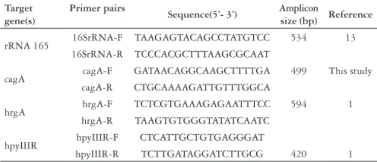
Target gene(s)
Primer pairs
Sequence(5’- 3’) Amplicon
size (bp) Reference
rRNA 165 16SrRNA-F TAAGAGTACAGCCTATGTCC 534 13 16SrRNA-R TCCCACGCTTTAAGCGCAAT
cagA cagA-F GATAACAGGCAAGCTTTTGA 499 This study cagA-R CTGCAAAAGATTGTTTGGCA
hrgA hrgA-F TCTCGTGAAAGAGAATTTCC 594 1 hrgA-R TAAGTGTGGGTATATCAATC
hpyIIIR hpyIIIR-F CTCATTGCTGTGAGGGAT
hpyIIIR-R TCTTGATAGGATCTTGCG 420 1
TABLE1. List of oligonucleotide primers used during the study
S. No Clinical status (n) cagA status (%) hrgA status (%)
1 Prepyloric ulcer (14) 14 (100%) 14 (100%) 2 Duodenal ulcer (15) 11 (73.3%) 15 (100%) 3 Gastric adenocarcinoma (14) 14 (100%) 14 (100%) 4 NUD (8) 6 (75%) 8 (100%) Total (51) 45(88.23%) 51 (100%)
TABLE 3. Comparison of the cagA and hrgA status in H. pylori positive subjects
Target gene Amplification condition
16S rRNA
Initial denaturation 950C for 5 min
Denaturation 940C for 30 sec
Annealing 560C for 1 min 40 cycles
Extension 720C for 1 min
Final extension 720C for 7 min
hrgA & cagA
Initial denaturation 950C for 5 min
Denaturation 940C for 30 sec
Annealing 520C for 1 min 40 cycles
Extension 720C for 1 min
Final extension 720C for 7 min
hpyIIIR
Initial denaturation 950C for 5 min
Denaturation 940C for 45 sec
Annealing 470C for 1 min 40 cycles
Extension 720C for 1 min
Final extension 720C for 7 min
TABLE 2. Amplification conditions used for the PCR in the present study
of significant preneoplastic and neoplastic lesions. Briefly, two sections (4 µM) were cut from each block: one section was stained with a modified Giemsa stain, and the other section was stained with hematoxylin-eosin (H-E) to assess the presence of intestinal metaplasia and dysplasia. A single pathologist (Z. A.) who was blinded to the patient’s clinical conditions evaluated all the histologic sections. Grading of the histological lesions was done according to the updated Sydney system of classification(3).
Statistical analysis
The data obtained was evaluated using Student Chi (χ2)
square test and probability values less than 5% (P<0.05) was considered statistically significant.
RESULTS
Out of 56 patients enrolled for the study, we categorized the patients according to their disease status (prepyloric ulcer-14, DU-18, non-ulcer dyspepsia-10, and gastric adenocarcinoma-14). H. pylori positivity was found in 36 (64.28%) by culture, 44 (78.57%) by RUT, 28 (50%) by histopathology, and 51 (91.07%) by biopsy DNA amplification.
Status of hrgA, wA and hpyIIIR gene locus
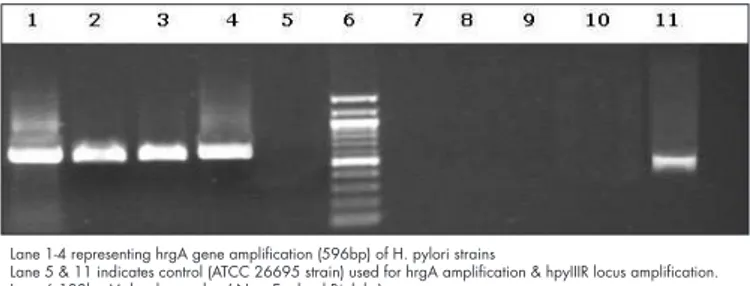
Of the 56 genomic DNA isolated (36 cultures and 51 biopsy samples), hrgA gene amplification was seen in all the samples screened giving a product size of 594bp. Besides this, the cagA gene was found in all (100%) the patients with pre-pyloric ulcers and gastric adenocarcinoma but only in 11 (61%) with DU and 6 (60%) with non ulcer dyspepsia (NUD), respectively. However, among the remaining 11 subjects with DU and NUD that did not amplify for the cagA gene, hrgA amplified giving the expected product (Figure 1 and Table 3). The prevalence of hrgA was then compared
twice to confirm the presence of hrgA gene and absence of
hpyIIIR locus. No mixed genotypes of hpyIIIR/hrgA were found in this study.
Histological assessment
Analysis of the sections from each biopsy from various patients showed chronic gastritis grade II among 10 (66.67%) subjects with DU, 8 (57.14%) with pre-pyloric ulcer, 3 (37.5%) with NUD, whereas chronic grade III gastritis with atrophic gastritis was seen in 2 (13.33%) with DU, 5 (35.71%) with pre-pyloric ulcers. H-E staining showed mild intestinal metaplasia (IM) in one (6.67%) subject with DU and one (7.14%) with pre-pyloric ulcer. Among 14 gastric carcinoma subjects, high grade dysplasia was seen in 8 (57.14%) and low grade dysplasia in 3 (21.42%) subjects, the remaining 3 (21.42%) subjects showed foci of chronic atrophic gastritis with moderate IM (Figures 3 and 4).
FIGURE 2. Corpus biopsy showing focal intestinal metaplasia with mild dysplasia
FIGURE 3. Invasion of stroma, dysplasia associated with invasive intestinal type cancer
FIGURE 1. Gel picture showing amplification of hrgA gene of Helicobacter
pylori from various subjects
Lanes 1 - 10 H. pylori DNA from various patients with different gastric diseases Lane M - 100bp Molecular weight DNA marker (New England Biolabs)
FIGURE 4. Gel photograph showing amplification of the hgrA and
hpyIIIR Status in H. pylori strains
Lane 1-4 representing hrgA gene amplification (596bp) of H. pylori strains
Lane 5 & 11 indicates control (ATCC 26695 strain) used for hrgA amplification & hpyIIIR locus amplification. Lane 6 100bp Molecular marker ( New England Biolabs)
Lane 7-10 represents hpyIIIR amplification in hrgA amplified strains.
Sequence analysis
Sequences obtained were compared using BLASTn with the
NCBI database. Sequencing analysis showed the presence of
hrgA gene of H. pylori with a homology of 99%.
DISCUSSION
The present study evaluated the presence of Helicobacter pylori restriction endonuclease-replacing gene (hrgA) among subjects with various gastric disorders. The results of this study showed the presence of hrgA gene in all the 51 (100%) H. pylori strains isolated from each subjects with different clinical presentations.
Even though number of bacterial virulence determinants such as the genes of the cag-pathogenicity island, vacA,
iceA, and babA, have been extensively studied in the past (in association with) the clinical status in various geographical regions. No significant association could be established with the presence of any of these genes with respect to the clinical outcome, the main reason being the highly plastic genome of
H. pylori(6, 12, 15), therefore in a continued pursuit to identify
novel bacterial markers of H. pylori, which could serve as a surrogate marker of disease progression, ANDO et al.(1) reported
the presence of hrgA to be high among patients with gastric cancer of the Asian population than those with non-cancerous or benign form of disease. However the present study could not find any such specific association of this gene with any of the disease conditions and found that the hrgA gene was present in all the subjects included in the study irrespective of their disease status. Further this study also found that the prevalence was not dependent on the presence of the cagA
gene as it was evenly distributed among the patients with
cagA positive and cagA negative H. pylori strains.
As evident from the results, we could not find any significant difference in the histological pattern among the subjects infected with cagA+ve/hrgA+ve H. pylori and those with cagA-ve/hrgA+ve. These findings suggest that development of overt disease such as ulceration or gastric carcinoma cannot be predicted based on the presence or absence of a one or two bacterial virulence factors. Rather development of severe form of disease is a result of complex molecular interactions between the host and the bacterial factors over a period of time that causes significant damage to the host. In addition, the site of the stomach where H. pylori colonizes also has an important bearing on the predisposition to develop pre-pyloric ulcer, DU and gastric cancer. Further, the environmental factors also play major role in disease manifestation besides the host and bacterial factors. This could have been one of the main reasons in ANDO et al.(1) study that could not establish a
correlation between hrgA gene and gastric cancer patients of the Western countries whereas the same was possible among the Asian gastric cancer patients.
The results obtained in the present study are in contrast to those obtained by ANDO et al.(1) thereby suggesting that,
though presence of hrgA had a higher predictive value for gastric carcinoma, our study however, could not hint at any such correlations, as hrgA gene was found to be present unequivocally in all the strains screened from various disease pathologies. The exploration for reasons responsible for these varied results warrants further investigations on large cohort population from different geographic areas. Besides this, an in depth molecular profile of the R-M systems and especially the functional role of hrgA gene would further be helpful to delineate the direct/ indirect role played by this gene in gastric carcinogenesis.
In conclusion, hrgA gene of H.pylori may not be used as a ideal surrogate marker for identifying individuals at higher risk of overt form of gastro-intestinal disorders among the South Indian population.
ACKNOWLEDGEMENTS
G M, Tiwari SK, SHAMA V,, Habeeb MA, Khan AA, CM H. Associação entre o hrgA (Helicobacter pylori restriction endonuclease-replacing gene) com as principais doença gastrointestinais. Arq Gastroenterol. 2008;45(3):225-9.
RESUMO – Racional e Objetivos – O Helicobacter pylori tem sido incriminado como causador de vários distúrbios digestivos, incluindo o adenocarcinoma gástrico. Diversos genes patogênicos (os genes do cag-PAI, vacA, iceA e babA), em combinação ou independentes, têm sido reportados como fatores de aumento de risco para ulceração/carcinoma gástrico, tendo o gene cagA forte valor preditivo. A procura da identificação de novos genes que possam vir a ser marcadores da progressão da doença levaram à descoberta do gene hrgA. Métodos – Cinqüenta e seis amostras de H. pylori provenientes de pacientes com diversas afecções gástricas foram examinadas para caracterizar a presença do hrgA juntamente ao cagA, usando iniciadores específicos da reação de cadeia da polimerase. Após amplificação, os produtos amplificados pela PCR foram seqüenciados para a identificação de variações específicas nas seqüências do
H. pylori isolado de diferentes doenças gastroduodenais. A análise histopatológica foi feita para assegurar qualquer mudança significativa nos escores dos indivíduos infectados com cagA+hrgA+ e cagA-/hrgA+. Resultados – Todas as 56 amostras (100%) foram amplificadas com iniciadores específicos para o hrgA, enquanto que 81,71% mostraram a presença do cagA. O seqüenciamento do produto amplificado pela PCR mostrou 99% de homologia. A histologia entre os grupos cagA+/hrgA+ e cagA-/hrgA+ não mostrou nenhuma diferença significante. Conclusão – O gene hrgA do H. pylori não é o marcador ideal para identificar indivíduos com alto risco de desenvolvimento de doenças gastrointestinais como a neoplasia de estômago.
DESCRITORES – Helicobacter pylori. Neoplasias gástricas. Adenocarcinoma. Proteínas de bactérias. Reação em cadeia da polimerase.
REFERENCES
Ando T, Wassenaar TM, Peek RM Jr, Aras RA, Tschumi AI, van Doom LJ, Kusugami 1.
K, Blaser MJ. A Helicobacter pylori restriction endonuclease-replacing gene, hrgA, is associated with gastric cancer in Asian strains. Cancer Res. 2002;62:2385-9. Aspholm-Hurtig M, Dailide G, Lahmann M, Kalia A, Ilver D, Roche N, Vikstrom 2.
S, Sjostrom R, Linden S, Backstrom A, Lundberg C, Arnqvist A, Mahdavi J, Nilsson UJ, Velapatino B, Gilman RH, Gerhard M, Alarcon T, Lopez-Brea M, Nakazawa T, Fox JG, Correa P, Dominguez-Bello MG,Perez-Perez GI, Blaser MJ, Normark S, Carlstedt I, Oscarson S, Teneberg S, Berg DE, Boren T. Functional adaptation of BabA, the H. pylori ABO blood group antigen binding adhesin. Science. 2004;305:519–22.
Dixon MF, Genta RM, Yardley JH, Correa P. Classification and grading of gastritis. 3.
The updated Sydney System. International Workshop on the Histopathology of Gastritis, Houston, 1994. Am J Surg Pathol. 1996;20:1161-81.
IARC (International Agency for Research on Cancer) Working Group on the Evaluation 4.
of Carcinogenic Risks to Humans. Helicobacter pylori. In: Schistosomes, liver flukes and Helicobacter pylori / World Health Organization, International Agency for Research on Cancer. Geneva: The Agency: Secretariat of the World Health Organization [distributor]; 1994. p.177-240. (IARC monographs on the evaluation of the carcinogenic risk of chemicals to humans, v. 61).
Israel DA, Salama N, Krishna U, Rieger UM, Atherton JC, Falkow S, Peek RM Jr. 5.
2001. Helicobacter pylori genetic diversity within the gastric niche of a single human host. Proc Natl Acad Sci U S A. 2001;98:14625–30.
Ladeira MS, Rodrigues MA, Salvadori V, Neto PP, Achilles P, Lerco MM, Rodrigues PA, 6.
Goncalves I Jr, Queiroz DM, Freire-Maia DV. Relationships between cagA, vacA, and iceA genotypes of Helicobacter pylori and DNA damage in the gastric mucosa. Environ Mol Mutagen. 2004;44:91-8.
Maeda S, Ogura K, Yoshida H, Kanai F, Ikenoue T, Kato N, Shiratori Y, Omata, M. 7.
Major virulence factors, vacA and cagA, are commonly positive in Helicobacter pylori isolates in Japan. Gut. 1998;42:338–43.
Mapstone NP. The detection of
8. Helicobacter pylori by the polymerase chain reaction. In: Clayton CL, Mobley HLT, editors. Methods in molecular medicine: Helicobacter pylori protocols. Totowa, NJ: Humana Press; 1997. p.31-6.
Parkin DM, Bray FI, Devesa SS. Cancer burden in the year 2000. The global picture. 9.
Eur J Cancer. 2001;37(Suppl 8):s4-s66.
Peek RM Jr. Events at the host-microbial interface of the gastrointestinal tract IV. 10.
The pathogenesis of Helicobacter pylori persistence. Am J Physiol Gastrointest Liver Physiol. 2005;289:g8--g12.
Pinto-Santini D, Salama NR. The biology of
11. Helicobacter pylori infection, a
major risk factor for gastric adenocarcinoma. Cancer. Epidemiol Biomarkers Prev. 2005;14:1853-8.
Sadakane Y, Kusaba K, Nagasawa Z, Tanabe I, Kuroki S, Tadano J. Prevalence and 12.
genetic diversity of cagD, cagE, and vacA in Helicobacter pylori strains isolated from Japanese patients. Scand J Gastroenterol. 1999;34:981–6.
Tiwari SK, Khan AA, Khan SA, Ali M, Habeeb A, Kauser F, Hussain MA, Ahmed 13.
N, Habibullah CM. PCR based analysis of the cag-PAI of Helicobacter pylori from saliva: an approach for rapid molecular genotyping in correlation with disease status. J Gastroenterol Hepatol. 2005;20:1560-8.
Vieth M, Stolte M. Elevated risk for gastric adenocarcinoma can be predicted from 14.
histomorphology. World J Gastroenterol. 2006;12:6109-14.
Zheng PY, Hua J, Yeoh KG, Ho B. Association of peptic ulcer with increased 15.
expression of Lewis antigens but not cagA, iceA, and vacA in Helicobacter pylori isolates in an Asian population. Gut. 2000;47:18–22.