CDKN2A
promoter methylation is related to the tumor
location and histological subtype and associated with
Helicobacter pylori fla
A(+) strains in gastric
adenocarcinomas
MARKEˆNIA KE´LIA SANTOS ALVES,1VALESKA PORTELA LIMA,1ADRIANA CAMARGO FERRASI,2MARIA APARECIDA RODRIGUES,2MARIA INEˆS DE MOURA CAMPOS PARDINI2
and SILVIA HELENA BAREM RABENHORST1
1Microbiology Section, Department of Pathology and Forensic Medicine, Federal University of Ceara´,
Porangabussu Campus, Ceara´; and2Molecular Biology Laboratory, Botucatu Hemocenter, School of Medicine, Paulista State University, Botucatu Campus, Sa˜o Paulo, Brazil
Alves MKS, Lima VP, Ferrasi AC, Rodrigues MA, de Moura Campos Pardini MI, Rabenhorst SHB. CDKN2Apromoter methylation is related to the tumor location and histological subtype and associ-ated withHelicobacter pylori flaA(+) strains in gastric adenocarcinomas. APMIS 2010; 118: 297–307.
Promoter hypermethylation ofCDKN2A(p16INK4Aprotein) is the main mechanism of gene inactiva-tion. However, its association withHelicobacter pyloriinfection is a controversial issue. Therefore, we examined a series of gastric adenocarcinomas to assess the association between p16INK4Ainactivation andH. pylorigenotype (vacA,cagA,cagE,virB11 andflaA) according to the location and histological subtype of the tumors. p16INK4Aexpression andCDKN2Apromoter methylation were found in 77 gas-tric adenocarcinoma samples by immunohistochemistry and methylation-specific PCR, respectively. Helicobacter pylori infection and genotype were determined by PCR. A strong negative correlation between immunostaining andCDKN2Apromoter region methylation was found. In diffuse subtype tumors, the inactivation of p16INK4A by promoter methylation was unique in noncardia tumors (p = 0.022). In addition, H. pylori-bearing flaA was associated with non-methylation tumors (p = 0.008) andH. pyloristrain bearingcagA orvacAs1m1 genes but withoutflaA was associated with methylated tumors (p = 0.022 and 0.003, respectively). Inactivation of p16INK4A in intestinal and diffuse subtypes showed distinct carcinogenic pathways, depending on the tumor location. Moreover, the process of methylation of theCDKN2Apromoter seems to depend on theH. pylorigenotype. The present data suggest that there is a differential influence and relevance ofH. pylorigenotype in gastric cancer development.
Key words: p16INK4A; gastric cancer; histological subtypes; tumor location; methylation;Helicobacter pylorigenotypes.
Markeˆnia Ke´lia Santos Alves, Rua Osvaldo Cruz, 3485, Dionı´sio Torres, Fortaleza, Ceara´ 60125-151, Brazil. e-mail: [email protected]; [email protected]
Gastric cancer (GC) is an important world-wide health problem because of its high frequency, poor prognosis, advanced stage at
the time of diagnosis and limited treatment options (1, 2).
There is much evidence that the carcinogene-sis process involves abnormalities of cell cycle regulators (3, 4). In GC, besides p53, p16INK4A (cyclin-dependent kinase inhibitor 2A-CDKN2A) Received 22 November 2009. Accepted 14 January
is often inactivated (3, 5–9). The p16INK4A protein is a cell cycle regulator involved in the inhibition of G1 (Gap1) phase progression, and although the common mechanism of its inacti-vation is through the hypermethylation of its promoter, few studies have evaluated this pro-cess in GC considering the distinct histological subtypes, tumor location (10, 11) and possible causes leading to this.
Helicobacter pylori is a major environmental factor associated with the development of GC (12). Its high genetic variability has been related to its virulence and consequently to disease outcome (13–15). Two well-established viru-lence factors are the presence of the cytotoxin-associated antigen A (cagA) gene, located within the right portion of thecagpathogenicity island (cag-PAI), and vacuolating cytotoxin A (vacA), mostly the vacAs1m1 allele (16, 17).
cag-PAI also hascagE andvirB11 genes, located in the right and left regions of the island, respec-tively. These genes are thought to play a role in constructing the type IV secretion system and inducing proinflammatory, pro-proliferative epithelial cell signaling (13, 18, 19). Another importantH. pylorivirulence factor is the pres-ence of flagella, codified by several genes, such as flaA and flaB, which are responsible for successful colonization of its unusual habitat. TheflaA gene is the main factor responsible for codification of the flagellar filament protein, flagellin (20).
Although the exact mechanism of H. pylori -associated gastric carcinogenesis remains un-known,H. pyloriinfection has been reported as an important factor causing promoter hyperme-thylation of tumor suppressor genes, such as the E-cadherin and CDKN2A genes (21, 22). How-ever, this is a controversial issue (23–25). More-over, studies associating H. pylori infection with CDKN2A methylation status have not accounted for p16INK4A expression, which pre-cludes a more comprehensive assessment of the involvement of this bacterium in the inactiva-tion of p16INK4A. Moreover, these studies are restricted to cases of gastritis and precancerous lesions (22, 23). So far, there are no studies relating H. pylori genotypes to p16INK4A inactivation in GC.
In this context, the present investigation aimed to evaluate the relationship between the
H. pylori genotype and p16INK4A inactivation.
As intestinal and diffuse tumors are different entities and there is evidence of different path-ways according to the tumor location (cardia and noncardia tumors), the data were also ana-lyzed considering the tumor histological subtype and location.
MATERIALS AND METHODS
Clinical specimens
The present study was approved by the Hospital Ethics Committee of Ceara´ Federal University and all subjects signed the informed consent form before inclusion. Samples from 77 patients with gastric ade-nocarcinoma who had undergone gastrectomy were collected from two hospitals in Ceara´ state, Brazil: Walter Cantı´deo Hospital (associated with Ceara´ Federal University) and Santa Casa de Miserico´rdia Hospital, both located in Fortaleza, the state capital. The histological classification was carried out by the team’s pathologists according to the Lauren classifi-cation. The clinical and histopathological features are shown in Table 2.
DNA extraction
Genomic DNA was extracted from frozen tumor tissue using the cetyltrimethyl ammonium bromide (CTAB) technique, adapted from the method of Foster and Twell (26). The DNA extraction was performed only in fragments that showed more than 80% of tumor cells, and the quality was analyzed by 1% agarose gel electrophoresis with ethidium bromide staining. The amount of DNA was deter-mined using a NanoDropTM 3300 fluorospectro-meter (Wilmington, DE, USA).
Sodium bisulfite treatment and methylation-specific PCR (MS-PCR)
The DNA of tumor tissue extracted was modified by sodium bisulfite to determine the methylation status of the CDKN2A gene by MS-PCR, as described previously by Herman et al. (27). The primers targeted to the promoter CDKN2A region were 5¢-TTATTAGAGGGTGGGGCGGATCGC-3¢
(sense) and 5¢- GACCCCGAACCGCGACCGTAA-3¢(antisense) for the methylatedCDKN2A(150 bp),
and 5¢-TTATTAGAGGGTGGGGTGGATTGT-3¢
(sense) and 5¢ -CAACCCCAAACCACAACCATAA-3¢ (antisense) for the unmethylated CDKN2A (151 bp). PCR was performed in 25ll reaction
primer set, 1 U of PlatinumTaqDNA Polymerase (Invitrogen, Foster, CA, USA) and 1ll of treated
DNA. DNA methylatedin vitroby Sss-I Methylase (New England Biolabs, Beverly, MA, USA) was used as a positive control, and water and DNA from peripheral lymphocytes of healthy donors were used as negative controls. The PCR products were sepa-rated in 6% non-denaturing polyacrylamide gel and subsequently submitted to silver staining.
Bisulfite sequencing analysis
For confirmation of the reaction specificity, MS-PCR products from CDKN2Agene analyzed were cloned using a TOPO TA Cloning Kit (Invitrogen) and sequenced using an ABI PRISM BigDye Terminator v.3.0 Cycle Sequencing Kit (Applied Biosystems, Foster, CA, USA) and ABI Prism 3100 DNA Sequen-cer (Applied Biosystems). Both the methylated and unmethylated PCR products were sequenced.
Detection ofH. pyloriand the presence ofvacA,cagA,
cagE,virB11 andflaA genes
Helicobacter pyloriinfection was detected by amplifi-cation of the ureC gene using primers for PCR, as described by Lage et al. (28). For theH. pylori -posi-tive samples, the presence of the vacA alleles,cagA, cagE,virB11 andflaA genes, was identified using the primer sets from the published literature (Table 1).
PCR mixtures, for amplification ofcagE andvirB11 genes, were prepared in a volume of 20 ll using
Mas-terMix(TaqDNA Polymerase, dNTPs and MgCl2)
according to the manufacturer’s instructions (Pro-mega, Madison, WI, USA), with addition of 0.8% Tween 20, 0.3lM of each primer (virB11 andcagE)
and 1ll of the DNA sample.
TheureC,cagA,vacA s1⁄s2, vacA m1 genes were amplified in a 25ll volume containing the following:
2.5ll of 10·PCR buffer (Invitrogen, Cergy Pontoise,
France); 1% Tween 20; 1.5 mM of MgCl2
(Invitro-gen); 200lM (each) of dNTPs(Invitrogen); 1 U of
Platinum Taq polymerase (Invitrogen); 0.4lM of
each of the primers for ureC,cagA,vacA s1⁄s2 and vacA m1; 0.3lM of primer forvacA m2; and 1ll of H. pyloriDNA. The PCR mixtures, for amplification of the flaA gene, were prepared in a 20ll volume
using Green MasterMix (Taq DNA Polymerase, dNTPs and MgCl2) according to the manufacturer’s
instructions (Promega), with addition of 0.8% Tween 20, 0.3lM of each primer and 1ll of the DNA
sample.
The PCR products were analyzed by 1% agarose gel electrophoresis with ethidium bromide staining. The size of the amplification product was used to confirm the identity of the PCR product. The sample was consideredH. pyloripositive when anureC frag-ment of 294 bp was amplified. For confirmation of the reaction specificity, PCR products from theureC gene were cloned using a TOPO TA Cloning Kit (Invitrogen, Carlsbad, CA, USA) and sequenced using an ABI PRISM BigDye Terminator v.3.0 Cycle
Table 1. PCR primer sets used for genotypingHelicobacter pylori
Gene Primer sequence Reference
number
Annealing (C) Size (bp) of the PCR product ureC F 5¢-AAGCTTTTAGGGGTGTTAGGGGTTT-3¢
R 5¢-AAGCTTACTTTCTAACACTAACGC-3¢
28 55 294
vacA
s1⁄s2 F 5¢-ATGGAAATACAACAAACACAC-3¢
R 5¢-CTGCTTGAATGCGCCAAAC-3¢
29 55 259⁄286
m1 F 5¢-GGTCAAAATGCGGTCATGG-3¢
R 5¢-CCATTGGTACCTGTAGAAAC-3¢
55 290
m2 F 5¢-GGAGCCCCAGGAAACATTG-3¢
R 5¢-CATAACTAGCGCCTTGCAC-3¢
52 192
cagA F 5¢-ATAATGCTAAATTAGACAACTTGAGCGA-3¢
R 5¢-TTAGAATAATCAACAAACATAACGCCAT-3¢
30 56 297
cagE F 5¢-TTGAAAACTTCAAGGATAGGATAGAGC-3¢
R -5¢GCCTAGCGTAATATCACCATTACCC3¢
14 50 509
virB11 F 5¢-TTAAATCCTCTAAGGCATGCTAC-3¢
R 5¢-GATATAAGTCGTTTTACCGCTTC-3¢
14 49 491
flaA F 5¢-TTCTATCGGCTCTACCAC-3¢
R 5¢-CTGACCGCCATTGACCAT-3¢
31 55 508
Sequencing Kit (Applied Biosystems) and an ABI Prism 3100 DNA Sequencer (Applied Biosystems). The vacA, cagA, cagE, virB11 andflaA genes were considered positive when a specific fragment was detected (Table 1). DNase-free water was used as a negative control. DNA preservation has also been confirmed by amplification of different genes using other approaches, which are underway in our labora-tory. Random samples were reanalyzed to confirm the results.
Immunohistochemistry
Immunohistochemical staining was performed using a CINtec p16INK4A Cytology Kit (K5340; Dako, Glostrup, Denmark), according to the manufacturer’s instructions. A confirmed case of p16-positive human breast carcinoma was used as a positive control.
Immunostaining analyses
The immunohistochemical evaluation was performed by two experienced analysts independently, using direct light microscopy. Any conflicting results were jointly considered for a consensual determination.
The protein expression was quantified through manual counting of at least 1000 tumor cells in 10 different fields at a magnification of 400x. The label-ing index (LI) expresses the percentage of nuclear or cytoplasmic positive cells in each tumor sample (32). Only cases with LI equal to or greater than 5% were considered positive. Also, the pattern of staining (focal, multifocal and diffuse) was observed and H-scored. The H-score takes into account the inten-sity of the cytoplasmic p16INK4A stain, expressed in values ranging from 0 to 3 (0 = no stain, 1 = weak, 2 = moderate and 3 = strong), following the meth-ods described by McCarty et al. (33).
Statistical analyses
The statistical analyses were carried out using the SPSS version 15.0 program (Chicago, IL, USA). Statistically significant differences were evaluated by the chi-square test or Fisher exact test. Correlations between immunostaining and CDKN2A promoter region methylation were analyzed by Spearman’s rank correlation coefficient. The results were consid-ered statistically significant when the p-values were less than 0.05.
Table 2. Relationship between p16INK4Aexpression,CDKN2Amethylation status and clinicopathological fea-tures in 77 gastric adenocarcinomas
Variable n (%) p16INK4Adetection CDKN2A
(+) (%) ()) (%) p-value M (%) U (%) p-value Gender
Male 49 (63.6) 18 (36.7) 31 (63.3) 0.404 22 (44.9) 27 (55.1) 0.666 Female 28 (36.4) 13 (46.4) 15 (53.6) 14 (50.0) 14 (50.0)
Age
<50 13 (16.9) 4 (30.8) 9 (69.2) 0.574 7 (53.8) 6 (46.2) 0.574 >50 64 (83.1) 27 (42.2) 37 (57.8) 29 (45.3) 35 (55.7)
Pathological type
Intestinal 50 (64.9) 19 (38.0) 31 (62.0) 0.582 25 (50.0) 25 (50.0) 0.437 Diffuse 27 (35.1) 12 (44.4) 15 (55.6) 11 (40.7) 16 (59.3)
Anatomical site
Cardia 17 (22.1) 3 (17.6) 14 (82.4) 0.031 8 (47.0) 9 (53.0) 0.977 Noncardia 60 (77.9) 28 (46.7) 32 (53.3) 28 (46.7) 32 (53.3)
Histological stage
I + II 20 (26.0) 10 (50.0) 10 (50.0) 0.302 10 (50.0) 10 (50.0) 0.735 III + IV 57 (74.0) 21 (36.8) 36 (63.2) 26 (45.6) 31 (54.4)
Tumor status
1 2 (2.6) 2 (100) 0 (0) 0.159 1 (50.0) 1 (50.0) 1.000
2–4 75 (97.4) 29 (38.7) 46 (61.3) 35 (46.7) 40 (53.3)
Lymph node status
0 16 (20.8) 9 (56.2) 7 (43.8) 0.143 7 (43.7) 9 (56.3) 0.787 1–4 61 (79.2) 22 (36.1) 39 (63.9) 29 (47.5) 32 (52.5)
RESULTS
The p16INK4A expression was observed in 40.3% (31⁄77) of the tumors. Positive gastric
tumor cells showed both nuclear and cytoplas-mic staining, mostly concomitantly (86.7%; 26⁄31), varying in intensity related to the
cyto-plasmic staining. Examples of immunostaining for p16INK4A are illustrated in Fig. 1. The median nuclear⁄cytoplasmic labeling score was
71.8% and the cytoplasmic H-score (HS) ran-ged from 12 to 294 (median = 213). The medians attributed to LI and H scores for the intestinal tumors (76.5%; 220, respectively) were higher than those observed in diffuse tumors (44.5%; 158). No statistical differences were found between the clinicopathological parameters analyzed, including gender, age,
histological stage, tumor invasion and lymph node status (p > 0.05) (Table 2).
Relationship between inactivation of p16INK4A, CDKN2A methylation status, histological type and tumor location
p16INK4A was negative in 59.7% (46⁄77) of the
tumors and methylation ofCDKN2A promoter was detected in 46.8% (36⁄77) (Table 2).
Among the methylated tumors, 88.9% (32⁄36)
of them were p16INK4Anegative, showing strong negative correlation between immunostaining and CDKN2A promoter region methylation (p = 0.000; r =)0.557).
No difference was observed between the intes-tinal and diffuse subtypes regarding the frequency of p16INK4A-negative cases and the
A B
D C
Fig. 1. Immunohistochemical staining for p16INK4Aprotein (A, C: 100·; B, D: 400·) in formalin-fixed
paraffin-embedded gastric adenocarcinomas. The p16INK4Aimmunostaining is seen as brown counterstained with Harry’s hematoxylin. (A, B) Intestinal subtype gastric cancer and (C, D) diffuse subtype gastric cancer: diffuse and intense cytoplasm and nuclear staining.
Table 3. Relationship between inactivation of p16INK4Aand histological subtypes according to tumor location (n = 46)
Variable n (%) Intestinal p16())
Diffuse p16())
Methylation Non-methylation
p-value Methylation Non-methylation
p-value
Cardia 14 (30.4) 8 3 0.873 0 3 0.022
Noncardia 32 (69.6) 14 6 10 2
CDKN2A promoter methylation status. How-ever, negative p16INK4A cases were significantly more frequent in the cardia tumors than in the noncardia tumors (p = 0.031), despite the similar frequency of CDKN2A promoter meth-ylation at both tumor sites (Table 2). Based on the histological subtype, it seems that this association was characteristic of the intestinal tumors because in these tumors, there was a strong tendency (85%; 11⁄13) for p16INK4A
-negative cases to be located in the cardia. However, this was not statistically significant (p = 0.051). Table 3 summarizes the distribu-tion of p16INK4A negativity in the intestinal and diffuse tumors considering the CDKN2A
promoter methylation status and the tumor location. This table shows that no difference was found related to the CDKN2A promoter methylation status in the intestinal tumors. On the other hand, in diffuse tumors, CDKN2A
promoter methylation was a relevant epige-netic alteration (p = 0.022) for the p16INK4A inactivation in noncardia tumors, although no significant difference was found in the
p16INK4A-negative cases according to the region (Table 2).
Relationship between detection of p16INK4A, CDKN2A methylation status,H. pyloriinfection andvacA,cagA,
cagE,virB11 andflaA genes
Infection by H. pylori was detected in 94.8% (73⁄77) of the cases. In 15 cases, it was not
possible to amplify the genes cagE, virB11 and
flaA. The most frequent H. pylori genes were
vacA s1m1 and flaA (Table 4). Among the genotypes of H. pylori observed, the most frequent was the H. pylori strain carrying all the genes studied (flaA+cagA+vacAs1m1+
cagE+virB11+).
The distribution of the cases considering the p16INK4Aexpression andCDKN2Amethylation status according to theH. pylorigenes is shown in Table 4. No relationship was found between
H. pylori genes alone or in association with p16INK4Anegativity. On the other hand, consid-ering the promoter methylation status, a signifi-cantly (p = 0.009) higher frequency offlaA was
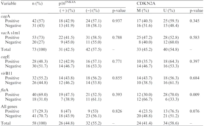
Table 4. Distribution of the p16INK4Adetection andCDKN2Amethylation according to theHelicobacter pylori genes
Variable n (%) p16INK4A CDKN2A
(+) (%) ()) (%) p-value M (%) U (%) p-value cagA
Positive 42 (57) 18 (42.9) 24 (57.1) 0.937 17 (40.5) 25 (59.5) 0.345 Negative 31 (43) 13 (41.9) 18 (58.1) 16 (51.6) 15 (48.4)
vacA s1m1
Positive 53 (73) 22 (41.5) 31 (58.5) 0.788 25 (47.2) 28 (52.8) 0.583 Negative 20 (27) 9 (45.0) 11 (55.0) 8 (40.0) 12 (60.0)
Total 73 (100) 31 (42.5) 42 (57.5) – 33 (45.2) 40 (54.8) –
cagE
Positive 28 (48.3) 12 (42.9) 16 (57.1) 0.771 10 (35.7) 18 (64.3) 0.397 Negative 30 (51.7) 14 (46.7) 16 (53.3) 14 (46.7) 16 (53.3)
virB11
Positive 32 (55.2) 14 (43.8) 18 (56.2) 0.855 14 (43.7) 18 (56.3) 0.684 Negative 26 (44.8) 12 (46.2) 14 (53.8) 10 (38.5) 16 (61.5)
flaA
Positive 40 (69.0) 19 (47.5) 21 (52.5) 0.393 12 (30.0) 28 (70.0) 0.009 Negative 18 (31.0) 7 (38.9) 11 (61.1) 12 (66.7) 6 (33.3)
All genes
Positive 17 (29.3) 8 (47) 9 (53) 0.826 4 (23.5) 13 (76.5) 0.076 Negative 41 (70.7) 18 (43.9) 23 (56.1) 20 (48.8) 21 (51.2)
found to be associated with non-methylated tumors. Also, there was a tendency between non-methylated tumors with the presence of
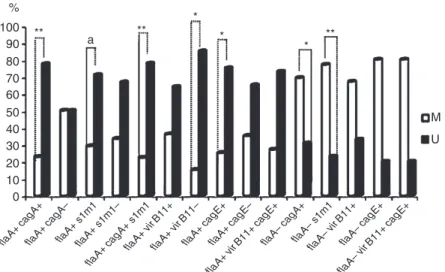
H. pylori carrying all studied genes; however, this was not statistically significant (p = 0.076). In addition, H. pylori strains bearing cagA or
vacAs1m1 genes but without flaA were associ-ated with methylassoci-ated tumors (p = 0.022 and 0.003, respectively) (Fig. 2).
According to the histological subtypes and tumor location, no association was found between the negativity of p16INK4A and
H. pylori strains. However, in intestinal tumors, the presence of the flaA gene, isolated or in combination with all other H. pylori
genes studied, was related to a tendency of non-methylation of CDKN2A, but was not statistically significant (p=0.071 and 0.071 respectively).
According to the location, in noncardia tumors, non-methylated CDKN2A was signifi-cantly more frequent in association with strains of H. pylori carrying all genes studied (p = 0.035) orflaA alone (p=0.036). This fact seems characteristic of noncardia intestinal tumors, becauseH. pyloristrains carrying all the genes studied were associated with non-methyla-tion tumors in this histological subtype, though without a statistical difference (p = 0.076).
To investigate the involvement of the
H. pylori strain in the p16INK4A inactivation process, we analyzed the p16INK4A-negative cases considering the inactivation pathway
(methylation or non-methylation) and the pres-ence of bacterial genes. From these analyses, we found a significant association between the inactivation of p16INK4A by a non-methylation pathway and theflaA gene (p = 0.029). Analy-sis of the inactivation of p16INK4A, according to histological subtype and location, considering
H. pylori strains, was not possible because of the small number of cases.
DISCUSSION
The importance of loss of the tumor suppressor p16INK4A in the carcinogenic process has been pointed out in many cancers (34–36). In GC, loss of expression of p16INK4A is common and hypermethylation of promoter regions of this gene is considered to be its main inactivation mechanism (7–9). In this study, the p16INK4A positivity (40.3%) and CDKN2A methylation promoter status (46.3%) are in accordance with other reports (24, 37, 38). Moreover, the predominantly nuclear⁄cytoplasmic staining
pattern observed was the same as that most often described in the literature. According to Liang et al. (39), cytoplasmic staining indicates the accumulation of this protein in the cyto-plasm, caused by its phosphorylation mediated by protein kinase B, oncogenetically activated, blocking the recognition of its sites by nuclear importation factors. However, the agreement between nuclear and cytoplasmic staining found 100
90 80 70 60 50 40 30 20 10 0 %
** **
*
*
* **
M
U
flaA+ cag
A+
flaA+ cag
A–
flaA+ s1m1flaA+ s1m1–
flaA+ cag
A+ s1m1
flaA+ virB11+
flaA+ virB11–
flaA+ cag
E+
flaA+ cag
E–
flaA– cag
A+
flaA– s1m1 flaA–
virB11+
flaA– vir
B11+ cag
E+
flaA– cag
E+
flaA+ vir
B11+ cag
E+
a
in this study suggests that the presence of p16INK4A protein in the cytoplasm reflects its nuclear expression.
In previous studies, p16INK4A positivity was associated with the absence of lymph node invasion (38, 40) and CDKN2A promoter methylation was related to older patients and significantly lower in well-differentiated tumors (24), but no correlation was found between these clinicopathological parameters and p16INK4A expression andCDKN2Apromoter methylation in the present study. Kishimoto et al. (41) and Mino et al. (42) did not find this association as well.
The strong correlation found in this study between p16INK4A negativity and promoter methylation is in agreement with the other studies (9, 24, 43). However, despite the indica-tion that CDKN2A hypermethylation is the main mechanism implicated in p16INK4A inacti-vation, it is not the only one, because 30.4% of the p16INK4A-negative cases were not methylat-ed. Other studies also have shown the same results (9, 24). The frequency found in this and the other studies cited was not so low and can-not be justified by methodology standardiza-tion error. Instead, it can be explained by alternative mechanisms of p16INK4A inactiva-tion, such as mutations and deletions. In GC, the frequency of p16 inactivation by homozy-gous deletions reported in the literature ranges from 0% to 9% and by mutation from 0% to 2% (9, 44, 45).
In light of studies demonstrating that genetic and epigenetic changes in gastric carcinoma differ, depending on the GC histological type (46, 47), we analyzed the data considering the adenocarcinoma subtypes separately. However, no differences in p16INK4A expression and
CDKN2A promoter methylation were observed between intestinal and diffuse tumors. Interest-ingly, regarding tumor location, there was a difference in the p16INK4Aexpression, but not in the CDKN2A promoter methylation status, as the cardia tumors had a significant number of p16INK4A-negative cases. The location of the tumors seems to be a relevant aspect for the tumorigenic pathway. Driessen et al. (48, 49) reported that the characteristics of cardia carci-noma, including clinical data and cytokeratin expression patterns, were closer to esopha-geal adenocarcinoma than to distal gastric
adenocarcinoma, suggesting the distinct nature of those tumors in the determination of tumori-genic pathways. The higher frequency of p16INK4A negativity in cardia tumors was also observed by Kim et al. (11), in a study compar-ing the various protein expression profiles between patients with cardia and noncardia car-cinomas, such as p53, E-cadherin, MGMT and p16INK4A. However, the authors did not evalu-ate this expression considering the histological subtypes.
Thus, in light of the differences in p16INK4A expression between cardia and noncardia tumors, we analyzed the data considering histo-logical subtypes and tumor location concomi-tantly. The higher number of p16INK4A-negative cases located in the cardia was characteristic of intestinal tumors, although these lesions did not differ regarding the CDKN2A methylation status according to the location. As in this study, Abbaszadegan et al. (24) also found no relationship between CDKN2A promoter methylation and tumor location or between
CDKN2A promoter methylation and histologi-cal subtypes.
Therefore, based on p16INK4A negativity,
CDKN2A promoter methylation and tumor location, it was possible to identify distinct pathways in intestinal and diffuse tumors. Although the number of cases of non-methy-lated tumors was small for both histological subtypes, in noncardia diffuse tumors, the meth-ylation ofCDKN2Apromoter was the only way by which the p16INK4A inactivation occurred. On the other hand, in intestinal tumors, this process occurred by two different routes inde-pendently of tumor location. This study is the first report in the literature regarding p16INK4A inactivation by promoter methylation according to histological subtypes and tumor location.
Although the p16INK4A inactivation is accepted as part of GC tumorigenesis, the mech-anism that triggers this process remains unknown. It is suggested that H. pylori has an important role in this process (22, 23). In the present study, we observed that the influence of
H. pylori carrying all the studied genes was observed. In addition, inactivation of p16INK4A by a non-methylation pathway was associated with theflaA gene. However, strains withcagA or vacA s1m1 without flaA genotype were related toCDKN2Apromoter methylation. It is possible that the H. pyloristrain carrying these genes acts by releasing reactive oxygen species, which can make DNA unstable, and nitric oxide, which can activate DNA methyltransfe-rases (50, 51). In addition, increasing evidence has shown that H. pylorican induce cell prolif-eration, and cell proliferation itself has been suggested as a promoting factor for DNA meth-ylation (52, 53). In spite of the lack of studies relating bacterium genotype to p16INK4A inacti-vation and promoter methylation, some studies have correlated H. pylori infection with p16INK4A methylation, but with controversial results. This can be explained by the variation in the genotypes of the H. pylori strains studied. Nevertheless, we cannot justify the presence of the flaA as a cut-off point for the CDKN2A
promoter methylation status. The flaA gene codifies the main flagellin protein of the flagella filament, responsible for the motility of
H. pylori in the gastric mucus and for the suc-cessful colonization in the gastric mucosa (20). Therefore, future studies are needed to explain the involvement of this gene associated with p16INK4A promoter methylation status with a larger number of cases.
In conclusion, inactivation of p16INK4A in histological subtypes of GC occurs by different routes depending on the location of the tumor. In diffuse subtype tumors, inactivation of p16INK4Aby methylation of the CDKN2A pro-moter occurs exclusively in noncardia tumors. On the other hand, in intestinal tumors, although promoter methylation is an important route for inactivation of p16INK4A, it occurs both in cardia and noncardia tumors. In addi-tion, this process of methylation of the
CDKN2A promoter depends on the H. pylori
genotype.
REFERENCES
1. Jemal ADVM, Siegel RMPH, Ward E, Murray T, Xu J, Smigal CMPH, et al. Cancer statistic, 2006. CA Cancer J Clin 2006;56:106–30.
2. Ferlay J, Bray F, Pisani P, Parkin DM. Globocan 2002: Cancer Incidence, Mortality and Prevalence Worldwide. IARC CancerBase no. 5, version 2.0. Lyon: IARC Press, 2004.
3. Tamura G. Alterations of tumor suppressor and tumor-related genes in the development and pro-gression of gastric cancer. World J Gastroenterol 2006;12:192–8.
4. Ortega S, Malumbres M, Barbacid M. Cyclin D-dependent kinases, INK4 inhibitors and can-cer. Biochim Biophys Acta 2002;1602:73–87. 5. Lima VP, de Lima MA, Andre´ AR, Ferreira MV,
Barros MA, Rabenhorst SH. H. pylori (CagA) and Epstein–Barr virus infection in gastric carci-nomas: correlation with p53 mutation and c-Myc, Bcl-2 and Bax expression. World J Gastroenterol 2008;14:884–91.
6. Lima EM, Leal MF, Burbano RR, Khayat AS, Assumpc¸a˜o PP, Bello MJ, et al. Methylation sta-tus of ANAPC1, CDKN2A and TP53 promoter genes in individuals with gastric cancer. Braz J Med Biol Res 2008;41:539–43.
7. Ding Y, Le XP, Zhang QX, Du P. Methylation and mutation analysis of p16 gene in gastric cancer. World J Gastroenterol 2003;9:423–6. 8. Zhao GH, Li TC, Shi LH, Xia YB, Lu LM,
Huang WB, et al. Relationship between inactiva-tion of p16 gene and gastric carcinoma. World J Gastroenterol 2003;9:905–9.
9. Shim YH, Kang GH, Ro JY. Correlation of p16 hypermethylation with p16 protein loss in spo-radic gastric carcinomas. Lab Invest 2000;80: 689–95.
10. Ksiaa F, Ziadi S, Amara K, Korbi S, Trimeche M. Biological significance of promoter hyperme-thylation of tumor-related genes in patients with gastric carcinoma. Clin Chim Acta 2009;404:128– 33.
11. Kim MA, Lee HS, Yang HK, Kim WH. Clinico-pathologic and protein expression differences between cardia carcinoma and noncardia carci-noma of the stomach. Cancer 2005;103:1439–46. 12. Peek RM Jr, Blaser MJ. Helicobacter pylori and
gastrointestinal tract adenocarcinomas. Nat Rev Cancer 2002;2:28–37.
13. Atherton JC. The pathogenesis of Helicobacter pylori-induced gastro-duodenal diseases. Annu Rev Pathol 2006;1:63–96.
14. Sozzi M, Tomasini ML, Vindigni C, Zanussi S, Tedeschi R, Basaglia G, et al. Heterogeneity of cag genotypes and clinical outcome of Helico-bacter pylori infection. J Lab Clin Med 2005; 146:262–70.
15. Covacci A, Telford JL, Del Giudice G, Parsonnet J, Rappuoli R. Helicobacter pylori virulence and genetic geography. Science 1999;284:1328–33. 16. Lu H, Wu JY, Kudo T, Ohno T, Graham DY,
with Helicobacter pylori. Mol Biol Cell 2005; 16:4954–66.
17. Wang HT, Li ZH, Yuan JP, Zhao W, Shi XD, Tong SQ, et al. Effect of Helicobacter pylori VacA on gene expression of gastric cancer cells. World J Gastroenterol 2005;11:109–13.
18. Krause S, Pansegrau W, Lurz R, Cruz F, Lanka E. Enzymology of type IV macromolecule secre-tion systems: the conjugative transfer regions of plasmids RP4 and R388 and the cag pathogenic-ity island of Helicobacter pylori encode struc-turally and functionally related nucleoside triphosphate hydrolases. J Bacteriol 2000;182: 2761–70.
19. Maeda S, Yoshida H, Ikenoue T, Ogura K, Kanai F, Kato N, et al. Structure of cag pathogenicity island in Japanese Helicobacter pylori isolates. Gut 1999;44:336–41.
20. Suerbaum S. The complex flagella of gastric Heli-cobacter species. Trends Microbiol 1995;3:168–70. 21. Chan AO, Huang C, Hui WM, Cho CH, Yuen MF, Lam SK, et al. Stability of E-cadherin meth-ylation status in gastric mucosa associated with histology changes. Aliment Pharmacol Ther 2006; 24:831–6.
22. Dong CX, Deng DJ, Pan KF, Zhang L, Zhang Y, Zhou J, et al. Promoter methylation of p16 asso-ciated with Helicobacter pylori infection in pre-cancerous gastric lesions: a population-based study. Int J Cancer 2009;124:434–9.
23. Tahara T, Arisawa T, Shibata T, Nakamura M, Yoshioka D, Okubo M, et al. Increased number of methylated CpG islands correlates with Heli-cobacter pylori infection, histological and sero-logical severity of chronic gastritis. Eur J Gastroenterol Hepatol 2009;21:613–9.
24. Abbaszadegan MR, Moaven O, Sima HR, Gha-farzadegan K, A’rabi A, Forghani MN, et al. p16 promoter hypermethylation: a useful serum mar-ker for early detection of gastric cancer. World J Gastroenterol 2008;14:2055–60.
25. Chan AO, Lam SK, Wong BC, Wong WM, Yuen MF, Yeung YH, et al. Promoter methylation of E-cadherin gene in gastric mucosa associated with Helicobacter pylori infection and in gastric can-cer. Gut 2003;52:502–6.
26. Foster G, Twell D. Plant Gene Isolation: Princi-ples and Practice. Chichester, UK: John Wiley & Sons, 1996. p. 426.
27. Herman JG, Graff JR, Myo¨ha¨nen S, Nelkin BD, Baylin SB. Methylation-specific PCR: a novel PCR assay for methylation status of CpG islands. Proc Natl Acad Sci USA 1996;93:9821–6.
28. Lage AP, Godfroid E, Fauconnier A, Burette A, Butzler JP, Bollen A, et al. Diagnosis of Helicob-acter pylori infection by PCR: comparison with other invasive techniques and detection of cagA gene in gastric biopsy specimens. J Clin Microbiol 1995;33:2752–6.
29. Atherton JC, Cao P, Peek RM, Tummuru MK, Blaser MJ, Cover TL. Mosaicism in vacuolating cytotoxin alleles of Helicobacter pylori. Associa-tion of specific vacA types with cytotoxin produc-tion and peptic ulceraproduc-tion. J Biol Chem 1995; 270:17771–7.
30. Domingo D, Alarcon T, Pietro N, Sanchez I, Lopez-Brea M. cagA and vacA status of Spanish Helicobacter pylori clinical isolates. J Clin Micro-biol 1999;37:2113–4.
31. Go¨ttke MU, Fallone CA, Barkun AN, Vogt K, Loo V, Trautmann M, et al. Genetic variability determinants of Helicobacter pylori: influence of clinical background and geographic origin of iso-lates. J Infect Dis 2000;181:1674–81.
32. Landberg G, Roos G. Proliferating cell nuclear antigen and Ki-67 antigen expression in human haematopoietic cells during growth stimulation and differentiation. Cell Prolif 1993;26:427– 37.
33. McCarty KS Jr, Miller LS, Cox EB, Konrath J, McCarty KS Sr. Estrogen receptor analyses. Correlation of biochemical and immuno-histochemical methods using monoclonal antire-ceptor antibodies. Arch Pathol Lab Med 1985; 109:716–21.
34. Zhou J, Yang D, Zhang L, Wang J, Yao Q, Su Z, et al. Study on the relationship of alteration and expression of p16 gene to pancreatic carcinoma. Zhonghua Yi Xue Yi Chuan Xue Za Zhi 2000; 17:399–403.
35. Goto T, Mizukami H, Shirahata A, Sakata M, Saito M, Ishibashi K, et al. Aberrant methyla-tion of the p16 gene is frequently detected in advanced colorectal cancer. Anticancer Res 2009;29:275–7.
36. Vidaurreta M, Maestro ML, Sanz-Casla MT, Maestro C, Rafael S, Veganzones S, et al. Inac-tivation of p16 by CpG hypermethylation in renal cell carcinoma. Urol Oncol 2008;26:239– 45.
37. Napieralski R, Ott K, Kremer M, Becker K, Boulesteix AL, Lordick F, et al. Methylation of tumor-related genes in neoadjuvant-treated gas-tric cancer: relation to therapy response and clini-copathologic and molecular features. Clin Cancer Res 2007;13:5095–102.
38. He XS, Rong YH, Su Q, Luo Q, He DM, Li YL, et al. Expression of p16 gene and Rb protein in gastric carcinoma and their clinicopathological significance. World J Gastroenterol 2005;11: 2218–23.
39. Liang J, Zubovitz J, Petrocelli T, Kotchetkov R, Connor MK, Han K, et al. PKB⁄Akt phosphory-lates p27, impairs nuclear import of p27 and opposes p27-mediated G1 arrest. Nat Med 2002; 8:1153–60.
suppressor and tumor-related proteins in differen-tiated carcinoma, undifferendifferen-tiated carcinoma with tubular component and pure undifferentiated car-cinoma of the stomach. Jpn J Clin Oncol 2005; 35:580–6.
41. Kishimoto I, Mitomi H, Ohkura Y, Kanazawa H, Fukui N, Watanabe M. Abnormal expression of p16(INK4a), cyclin D1, cyclin-dependent kinase 4 and retinoblastoma protein in gastric carcinomas. J Surg Oncol 2008;98:60–6.
42. Mino A, Onoda N, Yashiro M, Aya M, Fujiwara I, Kubo N, et al. Frequent p16 CpG island hyper-methylation in primary remnant gastric cancer suggesting an independent carcinogenic pathway. Oncol Rep 2006;15:615–20.
43. Zhao YF, Zhang YG, Tian XX, Juan Du, Zheng J. Aberrant methylation of multiple genes in gas-tric carcinomas. Int J Surg Pathol 2007;15:242– 51.
44. Wu Q, Possati L, Montesi M, Gualandi F, Rimessi P, Morelli C, et al. Growth arrest and suppression of tumorigenicity of bladder-carcinoma cell lines induced by the p16⁄CDKN2 (p16INK4A, MTS1) gene and other loci on human chromosome 9. Int J Cancer 1996;65:840–6.
45. Toyota M, Ahuja N, Suzuki H, Itoh F, Ohe-Toyota M, Imai K, et al. Aberrant methylation in gastric cancer associated with the CpG island methylator phenotype. Cancer Res 1999;59:5438– 42.
46. Tahara E. Genetic pathways of two types of gas-tric cancer. IARC Sci Publ 2004;157:327–49. 47. Tahara E, Kuniyasu H, Yasui W, Yokozaki H.
Gene alterations in intestinal metaplasia and gas-tric cancer. Eur J Gastroenterol Hepatol 1994;6: S97–102.
48. Driessen A, Nafteux P, Lerut T, Van Raemdonck D, De Leyn P, Filez L, et al. Identical cytokeratin expression pattern CK7+⁄CK20)in esophageal
and cardiac cancer: etiopathological and clinical implications. Mod Pathol 2004;17:49–55.
49. Driessen A, Van Raemdonck D, De Leyn P, Filez L, Peeters M, Winnepenninckx V, et al. Are carci-nomas of the cardia oesophageal or gastric adeno-carcinomas? Eur J Cancer 2003;39:2487–94. 50. Obst B, Wagner S, Sewing KF, Beil W.
Helico-bacter pylori causes DNA damage in gastric epithelial cells. Carcinogenesis 2000;21:1111–5. 51. Hmadcha A, Bedoya FJ, Sobrino F, Pintado E.
Methylation-dependent gene silencing induced by interleukin 1beta via nitric oxide production. J Exp Med 1999;190:1595–604.
52. Suzuki H, Hibi T, Marshall BJ. Helicobacter pylori: present status and future prospects in Japan. J Gastroenterol 2007;42:1–15.