AR
TIGO ORIGINAL/ ORIGINAL AR
TICLE
IMPACT OF PROLONGED 48-H WIRELESS
CAPSULE ESOPHAGEAL pH MONITORING
ON DIAGNOSIS OF GASTROESOPHAGEAL
REFLUX DISEASE AND EVALUATION OF THE
RELATIONSHIP BETWEEN SYMPTOMS AND
REFLUX EPISODES
Gerson Ricardo de Souza
DOMINGUES
1, Joaquim Prado
P. MORAES-FILHO
2and
Aline Gonçalves Leite
DOMINGUES
1ABSTRACT – Context - Gastroesophageal relux disease is one of the most common digestive diseases and an important cause of
distress to patients. Diagnosis of this condition can require ambulatory pH monitoring. Objectives - To determine the diagnostic yield of a wireless ambulatory pH monitoring system of 48-hours, recording to diagnose daily variability of abnormal esophageal acid exposure and its symptom association. Methods - A total of 100 consecutive patients with persistent relux symptoms underwent wireless pH capsule placement from 2004 to 2009. The wireless pH capsule was deployed 5 cm proximal to the squamocolumnar junction after lower esophageal sphincter was manometrically determined. The pH recordings over 48-h were obtained after uploading data to a computer from the receiver that recorded pH signals from the wireless pH capsule. The following parameters were analyzed: (1) percentual time of distal esophageal acid exposure; (2) symptom association probability related to acid relux. The results between the irst and the second day were compared, and the diagnostic yield reached when the second day monitoring was included. Results - Successful pH data over 48-h was obtained in 95% of patients. Nearly one quarter of patients experienced symptoms ranging from a foreign body sensation to chest pain. Forty-eight hours pH data analysis was statistically signiicant when compared to isolated analysis of day 1 and day 2. Study on day 2 identiied seven patients (30.4%) that would be missed if only day 1 was analyzed. Three patients (18.7%) out of 16 patients with normal esophageal acid exposure on both days, showed positive symptom association probability, which generated an increase in diagnostic yield of 43.4%. Conclusion - Esophageal pH monitoring with wireless capsule is safe, well tolerated, does not require sedation. The extended 48-h period of study poses an increased yield to diagnose gastroesophageal relux disease patients.
HEADINGS – Gastroesophageal relux. Esophageal pH monitoring. Gastric acidity determination. Capsule endoscopes.
INTRODUCTION
Gastroesophageal relux disease (GERD) is one of the most prevalent diseases in gastroenterology(16).
In Brazil, a population based study looking for relux typical symptoms interviewed 13,959 individuals showed GERD prevalence of 12%(17).
Ambulatory catheter-based esophageal pHmetry is considered the gold-standard for diagnosing GERD because it quantiies esophageal acid exposure and symptom association(13). Traditionally, the catheter which
has an antimonium pH sensor is passed transnasally and its tip is positioned 5-cm above the upper border
1GRD – Gastro Resolução em Diagnóstico, Laboratório de Motilidade Digestiva, Rio de Janeiro, RJ, Brazil; 2 Departamento de Gastroenterologia, Faculdade de Medicina da Universidade de São Paulo, São Paulo, SP, Brazil.
Correspondence: Dr. Gerson R. S. Domingues. Av. Ayrton Senna, 1850/224 - Barra da Tijuca – 22775-003 - Rio de Janeiro, RJ, Brazil. E-mail: [email protected]
of the lower esophageal sphincter (LES) to monitor intra-esophageal pH. The study has 24-h duration(7)
and a sensitivity of 75% and a speciicity of 90%(11).
This relatively low sensitivity is due to changes in activities and behavior of patients during the study. The pharyngeal discomfort and social constraint lead patients to assume a more sedentary lifestyle, decreasing relux-provoking activities(6). In addition, the
day-to-day variability in intensity of gastroesophageal relux may inluence the results of the esophageal pH monitoring(8). Johnsson et al.(12) showed that only 77%
Recently, aiming to overcome these limitations, it was developed a new ambulatory esophageal pH monitoring device using a wireless system (Bravo system®)(18) that collects
data during an extended period of time (48 hours). The data are captured by a small capsule attached to distal esophageal mucosa that transmits the signals via radio telemetry to a receiver worn by the patient(18).
Therefore, the aim of this study was to retrospectively determine the advantages of a 48-h study using a wireless ambulatory pH monitoring system in terms of diagnostic yield in consecutive patients referred to a motility laboratory to investigate GERD.
METHODS
Patients
This is a retrospective study of diagnostic method. One hundred consecutive patients were selected, 39 female, mean age 53 years (range 18 to 81 years) between 2004 and 2009 referred to a gastrointestinal motility laboratory to investigate GERD with typical and/or atypical symptoms, persistent GERD symptoms on medical therapy, recurrent GERD symptoms after surgical fundoplication procedure.
The exclusion criteria were: patients aged less than 18 years or with history of bleeding tendency or coagulopathy; signiicant concomitant medical co-morbidities; severe gastrointestinal bleeding within the past 6 months; a past history of upper gastrointestinal surgery that could interfere with acid esophageal proile such as total or partial gastrectomy and esophagectomy; patients on medication with proton pump inhibitors (PPI); presence of esophageal varices; Barrett’s esophagus; esophageal stenosis; erosive esophagitis; pacemaker or implantable cardiac deibrillator in situ(20).
This study was approved by the Ethic Committee of University of Santo Amaro - UNISA, São Paulo, SP, Brazil.
Before the 48-hour wireless test was performed, patients were instructed to discontinue all antiacid and antisecretory medications for a minimum of 7 days. The only exception occurred in patients being evaluated for whom referring physicians speciically requested the study be performed on medications. Diet restriction included nothing by mouth at least 6 hours before procedure.
Study protocol
All patients underwent previous upper endoscopy after an overnight fast to discard counter indications to capsule attachment.
Initially, 98 patients underwent standard transnasal esophageal manometry to identify the proximal border of the LES. Two patients underwent upper endoscopy to determine the position of LES(20). Thereafter, to those patients that
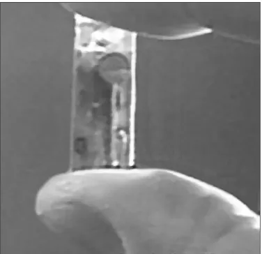
performed transnasal manometry, a validated conversion factor of 4 cm was applied in order to permit accurately transoral placement of Bravo capsule(15) (Figure 1). The Bravo
capsule with its delivery catheter was introduced through the mouth and positioned 5 cm above the upper border of LES determined by transnasal manometry or 6 cm above
the LES to those two patients who performed endoscopy(15).
A vacuum pump was turned on to apply a suction pressure of >500 mm Hg to a well within the wireless pH capsule(18).
After the optimal pressure has been reached a small amount of esophageal mucosa was drawn into the wireless pH capsule. Subsequently, an activation button was twisted clockwise (90°) and re-extended resulting in release of the
delivery system from the capsule(18). A remote data recorder
was worn around the waist of the patient and recording was carried out for 48-h. Patients were instructed to keep a diary, record meal times, position changes, the time and type of their symptoms and symptoms that could be related to the capsule, such as chest discomfort, chest pain and dysphagia. Patients were encouraged to pursue their every day activities and usual diet. During bath, sleep and physical exercises(19),
patients were instructed to put the recorder apart the body to a distance of no more than 2 meters. Patients were informed to refrain from a magnetic resonance imaging study within 30 days of the Bravo procedure and a 10-days period after the study ended, patients underwent a chest X-ray to conirm capsule detachment(21).
Data analysis
Data analysis was performed using standard commercially available computer software. Relux was deined as pH <4 and relux time as the interval until pH >4. The characteristics of esophageal acid exposure during the 48-h study period to consider a study positive are as follow(29): (1) % total time
acid exposure >4.4%; (2) % total time in upright position >6.3; (3) % total time in supine position >3.7; (4) symptom association probability (SAP) to express the relationship between symptoms and acid relux episodes(1). The SAP is
considered positive when the association is superior to 95%.
Adverse effects n (patients) % Mild foreign
body sensation
17 22.9
Chest pain during meal periods
2 2.7
Total 19 25.6
TABLE 1. Adverse effects reported during Bravo system procedure
Day 48-h P
n* (%) n (%)
Day 1 51 (68.9) 58 (78.3) 0.023 Day 2 49 (70.2) 58 (78.3) 0.007 TABLE 2. Comparison between the results of abnormal pHmetry on day 1 and 48-h and on day 2 and 48-h
* n represents the number of patients with abnormal pH data
SAP+ SAP+ 48-h P
n* (%) n (%)
Day 1 19 (44.1) 27 (62.7) 0.026 Day 2 17 (40.4) 26 (61.9) 0.007 TABLE 3. Comparison between the results of SAP+ on day 1 and 48-h
and on day 2 and 48-h
* n represents the number of patients with SAP+
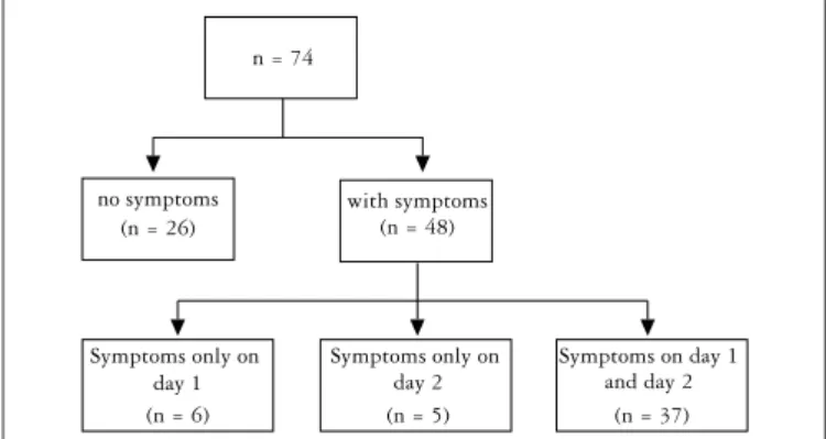
no symptoms (n = 26)
n = 74
with symptoms (n = 48)
Symptoms only on day 1 (n = 6)
Symptoms only on day 2 (n = 5)
Symptoms on day 1 and day 2
(n = 37)
FIGURE 2. Algorithm of symptom distribution on study day 1 and day 2
The results found on the 1st and 2nd days of the study were compared with those of the 48-h study.
The premature detachment of the capsule from the esophageal mucosa was deined as sudden prolonged drop in pH representing the capsule in the stomach (pH <4.0) and than the sharp rise as the capsule enters the small intestine through the pylorus (pH >7.0)(20).
Statistical analysis
The results are reported as percentages unless otherwise stated. The McNemar test was used to compare the pH data from the 1st or the 2nd day of the study with the data from the 48-h study. The SPS 11.0 was used for all statistical analyses. Alpha <0.05 deined statistical signiicance.
RESULTS
Eighty-two patients (82%) were referred to diagnosis GERD and 18 patients were referred due to persistent symptoms despite PPI use.
Successful capsule attachment to distal esophageal mucosa was seen in 95 patients (95%). Mean duration of the study was 47 hours.
Twenty-six patients were excluded from data analysis: (a) technical laws: failure to attach the capsule (1 patient), premature detachment of the capsule (2 patients), failure to capture the signal (2 patients); (b) only 24-h study (2 patients); (c) downloaded data lost (2 patients); (d) on PPI therapy (10 patients); (e) and with fundoplication (8 patients). One patient on PPI use also performed only 24-hour study. Thus, 74 patients composed the study group.
Complications related to capsule attachment were generally mild, reported by 19 patients (25.6%). Seventeen patients (22.9%) noted a mild foreign body sensation, and 2 patients (2.7%), chest pain during meal periods, mainly on day 1 of the study (Table 1).
On day 1 pH data were abnormal in 51 patients (68.9%) and on day 2 pH data were abnormal in 49 patients (70.2%). Analysis of 48-h study showed abnormal pH data in 58 patients (78.3%). Table 2 shows that there was statistical signiicant difference between pH data of the 48-h study and day 1 (P = 0.023) and day 2 (P = 0.007).
Forty-eight patients (64.8%) reported symptoms related to GERD. Six patients only on day 1, 5 patients only on day 2, and 37 patients reported symptoms on both days of the study (Figure 2). SAP was positive during day 1 in
19 patients (44.1%) and positive during 48-h in 27 patients (62.7%). There was statistical signiicant difference (P = 0.026) between SAP of the day 1 and the SAP of 48-h study. During day 2, SAP was positive in 17 patients (40.4%) and during 48-h in 26 patients (61.9%). There was also statistical signiicant difference (P = 0.007) between SAP of the day 2 and the SAP of 48-h study. The difference of one patient in the total number of SAP positive patients in the 48-h period from day 1 to day 2 is due to the methodology intrinsically used to calculate SAP. Thus, when SAP was calculated based on the relationship between symptoms and relux episodes during day 1, it was positive. However, when data of 48-h study period was taken into account, SAP showed a negative result (Table 3).
DISCUSSION
In this study, 48-h ambulatory wireless capsule intraesophageal pH monitoring was successful in 95% of patients. Failure to attach the capsule to distal esophageal mucosa occurred in only one patient, evincing that the period of the learning curve is short. The 48-h extended period of study revealed signiicant day-to-day variability in esophageal acid exposure and symptom perception. This represented an increase in GERD diagnostic yield of 43.4%.
Traditional 24-h catheter-based esophageal pHmetry has been considered the gold-standard method to diagnose GERD. However, Bortolotti et al.(3) monitored esophageal pH
of individuals with the Bravo system and a standard probe-based device simultaneously and showed both techniques had similar results to detect acid relux in distal esophagus. Subsequently, it was demonstrated that the Bravo system is capable of distinguish between GERD patients and controls with sensitivity of 84% and speciicity of 85%(18).
Acid exposure is greater just above the squamocolumnar junction(22), however, by convention, in catheter-based
pHmetry the tip of the probe is positioned 5 cm above the LES to prevent migration of the pH sensor into stomach, due to esophageal shortening during swallows, which in turn could lead to record false episodes of acid relux(8).
Bravo system monitors pH with a capsule attached to esophageal mucosa, therefore, in a ixed position in relation to LES, overcoming this limitation seen with catheter-based pHmetry(21). To compare our results with previous studies
and due to a lack of normal values to acid exposure just above the squamocolumnar junction(10) we decided to attach
the capsule 5 cm above the LES.
In general, capsule spontaneously falls off within 5 days. Endoscopic removal is reserved for patients who do not respond to conservative management and/or continue to have symptoms for more than 48 h. To remove the capsule one should use a cold snare placed loosely around the base of the attachment area between the capsule and the esophageal wall and than apply gentle traction in a to-and-fro movement(20). In our study the spontaneous dislodgement
of the capsule occurred in all patients within 5 to 10 days, with no complications.
The most common complication of Bravo system was foreign body sensation reported by 22.9% of patients, the second most common complication was chest pain observed in only 2.7% of patients, similar results were found by other authors(18, 31). Pandolino et al.(18) reported 3 out of 85 patients
with chest pain, 2 patients requiring endoscopic removal of the capsule. In this study, two patients reported chest pain during meal period, although endoscopic removal of the capsule was not necessary. The proposed mechanism to chest pain in these patients probably is related to abnormalities in central or peripheral visceral nociception, in particular, decreased threshold in generation or perception of symptoms(9).
Similar to other authors(10, 31) patients in this study did not
complain of pharyngeal discomfort or interference in their daily activities.
Limitations to Bravo system also occur. Laryngeal and/ or proximal esophageal relux monitoring is not feasible with capsule technology and in this direction, symptoms like globus sensation can not be suitably evaluated(23). Esophageal
impedance studies point to the height reached by the reluxate as an important factor in generation of globus sensation(14).
In patients with Barrett’s esophagus, capsule attachment to esophageal mucosa can be dificult(20). Recently, discussion
has been aroused around the role of non-acid relux and its relationship with generation of symptoms or persistence of symptoms despite antisecretory medical therapy(25). With
Bravo system is not possible to capture non-acid relux in order to correlate with symptoms. Other limitation of this system is that capsule is expensive.
Acid exposure in distal esophagus shows day-to-day variability and it can affect 24-h pHmetry accuracy(2). Factors such as
diet, body position, physical activity, social inconvenience and intraesophageal catheter position alter intensity of gastroesophageal relux(6). On the other hand, Bravo system
has inherent advantages, as patients report signiicant less discomfort during the procedure when compared to catheter-based pHmetry(31). Furthermore, there is less impairment in
daily activities and diet modiications and patients monitored by Bravo system can have normal daily routine(30), including
physical exercises practice(19). A study in patients that did
not tolerate catheter-based pHmetry and underwent Bravo system esophageal pH monitoring, showed that patients were highly tolerant and pleased during the study(27).
Our study showed increased yield to diagnose GERD when compared day 1 or day 2 with the extended 48-h period study. Three patients with only positive SAP but normal distal esophageal acid exposure were included in the results because according to Rome III criteria, these patients are considered sensitive to normal distal esophageal acid exposure, and therefore, they should be included in the same spectrum of GERD disease(7). Thus, in total, additional 10
patients were diagnosed as GERD (43.4%) among 23 patients that had no diagnosis of GERD when the irst 24-h of the study was analyzed. Increased GERD diagnostic yield with 48-h extended monitoring was also observed in a Mexican study(26). Pandolino et al.(18) observed daily variability with
Bravo system in normal individuals, patients with erosive and non-erosive GERD. Roughly 27% of patients with GERD diagnosis showed different results considering both days of monitoring, for instance, abnormal acid exposure on day 1 and normal on day 2 and vice-versa(18). In patients with erosive
esophagitis, the sensitivity of the Bravo system increased from 74% to 100% when the worst day was recruited and compared with the recording of the irst 24-h(18).
The extended time period to 48-h increases the probability of patients have symptoms in order to be associated with acid relux episodes, reducing the possibility of this association occurs by chance(24).
Results of Bravo system increases GERD diagnostic yield and can substantially help in decision making, for instance, in selection of candidates to fundoplication surgery(4). Good
DominguesGRS, Moraes-Filho JPP, Domingues AGL. Impacto da monitorização prolongada do reluxo por pHmetria sem cateter por 48 horas, no diagnóstico da doença do reluxo gastroesofágico e no estudo da relação entre os sintomas e o reluxo. Arq Gastroenterol. 2011;48(1):24-9 RESUMO – Contexto - A doença do reluxo gastroesofágico é uma das doenças digestivas mais comuns e importante causa de desconforto para os
pacientes. O diagnóstico desta condição clínica pode requerer monitoramento ambulatorial do pH esofágico. Objetivos - Determinar o espectro diagnóstico do sistema de monitoramento ambulatorial do pH esofágico com cápsula telemétrica por um período de 48 horas no diagnóstico da variabilidade diária da exposição ácida anormal e sua associação com sintomas. Métodos - Foram incluídos 100 pacientes adultos, consecutivos, com sintomas relacionados com a doença do reluxo gastroesofágico, que realizaram pHmetria com cápsula telemétrica por 48 horas entre 2004 e 2009. A cápsula foi posicionada e implantada a 5 cm da borda superior do esfíncter esofágico inferior, deinida pela manometria esofágica. Foram analisados os seguintes parâmetros: (1) tempos percentuais de exposição ácida no esôfago distal; (2) probabilidade de associação dos sintomas com o reluxo ácido. Foram comparados os resultados entre o 1º e o 2º dia de monitoramento, assim como o eventual ganho diagnóstico obtido após a inclusão do 2º dia no monitoramento. Resultados - Sucesso na obtenção dos dados do pH esofágico durante 48 horas foi obtido em 95% dos pacientes. Aproximadamente 25% dos pacientes apresentaram sintomas relacionados à implantação da cápsula, variando entre sensação de presença de corpo estranho à dor torácica. A análise dos resultados do estudo de 48 horas mostrou-se estatisticamente signiicante quando comparada com as análises isoladas do dia 1 e do dia 2. O estudo do dia 2 identiicou sete pacientes (30.4%) que teriam sido perdidos se somente o dia 1 fosse analisado. Três pacientes (18.7%) dos 16 pacientes com exposição ácida normal no esôfago distal em ambos os dias, apresentaram probabilidade de associação com o sintoma positivo, que gerou incremento no ganho diagnóstico com este método diagnóstico de 43.4%. Conclusões - O monitoramento do pH esofágico com a cápsula telemétrica é seguro, bem tolerado e não requer sedação. A extensão do período de estudo para 48 horas representa signiicativo aumento no ganho diagnóstico em pacientes com doença do reluxo gastroesofágico.
DESCRITORES – Reluxo gastroesofágico. Monitoramento do pH esofágico. Determinação da acidez gástrica. Cápsulas endoscópicas.
REFERENCES
1. Aanen MC, Bredenoord AJ, Numans ME, Samson M, Smout AJ. Reproducibility of symptom association analysis in ambulatory relux monitoring. Am J Gastroenterol. 2008;103:2200-8.
2. Ayazi S, Lipham JC, Portale G, Peyre CG, Streets CG, Leers JM, Demeester SR, Banki F, Chan LS, Hagen JA, Demeester TR. Bravo catheter-free pH monitoring: normal values, concordance, optimal diagnostic thresholds, and accuracy. Clin Gastroenterol Hepatol. 2009;7:60–7.
3. Bortolotti M, Gatta L, Vakil N. Simultaneous esophageal pH recording with the Bravo system and a standard probe-based device. Gastroenterology. 2004;126(suppl 2):a320.
4. Campos GM, Peters JH, DeMeester TR, Oberg S, Crookes PF, Tan S, DeMeester SR, Hagen JA, Bremner CG. Multivariate analysis of factors prediciting outcome after laparoscopic Nissen fundoplication. J Gastrointest Surg. 1999;3:292-300. 5. Diaz S, Aymerich R, Clouse RE. The symptom association probability is superior
to the symptom index for attributing symptoms to gastroesophageal relux: validation using outcome from laparoscopic antirelux surgery. Gastroenterology. 2002;122(suppl):a75.
6. Fass R, Hell R, Sampliner RE, Pulliam G, Graver E, Hartz V, Johnson C, Jaffe P. Effect of ambulatory 24-hour esophageal pH monitoring on relux-provoking activities. Dig Dis Sci. 1999;44:2263–9.
7. Galmiche JP, Clouse RE, Bálint A, Cook IJ, Kahrilas PJ, Paterson WG, Smout AJ. Functional esophageal disorders. Gastroenterology. 2006;130:1459-65.
8. Hirano I, Richter JE; Practice Parameters Committee of the American College of Gastroenterology. ACG practice guidelines: esophageal relux testing. Am J Gastroenterol. 2007;102:668-85.
9. Hobson AR, Furlong PL, Aziz Q. Oesophageal afferent pathway sensitivity in non-erosive relux disease. Neurogastroenterol Motil. 2008;20:877-83. 10. Hong RKS, Vaezi MF. Gastroesophageal relux monitoring: pH (catheter and
capsule) and impedance. Gastrointest Endosc Clin N Am. 2009;19:1–22. 11. Jamieson JR, Stein HJ, DeMeester TR, Bonavina L, Schwizer W, Hinder RA,
Albertucci M. Ambulatory 24-h esophageal pH monitoring: normal values, optimal thresholds, speciicity, sensitivity, and reproducibility. Am J Gastroenterol. 1992;87:1102–11.
12. Johnsson F, Joelsson B. Reproducibility of ambulatory oesophageal pH monitoring. Gut. 1988;29:886–9.
13. Kahrilas PJ. Clinical practice. Gastroesophageal relux disease. N Engl J Med. 2008;359:1700-7
14. Kahrilas PJ, Sifrim D. High-resolution manometry and impedance-pH/manometry: valuable tools in clinical and investigational esophagology. Gastroenterology. 2008;135:756-69.
15. Lacy BE, O’Shana T, Hynes M, Kelley MR Jr, Weiss JE, Paquette L, Rothstein RI. Safety and tolerability of transoral Bravo capsule placement after transnasal manometry using a validated conversion factor. Am J Gastroenterol. 2007;102:24-32.
16. Locke GR 3rd, Talley NJ, Fett SL, Zinsmeister AR, Melton LJ 3rd. Prevalence and clinical spectrum of gastroesophageal relux: a population based study in Olmsted County, Minnesota. Gastroenterology. 1997;112:1448–56.
of good response to fundoplication for those patients with abnormal acid GERD. However, to a certain extent, pH data can also help to predict favorable response to treatment. Diaz et al.(5) studied the role of pre-operative SAP to predict
symptom response after laparoscopic fundoplication in GERD patients with abnormal traditional esophageal pHmetry. During follow-up in the post-operative period, it was observed that in patients main symptom associated with a pre-operative positive SAP improved. The authors concluded that SAP was retained as an independent predictor of change in principal symptom and the SAP complements response to medical therapy, in selecting patients to laparoscopic antirelux surgery(5).
CONCLUSION
17. Moraes-Filho JP, Chinzon D, Eisig JN, Hashimoto CL, Zaterka S. Prevalence of heartburn and gastroesophageal relux disease in the urban Brazilian population. Arq Gastroenterol. 2005;42:122-7.
18. Pandolino JE, Richter JE, Ours T, Guardino JM, Chapman J, Kahrilas PJ. Ambulatory esophageal pH monitoring using a wireless system. Am J Gastroenterol. 2003;98:740-9.
19. Pandolino JE, Bianchi LK, Lee TJ, Hirano I, Kahrilas PJ. Esophagogastric junction morphology predicts susceptibility to exercise-induced relux. Am J Gastroenterol. 2004;99:1430-6.
20. Pandolino JE. Bravo capsule pH monitoring. Am J Gastroenterol. 2005;100: 8-10.
21. Pandolino JE, Kahrilas PJ. Prolonged pH monitoring: Bravo capsule. Gastrointest Endosc Clin N Am. 2005;15:307-18.
22. Pandolino JE, Lee TJ, Schreiner MA, Zhang Q, Roth MP, Kahrilas PJ. Comparison of esophageal acid exposure at 1 cm and 6 cm above the squamocolumnar junction using the Bravo pH monitoring system. Dis Esophagus. 2006;19:177-82. 23. Pohl D, Tutuian R. Relux monitoring: pH-metry, bilitec and oesophageal impedance
measurements. Best Pract Res Clin Gastroenterol. 2009;23:299–311. 24. Prakash C, Clouse RE. Value of extended recording time with wireless pH
monitoring in evaluationg gastroesophageal relux disease. Clin Gastroenterol Hepatol. 2005;3:329-34.
25. Pritchett JM, Aslam J, Slaughter C, Ness RM, Garrett CG, Vaezi MF. Eficacy of esophageal impedance/pH monitoring in patients with refractory gastroesophageal relux disease, on and off therapy. Clin Gastroenterol Hepatol. 2009;7:743-8.
26. Remes-Troche JM, Ibarra-Palomino J, Carmona-Sánchez RI, Valdovinos MA. Performance, tolerability, and symptoms related to prolonged pH monitoring using the Bravo system in Mexico. Am J Gastroenterol. 2005;100:2382-6. 27. Sweis R, Fox M, Anggiansah R, Anggiansah A, Basavaraju K, Canavan R, Wong T.
Patient acceptance and clinical impact of Bravo monitoring in patients with previous failed catheter-based studies. Aliment Pharmacol Ther. 2009;29:669-76. 28. seng D, Rizvi AZ, Fennerty B, Jobe BA, Diggs BS, Sheppard BC, Gross SC,
Swanstrom LL, White NB, Aye RW, Hunter JG. Forty-eight-hour pH monitoring increases sensitivity in detecting abnormal esophageal acid exposure. J Gastrointest Surg. 2005;9:1043-52.
29. Wenner J, Johnsson F, Johansson J, Oberg S. Wireless oesophageal pH monitoring: feasibility, safety and normal values in healthy subjects. Scand J Gastroenterol. 2005;40:768-74.
30. Wenner J, Johnsson F, Johnsson J, Oberg S. Wireless esophageal pH monitoring is better tolerated than the catheter-based technique: results from a randomized cross-over trial. Am J Gastroenterol. 2007;102:239-45.
31. Wong WM, Bautista J, Dekel R, Malagon IB, Tuchinsky I, Green C, Dickman R, Esquivel R, Fass R. Feasibility and tolerability of transnasal/per-oral placement of the wireless pH capsule vs. traditional 24h oesophageal pH monitoring – a randomized trial. Aliment Pharmacol Ther. 2005;21:155-63.