Arq Neuropsiquiatr 2009;67(4):1017-1022
Study of oligoclonal bandS reStricted
to the cerebroSpinal fluid in multiple
ScleroSiS patientS in the city of São paulo
Paulo Diniz da Gama
1,2, Luís dos Ramos Machado
3, José Antonio Livramento
3,
Hélio Rodrigues Gomes
4, Tarso Adoni
2,4, Angelina Maria Martins Lino
3, Paulo Eurípedes Marchiori
3,
Rogério de Rizo Morales
4, Marco Aurélio Lana-Peixoto
5, Dagoberto Callegaro
6abstract – The frequency of oligoclonal bands (OCB) restricted to the cerebrospinal fluid (CSF) from patients with multiple sclerosis (MS) varies widely in different populations. The objective of this study was to determine the frequency of these OCB in a group of MS patients in the city of São Paulo. Techniques used to detect OCB consisted of isoelectric focusing followed by immunoblotting. Oligoclonal bands were found in 49 (54.4%) out of 90 patients with clinically definite MS; in (31.2%) of the 16 patients with clinically isolated syndrome; in 7 (17.9%) of 39 patients with inflammatory disorders of the central nervous system (IDCNS), and in none of the individuals with no neurological condition (control group). The specificity of the method was 100% when compared to the control group and 82.1% when compared to the IDCNS group. These results suggest that the frequency of CSF OCB is much lower in Brazilian MS patients from São Paulo city than that reported in MS series from Caucasian populations.
KEY WORDS: multiple sclerosis, cerebrospinal fluid, oligoclonal bands, Brazilian population.
estudo de bandas oligoclonais restritas ao líquido cefalorraquidiano em pacientes com esclerose múltipla na cidade de São paulo
resumo – A frequência da detecção de bandas oligoclonais (BOC) restritas ao líquido cerebrorraquidiano (LCR) em pacientes com esclerose múltipla (EM) varia amplamente em diferentes populações. O objetivo deste estudo foi determinar a frequência destas BOC em pacientes com EM em amostra de população da cidade de São Paulo. A técnica utilizada para a detecção das BOC foi a focalização isoelétrica, seguida do immunoblotting. BOC foram detectadas: em 49 (54,4%) de 90 pacientes com EM clinicamente definida; em 5 (31,2%) de 16 pacientes com síndrome clínica isolada; em 7 (17,9%) de 30 pacientes com doenças inflamatórias do sistema nervoso central (DISNC); e em nenhum indivíduo sem doença neurológica. A especificidade do método foi 100% quando comparada ao grupo controle e 82,1% quando comparada ao grupo de DISNC. Estes resultados sugerem que a freqüência de BOC no LCR é mais baixa em pacientes da cidade de São Paulo portadores de EM do que aquelas descritas em populações caucasianas.
PALAVRAS-CHAVE: esclerose múltipla, líquido cefalorraquidiano, bandas oligoclonais, população brasileira.
Department of Neurology and Neurosurgery of the Faculty of Medicine of University of São Paulo (USP), São Paulo SP, Brazil: 1Assistant Professor,
Pontiical Catholic University of São Paulo, São Paulo SP, Brazil; 2Post-Graduate Student of Neurology, USP; 3Associate Professor of Neurology, USP; 4Neurologist, USP; 5Associate Professor of Neurology and Ophthalmology, Federal University of Minas Gerais, Belo Horizonte MG, Brazil; 6Associate
Professor and Responsible for the Demyelinating Diseases Reference Center of the Clinical Hospital of the Faculty of Medicine University of São Paulo and the Brazilian Committee for Treatment and Research in Multiple Sclerosis (BCTRIMS), Belo Horizonte MG, Brazil.
Received 24 May 2009, received in inal form 20 July 2009. Accepted 7 August 2009.
Dr. Paulo Diniz da Gama – Rua Conde Francisco Matarazzo 58 - 18030-010 Sorocaba SP - Brasil. E-mail: [email protected] Multiple sclerosis (MS) is an inlammatory central
ner-vous system (CNS) disorder, increasingly diagnosed in trop-ical countries. The anatomtrop-ical substrate is the inding of inlammation, demyelination, axonal degeneration and gli-osis. Although its etiology still remains a mystery,
autoim-mune mechanisms must play an essential role in its patho-genesis1-4. MS diagnosis is still largely based upon clinical
le-sions, characterization of the local immune response and differential diagnosis with other neurological diseases2,5,6.
CNS inlammation in MS can be estimated by intrathe-cal production of immunoglobulins (IgG). This production can be assessed by the inding of IgG bands in the CSF sys-tem without a clear recognition of the same bands in the serum, i.e. CSF oligoclonal bands (OCB)1,6.
Different MS populations living in different countries appear to have different prevalence rates of CSF OCB. The prevalence rate is as high as 90 to 95% in Scandinavia7-9,
and as low as 30 to 60% in China, Japan, Lebanon and In-dia10-13. We describe the frequency of OCB in the CSF of a
group of MS patients in the city of São Paulo, Brazil.
method
The cohort comprised 164 patients seen at the Demyelinat-ing Disorders Outpatient Clinics of the University of São Paulo Medical School. All patients or their legal guardians signed the informed consent agreement. The study was authorized by the Research Ethics Committee of the institution.
The patients’ history, physical examination and analysis of the MRI were all done at the irst visit, and then re-evaluated by a second researcher. Both researchers were neurologists with ex-pertise in MS diagnosis. Diagnosis was based upon the interna-tional criteria of McDonald, revised in 200514.
Serological tests (hemogram; erythrocyte sedimentation rate; urea and creatinine; transaminases; total and fractionat-ed bilirubin; glycemia; calcium; free thyroxin; thyroid-stimulat-ing hormone; C-reactive protein; anti-nuclear factor; latex test, fractions of the complement, anticardiolipin antibodies; cyano-cobalamin and folate dosage; immunological reactions for syph-ilis, hepatitis B and C, HIV and HTLV) were requested to exclude diseases that could have differential diagnosis with MS.
No OCB investigation was made previously in CSF from these patients. So, the results of the CSF analysis were not used to establish the diagnosis or to include patients in this study.
We divided subjects into four different groups. Group 1 comprised 90 patients with clinically-deinite multiple sclero-sis (CDMS) according to McDonald’s criteria14: 61 with relapsing remitting form (RR); 11 with secondary-progressive form (SP); 16 with primary-progressive form (PP); 2 with progressive relapsing form (PR). Group 2 comprised 16 individuals who presented a clinically isolated syndrome (CIS) indicative of a CNS irst demy-elinating event. Group 3 comprised 39 patients with a variety of chronic inlammatory disorders of the CNS. Group 4 comprised 19 subjects with neither neurological complaints nor infectious conditions, whose CSF sample was collected during anesthetic procedure for minor surgery.
Blood serum and CSF analysis were carried out simultaneous-ly. The CSF analysis included the classical routine tests (cytomor-phological proile; determination of total protein content; dos-age of glucose) and immunological reactions for the infectious diseases more frequently observed in tropical countries2,3,6.
The technique used to assess the OCB was isoelectric
fo-cusing (IF) in polyacrylamide gel (ETC – Elektrophorese – Tech-nok Bahnhofstr). Each paired sample of CSF and blood serum was submitted to IF (CWP-400 Isolab Inc) always with a positive control. IF was transferred to a nitrocellulose membrane (Bio Agency), followed by immunoblotting using a primary antibody (Goat-anti-human IgG, Sigma) and a polyclonal rabbit anti-goat immunoglobulin (Dako Cytomation) as a secondary antibody. Subsequently, the reaction was developed using 3-amino-9-eth-ylcarbazole (Sigma). OCB were considered recognized when two or more bands were found in the CSF, but absent in the serum. All IF were examined by two specialists who were not aware to which group the samples belonged. In a case of discrepant or du-bious interpretation of results, the sample was processed again. Quantitative intrathecal immunoproduction of IgG was car-ried out in parallel. Concentrations of IgG and albumin both in the serum and CSF were measured by nephelometry. This al-lowed to determine: (1) the CSF/serum ratio for IgG or IgG quo-tient (IgGQ) and the CSF/serum ratio for albumin or albumin quocient (AQ); (2) IgG index (the ratio between IgGQ and AQ); (3) Reiber and Felgenhauer nomogram.
AQ values were considered increased for values of 6,5 × 10⁻³ or higher. AQ over 8 x 10⁻³ in patients with more than 40 years are considered normal. IgG index was considered increased for values ≥0.8.
reSultS
Table 1 shows the demographic characteristics of the patients.
Out of the 90 patients with CDMS, 49 (54.4%) present-ed OCB restrictpresent-ed to the CSF. These OCB were detectpresent-ed in 21 out of 41 CDMS patients (51.2%) who were in use of immunomodulatory agents. Fig 1 shows the frequencies of positive CSF OCB according to the clinical course of MS (27 RR, 6 SP, 14 PP and 2 PR).
The sensitivity of OCB in patients with MS in our study was 54.4%. The speciicity was 100% when patients with-out neurological disease were used as controls; 82.1% when patients with inflammatory diseases of the CNS were used as the control. These data, together with the accuracy and the positive and negative predictor values are shown in Table 2.
As shown in Fig 1, OCB were more frequent in progres-sive forms (PP, SP and PR) than in RR form of MS (p=0.009).
In the group of patients with CIS, positive CSF OCB were found in 5 (31.2%) out of 16 patients. There was no statistical difference between the frequency of OCB in pa-tients with CIS when compared to papa-tients with RR form of MS (p=0.35) (Fig 1).
OCB were found in 7 (17.9%) out of 39 patients with in-lammatory diseases of the CNS. This frequency is much smaller than that observed for the pool of MS patients (p=0.00002) and for the subgroup of patients with RR form of MS (p=0.001).
Table 3 shows the frequency of positive OCB in the CSF of patients with a variety of inlammatory diseases of the CNS. The frequencies of OCB in the CSF of MS pa-tients in different populations worldwide are shown in Table 4.
The IgG index was abnormal in 42 of the 90 patients with CDMS (46.6%), with a median of 0.8, media of 1.02
Table 1. Demographic characteristics of the studied population.
Groups N Age media
Gender F/M
Ethnical origin
European African
MS 90 35.6±12.1 2.9/1 72% 18%
CIS 16 25.4± 8.2 2.2/1 81% 19%
ID 39 31.5±18.4 2.3/1 54% 46%
NNC 19 39.2±16.2 2.2/1 63% 37%
N: number; MS: multiple sclerosis; CIS: clinical isolated syndrome; ID: inlammatory disease of the central nervous system; NNC: no neurological complain.
Table 2. Indicators of signiicance in the detection of OCB for the diagnosis of multiple sclerosis, using the isoelectric focusing technique followed by immunoblotting.
OCB in multiple sclerosis
Control group (%)
ID group (%)
Sensitivity 54.4 54.4
Speciicity 100 82.1
Accuracy 77.2 68.2
Positive predictor value 100 75.2
Negative predictor value 68.6 64.2
OCB: oligoclonal bands; control group: patients submitted to spinal tap for anesthesia; ID group: inlammatory diseases of the central nervous system.
31.2
44.3
54.5
87.5
100
68.8
55.7
45.5
12.5
0% 10% 20% 30% 40% 50% 60% 70% 80% 90% 100%
CIS n=16 RR n=61 SP n=11 PP n=16 PR n=02
OCB + OCB –
Fig 1. Correlation between the clinical forms of multiple sclerosis and analysis of cerebrospinal luid. CIS: clinically isolated syndrome; RR: relapsing remitting; SP: secondary-progressive; PP: primary-progres-sive; PR: progressive relapsing; OCB: oligoclonal bands by isoelectric focusing and immunoblotting. No-association test for CIS and RR: p=0.35 and RR to progressive forms: p=0.009.
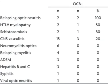
Table 3. Frequency of restricted oligoclonal bands to the cerebrospinal luid in patients with inlammatory diseases of the central nervous system.
OCB+
n n %
Relapsing optic neuritis 2 2 100
HTLV myelopathy 2 1 50
Schistosomiasis 2 1 50
CNS vasculitis 15 3 20
Neuromyelitis optica 6 0 0
Relapsing myelitis 4 0 0
ADEM 3 0 0
Hepatitis B and C 3 0 0
Syphilis 1 0 0
Viral optic neuritis 1 0 0
OCB+: presence of oligoclonal bands by isoelectric focusing and immunoblotting); n: number of patients; CNS: central nervous system; ADEM: acute disseminated encephalomyelitis.
(SD 0.67). Sensitivity of the IgG index for the diagnosis of MS was 46.6%; specificity was 94.8% when patients without neurological disease were used as controls, and 64.2% when patients with inlammatory diseases of the CNS were used as controls.
There was concordance between the presence of OCB and increased IgG index in 37 patients with MS (41.1%). De-tection of OCB and/or increased values of IgG index may allow to characterize the CSF immunoproduction of IgG in 57 out of 90 patients (63.3%).
In the group of patients with inlammatory diseases of the CNS, increased IgG index was found in 14 of 39 pa-tients (35.8%).
In the control group, one of the 19 patients present-ed a slight increase in IgG index (5.2%).
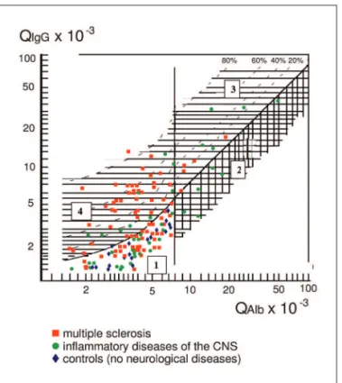
Reiber and Felgenhauer nomogram for quantitative evaluation of immunoproduction is shown in Fig 2. The comparative study of qualitative (OCB) and quantitative IgG immunoproduction (IgG index) is shown in Fig 3.
Pleocytosis up to 30 cells/mm3 (mononuclear cells) was
seen in 51.7% of the patients. In 98.1% of the patients with MS, the total protein content (mean: 31.3 mg/dl, SD: 7.2, median: 29 mg/dL) was below 72 mg/dl and the total cell count was below 30 cells/mm3. Glucose concentration
was normal in all patients. QA was high in 14.1%, suggesting some degree of dysfunction of the blood-CSF barrier.
All immunological tests showed either normal results or slight alterations without clinical signiicance.
diScuSSion
Our study showed that 54.4% of the patients with CDMS had OCB restricted to the CSF. The sensitivity of this inding was 100% when compared to patients with-out neurological diseases, and 82.1% when compared to patients with inlammatory diseases of the CNS.
The frequency of OCB found in this study is low in comparison with both the prevalence observed in Euro-pean and North-America MS patients and other previous Brazilian studies which found a frequency of 85%17 and
91%18. However, this low rate is similar to that observed in
other regions of the world (Table 4).
Three main hypotheses may be postulated to explain the lower results for OCB detection in our study: (a) there was a bias in clinical evaluation of the patients included; (b) there were problems in methodology for CSF
analy-sis of these patients; (c) OCB does occur in a smaller per-centage in São Paulo city by unknown reasons.
As for the irst hypothesis, a special and intensive care was taken to include only patients with MS clinical def-inite. The inclusion criteria strictly followed recommen-dations for MS diagnosis, widely discussed in the interna-tional panel of McDonald revised in 200514. These patients
were carefully and independently examined by two expe-rienced neurologists, with expertise in MS diagnosis and treatment. Therefore, this hypothesis is highly unlikely to explain this inding.
As for the second hypothesis, some considerations must be made: (1) the results of CSF analysis were not used as an inclusion criteria; (2) our study was carried out in an academic reference laboratory with a very experi-enced research staff in CSF analysis, mainly in electro-phorectic methods. The CSF sample was processed us-ing high standards accordus-ing to CSF Analysis Consensus of Freedman MS et al.6. This included the use of the most
up-to-date internationally accepted technology and strict adherence to the criteria of reproducibility ensuring high levels of idelity of the results; (3) the IF method followed by immunoblotting used in our patients is the most sensi-tive and suitable technique for detecting OCB when MS is suspected6. Nevertheless, the use of this technique is not
a universal practice: according to the College of American Pathologists Survey16, only 10% of the 235 laboratories in
the USA that conduct CSF analysis use this technique to detect OCB. This clearly shows that, unlike our group, in practice, the most suitable method for investigating OCB has been underutilized in many research centers, even in the most developed countries; (4) the analysis of paired CSF and serum electrophoresis was made by two skilled Fig 2. Quotient of immunoglobulin plotted against the quotient of
bumin for the three groups of patients. QIgG: IgG quocient; QAlb: al-bumin quocient; region 1: normal; region 2: blood-brain barrier break-down; region 3: blood-brain barrier breakdown associated with local synthesis; region 4: local synthesis.
Fig 3. Comparison of the frequency of positive oligoclonal bands and IgG index ≥0.8 in the cerebrospinal luid of patients with different central nervous system disorders and controls. MS: multiple sclero-sis; CIS: clinically isolated syndrome; ODD: other demyelinating dis-ease, include neuromyelitis optica, acute disseminated encephalo-myelitis, relapsing myelitis and relapsing optic neuritis; controls: pa-tients submitted to spinal tap for anesthesia.
54%
31%
20% 20%
11%
0% 47%
38%
14% 13%
38%
5%
0% 10% 20% 30% 40% 50% 60%
MS CIS ODD vasculitis infections controls
researchers unaware of the diagnosis of the patients. So, the low frequency of OCB founded in this study may not be ascribed to problems in CSF analysis.
Apart from the relatively low sensitivity, the detection of OCB seemed to have a low speciicity, mainly when a blind method of analysis was used7,8. OCB have been
de-scribed in other inlammatory diseases, their frequency of detection ranging from 25 to 80%5,7,19, as well as in
non-inflammatory diseases of the CNS (in up to 17% of pa-tients15). However, there is no consensus about this low
speciicity of OCB in MS.9,20,21.
In the MS group, OCB were observed more frequent-ly in progressive forms than in RR form. This may be ex-plained by the continuous course of the disease and a consequent more effective exposition to immunological mechanisms in those forms of MS.
On the other hand, the frequency of OCB observed in patients with CIS is similar to that observed in patients with RR form of MS. This observation may give rise to two comments: (a) CIS is a true form of MS; (b) our clin-ical appraisal of patients included in this study was ful-ly satisfactory.
The quantitative determination of immunoproduction of IgG must be made in addition to OCB search. This com-plementary method seems to improve the sensitivity of CSF diagnosis in MS, since 8.9% of patients with deinite MS diagnosis presented increased IgG index without OCB in CSF. However, it remains to be explained why this dif-ference occurs and what is the meaning of this inding.
The relatively low frequency of increased AQ observed
in CSF samples studied suggests that our MS patients do not often have impairment of the blood-brain-barrier (BBB) function.
The Reiber and Felgenhauer nomogram is a very inter-esting quantitative method to evaluate CSF intrathecal IgG production. It permits to recognize: (1) the patients in which no IgG immunoproduction nor BBB breakdown is happening (region 1 of Fig 2); (2) the occurrence of BBB breakdown, more usually seen in inlammatory diseases other than MS (region 2 of Fig 2); (3) the association of immunoproduction and BBB breakdown as it is shown in the region 3 of the Fig 2. This characteristic may be im-portant in patients with MS, since it is probably the only complementary diagnostic method that allows
recogni-Table 4. Frequency of oligoclonal bands restricted to the cerebrospinal luid of patients with multiple sclerosis in different countries.
Country Method
% OCB +
References
MS ID Control
Norway IEF IF 100% 9% Lunding J et al. 20009
Slovenia IEF IF 95% Rot et al. 20088
Norway IEF IB 93% 67% 4% Mygland A et al. 20077
USA IEF IB 90% 6% Fortini AS et al. 200320
Canada IEF IB 89% Siritho S et al. 200926
Italy IEF 89% Annunziata P et al. 200627
Spain IEF IB 87% 52% 17% Falip M et al. 200115
France IEF 85% 21% 8% Bourahoui A et al. 200428
Portugal IEF IF 82% 40% 3% Sá MJ et al. 200519
Czech Rep. IEF IB 81% 7% Bednarova et al. 200529
Holland IEF IF 76% Koch M et al. 200730
France IEF IF 76% 7% Fromont A et al. 200521
China IEF SS 63% 47% 4% Li B et al. 200710
Japan IEF IF SS 56% Fukazawa et al. 199811
China NR 40% Lau KK et al. 200223
Lebanon NR 40% Yamout B et al. 200812
Japan IEF 33% Tanaka K et al. 200524
India NR 30% Syal P et al. 199913
Taiwan IEF 7% 0% Chang KH et al. 200631
brazil ief ib 54% 17% 0% present study
tion of the acute phase phenomena (BBB breakdown) tak-ing place over an older chronic immunoproductak-ing pro-cess. This information may be helpful for distinction when images seen at MRI may correspond to acute lesions or only to cicatricial demyelination; (4) the graphic represen-tation of samples from patients with IgG immunoproduc-tion is displayed in a similar way to that of the Link’s IgG index analysis (region 4 of Fig 2).
Although CSF analysis in MS aims to characterize the local immune response in the CNS, a number of other features may differentiate MS from other diseases of the CNS, such as: (1) in MS, there is usually a high number of OCB; (2) in MS, the simultaneous presence of OCB in CSF and in the blood serum is uncommon, even if a high num-ber of bands have been identiied in the CSF; (3) there usu-ally is a normal number of cell count or a slight pleocyto-sis, usually below 30 cells/mm3, with an absolute
predom-inance of lymphomononuclear cells; (4) the total protein content is usually normal or slightly above normal, gen-erally below 70 mg/dL; (5) the concentration of glucose should be within reference limits2-4,20.
These data show the importance of the CSF analysis in all MS patients, mainly in those with initial clinical symp-toms of demyelinating diseases. The detection of this CSF proile may be helpful to suggest the occurrence of an inlammatory process in the CNS, especially in patients whose clinical and imaging abnormalities are not clear-cut, thus providing the possibility of early diagnosis and treatment8,25. This is more important in progressive forms
of MS, mainly in the primary progressive form.
In conclusion, the expected results for the frequency of OCB in MS patients in Brazil cannot be estimated by simple extrapolation from the data obtained in European or North American populations. Possibly, variations in the clinical presentation and genetic susceptibility may deter-mine the occurrence of signiicant variations11,22-24.
referenceS
1. Reiber H. Cerebrospinal luid: physiology, analysis and interpretation of protein patterns for diagnosis of neurological diseases. Mult Scler 1998;4:99-107.
2. Link H, Huang Y-M. Oligoclonal band in multiple sclerosis cerebro-spinal luid: an update on methodology and clinical usefulness. J Neu-roimmunol 2006;180:17-28.
3. Zettl UK, Tumani H. Multiple sclerosis & cerebrospinal luid. Navarra Spain: Blackwell Publishing Ltd, 2005: 1-116.
4. Correale J. Oligoclonal bands and antibody responses in multiple scle-rosis. J Neurol 2002;249:375-389.
5. Reske D, Petereit H-F, Heis W-D. Dificulties in the differentiation of chronic inlammatory diseases of the central nervous system: value of cerebrospinal luid analysis and immunological abnormalities in the diagnosis. Acta Neurol Scand 2005;112:207-213.
6. Freedman MS, Thompson EJ, Deisenhammer F, et al. Recommended standard of cerebrospinal luid analysis in the diagnosis of multiple sclerosis: a consensus statement. Arch Neurol 2005;62:865-870. 7. Mygland A, Trydal T, Vinje BU, Vedeler C. Isoelectric focusing is
supe-rior to immunoixation eletrophoresis in diagnosing CNS inlamma-tion. Acta Neurol Scan 2007;115:122-125.
8. Rot U, Ledinek AH, Jazbec SS. Clinical, magnetic resonance imaging, cerebrospinal luid and electrophysiological characteristics of the ear-liest multiple sclerosis.Clin Neurol Neurosurg 2008;110:233-238. 9. Lunding J, Midgard R, Vedeler CA. Oligoclonal bands in
cerebrospi-nal luid: a comparative study of isoelectric focusing, agarose gel elec-trophoresis and IgG index. Acta Neurol Scand 2000;102:322-325. 10. Li B, Dong H, Zhang J, Song X, Guo L. Cerebrospinal luid IgG proiles
and oligoclonal bands in Chinese patients with multiple sclerosis. Acta Neurol Scand 2007;115:319-324.
11. Fukazawa T, Kikuchi S, Sasaki H, et al. The signiicance of oligoclonal bands in multiple sclerosis in Japan: relevance of immunogenetic back-grounds. J Neurolo Sci 1998;158:209-214.
12. Yamout B, Barada W, Tohme RA, Mehio-Sibai A, Khalifeh R, El-Hajj T. Clinical characteristics of multiple sclerosis in Lebanon. J Neurol Sci 2008;270:88-93.
13. Syal P, Prabhakar S, Thussu A, Sehgal S, Khandelwal N. Clinical proile of multiple sclerosis in north-west India. Neurol India 1999;47:12-17. 14. Polman D, Reingold SC, Edan G, et al. Diagnostic criteria for multiple
sclerosis: 2005 revisions to the “McDonald Criteria”. Ann Neurol 2005; 58:840-846.
15. Falip M, Tintoré M, Jardi R, Duran I, Link H, Montalbán S. Utilida clíni-ca de las bandas oligoclonales. Rev Neurol 2001;32:1120-1124. 16. Keren DF. Optimizing detection of oligoclonal band in cerebrospinal
luid by use of isoelectric focusing with IgG immunoblotting. Am J Clin Pathol 2003;120:649-651.
17. Puccioni-Sohler M, Lavrado FP, Bastos RRG, Brandão CO, Papaiz-Al-varenga R. Esclerose múltipla: correlação clínico-laboratorial. Arq Neu-ropsiquiatr 2001;59:89-91.
18. Brandão CO, Ruocco HH, Farias AS, et al. Cytokines and intrathecal IgG synthesis in multiple sclerosis patients during clinical remission. Arq Neuropsiquiatr 2005;63:914-919.
19. Sá MJ, Sequeira L, Rio ME, Thompson EJ. Oligoclonal IgG bands in the cerebrospinal luido f portuguese patients with multiple sclerosis: nega-tive results indicate benign disease. Arq Neuropsiquiatr 2005;63:375-379. 20. Fortini AS, Sanders EL, Weinshenker BG, Katzmann JA. Cerebrospinal
luid oligoclonal bands in the diagnosis of multiple sclerosis. Am J Clin Pathol 2003;120:672-675.
21. Fromont A, Couvreur G, Guiguet M, Giroud M, Caudie C, Moreau T. Comparaison de l´immunofixation après électrophorèse sur gel d´agarose avec la focalisation isoélectrique dans la détection des bande oligoclonales d´IgG du liquide céphalo-rachidien de patients atteints de sclérose en plaques. Rev Neurol (Paris) 2005;161:1183-1190. 22. Callander M, Haghighi S, Landtblom AM, et al. Multiple sclerosis
im-munopathic trait and HLA-DR(2)15 as independent risk factors in mul-tiple sclerosis. Mult Scler 2007;13:441-445.
23. Lau KK, Wong LKS, Li LSW, Chan YW, Li HL, Wong V. Epidemiolog-ical study of multiple sclerosis in Hong Kong Chinese: questionnaire survey. HKMJ 2002;8:77-80.
24. Tanaka K, Kujuro Y, Suzuki S, et al. Clinical and laboratory features of in-patients with multiple sclerosis in a University Hospital in Tokyo from 1988-2002. Inter Med 2005;44:560-566.
25. Tintoré M, Rovira A, Rio J, et al. Do oligoclonal bands add information to MRI in ist attacks of multiple sclerosis? Neurology 2008;70:1079-1083. 26. Siritho S, Freedman MS. The prognostic signiicance of cerebrospinal
luid in multiple sclerosis. J Neurol Sci 2009;279:21-25.
27. Annunziata P, Giorgio A, Santi L, et al. Absence of cerebrospinal luid oligoclonal bands is associated with delayed disability progression in relapsing-remitting MS patients treated with interferon-B. J Neurol Sci 2006;244:97-102.
28. Bourahoui A, Seze J, Guttierez R, et al. CSF isoelectrofocusing in a large co-hort of MS and other neurological diseases. Eur J Neurol 2004;11:525-529. 29. Bednarova J, Stourac P, Adam P. Relevance of immunological variables
in neuroborreliosis and multiple sclerosis. Acta Neurol Scand 2005;112: 97-102.
30. Koch M, Heersema D, Moster J, Teelken A, De Keyser J. Cerebrospinal luid oligoclonal bands and progression of disability in multiple scle-rosis. Eur J Neurol 2007;14:797-800.