Mass Spectrometry of Biomolecules:
From PDMS to MALDI
RonaldD. Macfarlane
Department of Chemistry Texas A&M University College Station, Texas, USA
May, 1999
Mother Nature (the Ultimate Chemist): South America holds some of the richest rain forests in the world where Nature, through the abundance of the essentials of life has produced a rich spectrum of biological molecules. The early inhabitants of these regions, over thousands of years, have, through experimentation, identied plants and animals that contain components that they have found to be benecial for their health or so toxic that they can be used as a weapon.
Do any of you know any examples of
drugs or toxins that have evolved to
com-mercial products that have had their
ori-gin from the experience of pre-modern
history South Americans?
While the knowledge of these important biological compounds was passed on through generations from stories and the practice of medicine by learned mem-bers of these native communities, until recently, these stories and \home remedies" were not recognized by the science community as having much validity. Long be-fore penicillin was discovered, the Native people knew that a bad wound would heal more quickly if moldy bread were layered over the wound.
Although we have reached a high level of sophis-tication in synthesizing biological compounds, Nature, through the experience of millions of years, has devel-oped synthesis pathways that are marvels in their com-plexity and ingenuity. Consequently, many scientists are not only isolating and identifying the active ingre-dients of a plant but also elucidating the pathways that Nature uses to produce that species. The countries of South America have been a rich natural laboratory for these activities.
Poisonous South American tree frogs
and mass spectrometry
When a plant or animal is identied as having pan of its chemical make-up, a species that has important physiological properties, that species is rst isolated and puried. The 3-dimensional chemical structure is elucidated utilizinga variety of spectroscopic tools. The most information rich of these methods are NMR, IR, and x-ray diraction of pieces of the molecule and the whole molecule. A structure is proposed that ts all of this information and a molecular mass determination is made that is a direct measure of the inventory of the atoms comprising the molecule. If the measured mass does not t the proposed structure, them something is wrong with the proposed structure and it must be modied. The molecular mass determination is made by converting molecules of the species into gas phase molecular ions and the mass is measured by the inter-action of these ions with magnetic or electric elds or a combination of these.
Figure 1. Proposed Structure of Tetrodotoxin.
This molecule interferes with the electrical circuitry of the heart and induces cardiac arrest. It is used in heart research to induced heart attacks in laboratory animals and to study the physiological action of this event. Attempts to obtain the molecular mass of this molecule by mass spectrometry in the early 1970's failed
because no gas phase molecular ions could be produced in the mass spectrometer. In fact, a large fraction of Nature's biomolecules could not be studied by mass spectrometry in that era because of this fundamental problem. The reason for this can be understood if the tetrodotoxin structure is examined in more detail. The highly polar groups on the structure result in a very strong dipole/dipole interaction between neighboring molecules in a sample of tetrodotoxin which means that it is dicult to pull them apart by thermal agitation. When the sample is heated to increase the intermolec-ular excitation, the thermal energy excites internal vi-brations of the molecule and it dissociates. This process is depicted below.
Figure 2. Thermal excitation of a tetrodotoxin matrix leading to decomposition.
A Chance Observation in a Nuclear
Physics Study Leads to a Solution to the
Problem
In 1972, our group at Texas A&M University was studying the emission of radioactive recoil ions from surfaces using time-of-ight mass spectrometry. In ad-dition to the expected ions, molecular ions of impuri-ties on the surface also appeared in the mass spectrum which were postulated to be due to the ionization of molecules on the surface of the sample by the nuclear radiation. To test this hypothesis, a 252Cf-source was
placed behind the sample and ssion fragments from the spontaneous of this nuclide irradiated the sample. Molecular ions of surface molecules were indeed pro-duced in high abundance and detected in the mass spec-trum verifying the origin of the molecular ions in the nuclear physics experiment. Key individuals partici-pating in this study in 1972 in our lab are participat-ing in the Desorption '98 meetparticipat-ing this week. They are
Back to the Poisonous Frog and the
Molecular Decomposition Problem
In 1973-4, when our studies on the use of ssion fragments of252Cf to generate mass spectra was
begin-ning to evolve, one of the students in my group began his graduate studies at Stanford University working for Harry Mosher, a renowned natural products chemist. Professor Mosher hid been studying the tetrodotoxin structure problem for several years and proposed the structure shown above. He was not able to verify
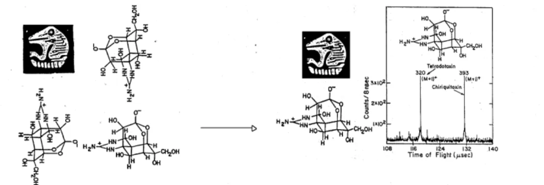
this structure by MW determination because of the in-volatility/decomposition problem. He had tried for sev-eral years using every new mass spectrometric method that had evolved but with no success. At the urging of his new student, he sent us a sample to run and we got a good result. At that time, we didn't know it was a tough problem and we also didn't know about the problem mass spectrometry was having with involatile biomolecules. The spectrum we got by shooting ssion fragments through a sample of this toxin is shown be-low.
Figure 3. First Mass Spectrum of tetrodotoxin obtained by PDMS
Naming the Method
Professor Mosher was very excited about this result. He suggested that we forget about this nuclear physics stu and really develop the method into a technology for natural product studies. He also insisted that we come up with a name for it, something organic chemists could pronounce and remember and, after several at-tempts, came up with252Cf PDMS or PDMS (Plasma
Desorption Mass Spectrometry).
So how do Fission Fragments solve the
Problem?
A ssion fragment is a high energy, highly-charged ion that is a highly destructive species. The surprise was that a component of its interaction with solids is gentle, resulting in the desorption and ionization of fragile molecules without decomposing them. Later studies showed that the combination of high energy density and short-excitation time was they key feature of the interaction that makes PDMS work and that even
very large biomolecules can survive the hostile environ-ment of the ssion track and become intact gas phase molecular ions.
PDMS Research, 1974-1984
amphotericin, and thiostreptin. Although we were suc-cessfull with these molecules, the mass spectrometry community did not embrace our endeavors for several reasons: fear of radioactivity, use of time-of-ight and not a magnet or quadrupole, low mass resolution, and you had to wait for more than a minute to obtain the mass spectrum. It was a time when editors of jour-nals were more sympathetic to our manuscripts than reviewers.
Accelerator Studies, 1976-present
During the time when we were actively involved in \running samples", my nuclear physics colleagues, who, like most physicists, generally distain chemistry, began to tackle the problem of mechanism of PDMS using their nuclear accelerators. It was from these experi-ments, that the properties of the incident ion were elu-cidated and the interaction of high energy ions with solids as a eld in its own began to emerge. These ac-tivities were initiated and sustained by Voit at Erlan-gen, Sundqvist at Uppsala, Le Beyec at Orsay, Wien at Darmstadt, da Silveira at Rio and Tombrello at Caltech. Intense and enduring friendships with these groups for the past 25 years have been one of the rich-est components of my professional and private life. We have maintained this closeness through the series of Desorption `xx meetings that have taken place in won-derful and interesting sites that include Brazil as a two-time favorite of this group. Over the years, the group has expanded as the eld has grown and developed new roots and branches. It is this feature that I wish to ad-dress next.
Secondary Ion Mass Spectrometry
(SIMS)
This method was well established before PDMS came along. Low energy ions bombard the surface of a sample producing gas phase ions of the matrix. The method was used extensively for characterizing the sur-faces of inorganic species before 1974 and continues to-day. The mechanism involves collisions of the incident ion with surface atoms that are ejected. Molecular ions were also present in the mass spectrum which were an \unwanted background". When PDMS was introduced, it was realized that a component of the SIMS process also was similar to PDMS, the rapid deposition of en-ergy, and molecular ions of involatile molecules could also be desorbed by these low energy ions. From 1976 to the present, SIMS has been a member of the Desorp-tion `xx family, led by Benninghoven at Munster and Standing in Winnipeg. A friendly rivalry emerged dur-ing this era where a series of papers were presented on SIMS catching up to PDMS in terms of number of ions desorbed and the mass range. In terms of acceptance by the mass spec. community,SIMS had the same problem as PDMS because it did not use sophisticated magnets and have high mass resolution. This acceptance issue comes up in several places here because it was a real dilemma. Thousands of mass spec. people were es-sentially ignoring the work of the PDMS/SIMS people despite the successes.
The Beginning of Being Accepted
The Competition Heats Up, Laser
Desorption is Introduced
It was generally accepted by the mass spectrome-try community that there must be a better way than PDMS to obtain mass spectra of biomolecules but us-ing the same general idea. In the late 1970's, Meuzze-lar in Holland began to obtain impressive mass spectra using laser excitation. When I heard one of his rst talks on his work, I had the feeling that the days of the monopoly that PDMS had on the eld were numbered. Laser desorption had so much going for it in terms of control of the excitation. The pulsed laser was a nat-ural for the TOF technique. However, the promise did not convert to reality in the next few years. One of the Desorption `xx participants, Franz Hillenkamp, took up the challenge of laser desorption at that time and for several meeting Hillenkamp kept us informed on the progress of the development of the method. The mass spectrometry community and commercial mass spec-trometer makers patiently waited for something more attractive.
Fast Atom Bombardment (FAB) makes
its debut
In the early 1980's, we were working with Kenneth Reinhart, a natural product chemist at Illinois, on a series of potentially important antibiotics derived from marine organisms. These were tough molecules to an-alyze. In the middle of the studies, Ken told us about a method he heard was just developed in England that was an important breakthrough for the analysis of in-volatile biomolecules. He was going to England imme-diately with his samples and would report back. When he returned, he told the story of a clandestine operation literally clocked in secrecy where he handed his samples
to an operator who then went behind a curtain, pre-pared the samples and inserted them into a mass spec-trometer. Out came high quality spectra and the big news was that a \proper" mass spectrometer was used
i. e. a magnetic sector instrument. The professor in
The Post-FAB era and the birth of MALDI
What kept PDMS going after FAB was introduced was the ability to study larger biomolecules. Largely through the work of the Uppsala group, the mass range limit was pushed up to close to 40, 000 for a protein. But it was hard work. At the same time, biotechnology and medicine was becoming more sophisticated and the desire to obtain accurate and precise molecular weights of biopolyimers was becoming more acute. It was ob-vious from our studies that there was a limit to the size of a protein that could be desorbed by a ssion fragment. This was a bit of disappointment because our own interests were heading in a direction in medi-cal research where we wanted to use PDMS to charac-terize proteins involved in human health and disease. It was clear that PDMS was not going to be used in these studies. In the late 1980's, at an International Science meeting in Bordeaux, Franz Hillenkamp gave a talk, imbedded deep in the program on the work he and Michael Karas were doing with laser desorption; a breakthrough and a new acronym: MALDI. You will hear more about this method from Franz in the next day or two. This time, my response to his talk was absolute excitement! I could see my desire to have a method available to accomplish my long term goal: to make a contribution to medicine, had a chance. I close this long set of lecture notes with an excerpt of dream that has become reality.