Giant Condyloma (Buschke-Loewenstein Tumor)
in a 16-year-old Patient: Case Report
Condiloma gigante (tumor de Buschke-Loewenstein) em
uma paciente de 16 anos: relato de caso
Caetano Galvão Petrini
1Patrícia Pereira dos Santos Melli
2Pedro Sérgio Magnani
2Laura Penna Rocha
3Francesca Maia Faria
4Geraldo Duarte
2Silvana Maria Quintana
21Hospital das Clínicas de Ribeirão Preto, Universidade de São Paulo (USP), Ribeirão Preto, SP, Brazil
2Gynecology and Obstetrics Department, Ribeirão Preto Faculty of Medicine, USP, Ribeirão Preto, SP, Brazil
3General Pathology Division, Biological and Natural Sciences Institute, Universidade Federal do Triângulo Mineiro, Uberaba, MG, Brazil
4Pathology and Forensic Medicine Department, Ribeirão Preto Faculty of Medicine, USP, Ribeirão Preto, SP, Brazil
Rev Bras Ginecol Obstet 2016;38:471–476.
Address for correspondence Caetano Galvão Petrini, MD,
Departamento de Ginecologia e Obstetrícia, Faculdade de Medicina de Ribeirão Preto, Universidade de São Paulo, Av. Bandeirantes, n° 3900, 8° andar. Monte Alegre, 14049-900. Ribeirão Preto, SP, Brazil (e-mail: [email protected]).
Keywords
►
giant condyloma
►
Buschke-Loewenstein
tumor
►
human
papillomavirus
►
HPV
►
surgical treatment
Abstract
The Buschke-Loewenstein tumor is characterized by excessive growth of verrucous
lesions on the genitals and/or perianal region. It is considered benign despite the high
rate of recurrence and the possibility of malignant transformation. It is commonly
associated with subtypes 6 and 11 of the human papillomavirus (HPV), and host
’
s
immunity plays an important role in the development of the disease. Surgical excision
is the recommended treatment in most cases. We present the case of a 16 years old
female patient with extensive vulvar lesions successfully treated surgically.
Palavras chave
►
condiloma gigante
►
tumor de
Buschke-Loewenstein
►
vírus do papiloma
humano
►
HPV
►
tratamento cirúrgico
Resumo
O tumor de Buschke-Loewenstein se caracteriza pelo crescimento excessivo de lesões
verrucosas na região genital e/ou perianal. É considerado benigno apesar da elevada
taxa de recorrência e da possibilidade de transformação maligna. Está comumente
associado aos sorotipos 6 e 11 do papiloma vírus humano (HPV) e a imunidade do
hospedeiro tem importante papel no desenvolvimento da doença. A excisão cirúrgica é
o tratamento recomendado na maioria dos casos. Apresentamos o caso de uma
paciente do sexo feminino, de 16 anos, com lesão vulvar de grande extensão tratada
cirurgicamente com sucesso.
received
March 16, 2016
accepted
September 12, 2016
DOIhttp://dx.doi.org/ 10.1055/s-0036-1593776. ISSN 0100-7203.
Copyright © 2016 by Thieme Publicações Ltda, Rio de Janeiro, Brazil
Introduction
In 1925, Abraham Buschke and Loewenstein Ludwig de-scribed a penis tumor that had intermediate characteristics between acuminated condyloma and squamous cell carcino-ma.1These authors denominated this tumor Buschke-Loe-wenstein Tumor (BLT) or giant condyloma. The BLT is included in the verrucous carcinoma category, which also comprises oralflorid papillomatosis, Ackerman tumor and cuniculatum epithelioma. However, although they provide considerable local destruction, these lesions do not produce distant metastases.2,3
The etiology of this tumor is an infection by Human Papillomavirus (HPV), usually the subtypes 6 and 11.4,5 Among the risk factors for the development of these lesions, we highlight smoking, having multiple sex partners, chronic genital infections, poor hygiene and immunodeficiencies.3 Moreover, rearrangements in the upstream regulatory re-gion (URR) of the HPV 6 DNA may occur naturally, leading to an additional risk factor to the host, as this HPV genome change predominates in histologically or clinically more aggressive lesions, as observed in some cases of BLT.6
In a review of 51 cases published in the literature, a higher frequency of the disease was observed in men (male/female rate 2.7:1), and in ages below 50 years old, a rate of 3.5:1. The average age of occurrence in women was 46.6 years old; however, with a tendency of occurrence at earlier ages.7 Moreover, the incidence of anogenital warts is increasing in children, which may be a reflection of the high incidence of this infection in adults. However, giant condyloma acumi-natum is an extremely rare clinical type of genital wart in the pediatric population.8
Case Presentation
KAAM, 16 years old, without previous diseases, reporting illicit drug use (marijuana and cocaine), alcoholism and homosexual and heterosexual relationships. Patient claimed beginning sexual activity at 14 years old. Serologic screening for HIV, Hepatitis B (HBsAg), hepatitis C (anti-HCV ELISA) and
syphilis (VDRL) did not reveal infections and patient had not mentioned any previous sexual diseases.
September 2013: appearance of verrucous lesions on the vulva. The medical evaluation diagnosed pelvic infl ammato-ry disease (PID) and condylomatous vulvar. It was prescribed ambulatory treatment for PID and podophyllotoxin 0.5% for condyloma. The patient underwent the treatment for PID, but did not use the local treatment for condyloma correctly and, consequently, the lesions increased.
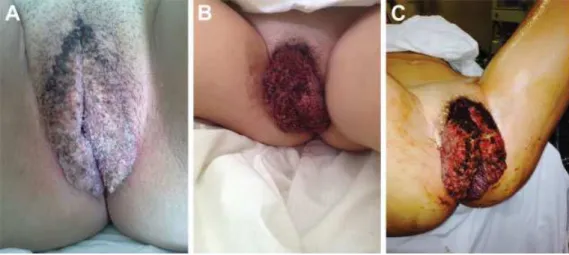
October 2013 (►Fig. 1A): A new assessment was per-formed and a significant growth of the vulvar lesions was observed. Imiquimod 5% ointment was prescribed, to be applied 3 times per week. However, there was no visible improvement.
November 2013: regression of the condyloma was not observed and the patient had secondary infection of the lesion. She was hospitalized and treated with intravenous antibiotics (►Fig. 1).
January 2014: patient is referred to the Department of Infectious Diseases in Obstetrics and Gynecology of the Ribeirão Preto Medical School, at Universidade de São Paulo, reporting appearance, since September of 2013, of condylo-ma lesions in the vulva that did not respond to prescribed treatments (4 months of evolution). The patient remained hospitalized for 30 days because lesions showed accelerated growth, making walking, urinating and defecating difficult, and also presenting secondary infection.
Gynecological examination in January of 2014: vegetating lesion, exophytic, occupying the entire length of the labia majora and perianal region, with areas of necrosis, erosion and fetid smell, measuring15 cm in length bilaterally and7cm thick;
it is not possible to perform speculum examination or vaginal examination because of the extent of the injury (►Fig. 1).
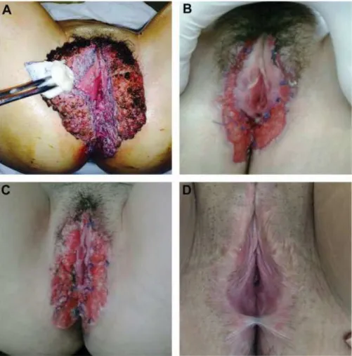
On January 24 of 2014, wide local excision was performed with radiofrequency waves surgery, leaving the wound open to heal by secondary intention. Patient remained hospital-ized, with daily dressing changes, for 4 days, and was dis-charged with surgical wound in healing process. The granulation tissue aspect showed no signs of local infection (►Fig. 2).
Result of the histopathological study of the lesion: giant condyloma, and no detectable malignant transformation in the samples. Chromogenic in situ hybridization (CISH) was positive for low-risk HPV (►Fig. 3).
First review performed 60 days after surgery (February 25, 2014): patient without complaints.
Colposcopy: satisfactory results. Squamocolumnar junc-tion (SCJ) in 0, type 2 transformajunc-tion zone, with minor findings of abnormal colposcopy.
Vagina: low-grade lesion in the anterior vaginal back-ground. Vulva without lesions (►Fig. 2D).
Colpocytology: satisfactory with atypical squamous cells of undetermined significance suggesting repair (ASCUS) Fig. 2 Before and after surgery: (A) preoperative; (B) immediate postoperative period; (C) fourth day postoperative; (D) 12 months postoperative.
Polymerase Chain Reaction (PCR) for the main HPV types– 6, 11, 16, 18 (HPV PCR): Positive.
Cervical and vaginal biopsy confirming cervical epithelial neoplasia grade I (CIN I) and vaginal intra-epithelial neoplasia grade I (VIN I) respectively, and opted for expectant management.
Second review (06/01/2015): A year after excision of giant condyloma and 12 months of expectant management for CIN I and VIN I.
Cervix: Normal colposcopy, with squamocolumnar junc-tion in zero, type 2 normal transformajunc-tion zone. Vagina: without lesions.
Vulva: without verrucous lesions, with retraction and discrete hypochromic at the site of excision of the lesion (►Fig. 2D).
Colpocytology in this date was satisfactory without atypia and the HPV PCR test was negative.
Discussion
affecting the vulva in women and balanopreputial area in men, but may affect the scrotum, rectum and bladder. This tumor is always preceded by acuminated condyloma, and host’s immunodeficiency has an important role for the development of the disease. Possible differential diagnosis is verrucous squamous cell carcinoma.9Patients with lesions with increased susceptibility to rapid progression and higher recurrence rates generally have various types of immunode-ficiency, which may lead to difficulty in the assessment of best treatment option.10
The biological behavior of BLT is intermediate between acuminated condyloma and squamous cell carcinoma and may undergo malignant transformation in 30–56% of cases, and rarely leads to metastasis.11 Histological evaluation allows the performance of differential diagnosis because the giant condyloma has high mitotic activity, acanthosis, important papillomatosis, thick edges and tumor infi ltra-tion of adjacent tissues not infiltrating the basement membrane.12
In the presented case, the history of large tumor occupy-ing the entire perineal area, together with thefindings and histopathological examination of marked papillomatosis, acanthosis, hyperkeratosis, parakeratosis, koilocytosis and increased presence of atypical mitosis confirm the suspected diagnosis of BLT (►Figs. 1and4).
The treatment of choice for BLT remains controversial, but surgery seems to be the best option. A wide surgical excision, healing by secondary intention, or radical local excision with reconstruction of skin defects have been the main treatments described.13 Other treatments with CO2 laser, radiation therapy, intralesional injection of interferon alfa or topical imiquimod can be used as an alternative treatment, espe-cially in cases where surgical resection can be mutilating in cases of locally advanced disease, which prevents the resec-tion with safety margins in patients without clinical con-ditions to perform surgery.14
A long postoperative follow-up is necessary because the disease has a high rate of recurrence, and early treatment of recurrent lesions must be performed.15 Moreover, biopsy should be performed in all cases because of the risk of malignant transformation.16There are some reports of suc-cessful long-term imiquimod treatments, avoiding major surgery or allowing realization of minor surgery for residual lesions.17However, in the case presented, the injury led to losses in essential functions such as urination and evacua-tion, as well as secondary infecevacua-tion, not allowing wait.
We present a case of a 16-year-old patient with alcohol consumption and use of illicit drugs, which are factors associated with immunosuppression.18,19 This case gets attention for fact that the disease in this patient, has appeared30 years earlier than the average age found in
the literature. Thisfinding is in accordance with Trombetta and Place7, who also observed a recent trend toward a younger presentation of the BLT.7
In this case the lesion had a rapid growth (4 months of evolution), which was successfully treated surgically, with-out occurrence of postoperative complications. Despite the high risk of recurrence of the disease (60–70%),20this case
presents no evidence of new lesions following 12 months post-resection. The fact that the patient had ceased alcohol consumption and the use of illicit drugs may have contrib-uted to the success of the treatment and the absence of recurrence.
Conclusion
The Buschke-Loewenstein tumor is a sexually transmitted disease caused by low-risk HPV, with benign histological features, but with an excessive local growth and high recur-rence rates. The overgrowth makes the hygiene difficult and increases the risk of secondary infections. In addition, the social and psychological damage brought up by the stigmas of the disease are difficult to measure. Therefore, a follow up with a multidisciplinary team is very important. Especially in cases such as the one presented, in which the patient was a teenager and had multiple risk factors for developing the disease, such as drug addiction and risky sexual behavior, a multidisciplin-ary approach is necessmultidisciplin-ary for a successful treatment.
References
1 Steffen C. The men behind the eponym–Abraham Buschke and Ludwig Lowenstein: giant condyloma (Buschke-Loewenstein). Am J Dermatopathol 2006;28(6):526–536
2 Schwartz RA. Verrucous carcinoma of the skin and mucosa. J Am Acad Dermatol 1995;32(1):1–21, quiz 22–24
3 Spinu D, Rădulescu A, Bratu O, ChecheriţăIA, Ranetti AE, Mischianu D. Giant condyloma acuminatum BuschkeLowenstein disease -a liter-ature review. Chirurgi-a (Bucur) 2014;109(4):445–450 4 Gissmann L, deVilliers EM, zur Hausen H. Analysis of human
genital warts (condylomata acuminata) and other genital tumors for human papillomavirus type 6 DNA. Int J Cancer 1982;29(2): 143–146
5 Dianzani C, Bucci M, Pierangeli A, Calvieri S, Degener AM. Associ-ation of human papillomavirus type 11 with carcinoma of the penis. Urology 1998;51(6):1046–1048
6 Rübben A, Beaudenon S, Favre M, Schmitz W, Spelten B, Grus-sendorf-Conen EI. Rearrangements of the upstream regulatory region of human papillomavirus type 6 can be found in both Buschke-Löwenstein tumours and in condylomata acuminata. J Gen Virol 1992;73(Pt 12):3147–3153
7 Trombetta LJ, Place RJ. Giant condyloma acuminatum of the anorectum: trends in epidemiology and management: report of a case and review of the literature. Dis Colon Rectum 2001;44-(12):1878–1886
8 Suárez-Ibarrola R, Heinze A, Sánchez-Sagástegui F, et al. Giant condyloma acuminatum in the genital, perineal and perianal region in a pediatric patient. Literature review and case report. Urol Case Rep 2016;7:14–16
9 Hicheri J, Jaber K, Dhaoui MR, Youssef S, Bouziani A, Doss N. Giant condyloma (Buschke-Löwenstein tumor). A case report. Acta Dermatovenerol Alp Panonica Adriat 2006;15(4):181–183 10 Niazy F, Rostami K, Motabar AR. Giant condyloma acuminatum of
vulva frustrating treatment challenge. World J Plast Surg 2015; 4(2):159–162
11 Bertram P, Treutner KH, Rübben A, Hauptmann S, Schumpelick V. Invasive squamous-cell carcinoma in giant anorectal condyloma (Buschke-Löwenstein tumor). Langenbecks Arch Chir 1995; 380(2):115–118
13 Gole GN, Shekhar T, Gole SG, Prabhala S. Successful treatment of buschke-löwenstein tumour by surgical excision alone. J Cutan Aesthet Surg 2010;3(3):174–176
14 Martin JM, Molina I, Monteagudo C, Marti N, Lopez V, Jorda E. Buschke-Lowenstein tumor. J Dermatol Case Rep 2008;2(4):60–62 15 Dinleyici M, Saracoglu N, Eren M, et al. Giant condyloma acumi-nate due to human papillomavirus type 16 in an infant success-fully treated with topical imiquimod therapy. Dermatol Rep 2015; 7(3):6134
16 Ganem NS, Silva BC, Nascimento MLFO, et al. [Giant condyloma acuminatum: a case report]. J Bras Doenças Sex Transm 2010; 22(4):222–224
17 Coelho FMP, Mano AL, Bacellar MS, Codes LMG, Souza ELQ, Azaro Filho EM. [Buschke-Lowenstein tumor: imiquimod therapy to esphincter preservation. Case report]. Rev Bras Colo-Proctol. 2008;28(3):342–346 Portuguese.
18 Szabo G. Consequences of alcohol consumption on host defence. Alcohol Alcohol 1999;34(6):830–841
19 Zhao H, Ye TH. [Drug abuse and infection]. Zhongguo Yi Xue Ke Xue Yuan Xue Bao 2006;28(6):858–861 Chinese.