Neurological Outcome in Fetuses with Mild and
Moderate Ventriculomegaly
Resultado neurológico em fetos com ventriculomegalia
leve e moderada
Gabriele Tonni
1Ida Vito
1Marcella Palmisano
1Wellington de Paula Martins
2Edward Araujo Júnior
31Department of Obstetrics & Gynecology, Guastalla Civil Hospital, AUSL Reggio Emilia, Reggio Emilia, Italy
2Department of Obstetrics and Gynecology, Faculdade de Medicina de Ribeirão Preto, Universidade de São Paulo (DGO-FMRP-USP), Ribeirão Preto, SP, Brazil
3Department of Obstetrics, Escola Paulista de Medicina, Universidade Federal de São Paulo (EPM-Unifesp), São Paulo, SP, Brazil
Rev Bras Ginecol Obstet 2016;38:436–442.
Address for correspondence Edward Araujo Júnior, PhD, Rua Napoleão de Barros, 875, São Paulo–SP, Brazil, CEP 04024-002 (e-mail: [email protected]).
Keywords
►
fetus
►
ventriculomegaly
►
ultrasound
►
magnetic resonance
imaging
►
neurodevelopmental
outcome
Abstract
Introduction
Ventriculomegaly (VM) is one the most frequent anomalies detected on
prenatal ultrasound. Magnetic resonance imaging (MRI) may enhance diagnostic
accuracy and prediction of developmental outcome in newborns.
Purpose
The aim of this study was to assess the correlation between ultrasound and
MRI in fetuses with isolated mild and moderate VM. The secondary aim was to report
the neurodevelopmental outcome at 4 years of age.
Methods
Fetuses with a prenatal ultrasound (brain scan) diagnosis of VM were
identi
fi
ed over a 4-year period. Ventriculomegaly was de
fi
ned as an atrial width of 10
–
15 mm that was further divided as mild (10.1
–
12.0 mm) and moderate (12.1
–
15.0
mm). Fetuses with VM underwent antenatal as well as postnatal follow-ups by brain
scan and MRI. Neurodevelopmental outcome was performed using the Grif
fi
ths Mental
Development Scales and conducted, where indicated, until 4 years into the postnatal
period.
Results
Sixty-two fetuses were identi
fi
ed. Ventriculomegaly was bilateral in 58% of
cases. A stable dilatation was seen in 45% of cases, progression was seen in 13%, and
regression of VM was seen in 4.5% respectively. Fetal MRI was performed in 54 fetuses
and was concordant with brain scan
fi
ndings in 85% of cases. Abnormal
neuro-developmental outcomes were seen in 9.6% of cases.
Conclusion
Fetuses in whom a progression of VM is seen are at a higher risk of
developing an abnormal neurodevelopmental outcome. Although brain scan and MRI
are substantially in agreement in de
fi
ning the grade of ventricular dilatation, a low
correlation was seen in the evaluation of VM associated with central nervous system
(CNS) or non-CNS abnormalities.
received April 10, 2016 accepted July 18, 2016 published online September 9, 2016
DOI http://dx.doi.org/ 10.1055/s-0036-1592315. ISSN 0100-7203.
Copyright © 2016 by Thieme Publicações Ltda, Rio de Janeiro, Brazil
Introduction
Ventriculomegaly (VM) is one of the most common brain abnormalities observed at prenatal ultrasound (brain scan).1 The prevalence of VM varies between 0.3 and 10 per 1,000 births, depending on the technique used for the evaluation of one or both ventricles.2Mild and moderate VM, defined as an atrial width of 10–15 mm between 15 and 40 weeks of gestation, may be associated with neural and extraneural malformations, fetal infections and chromosomal anoma-lies.3Traditionally, the diagnosis of VM is based on prenatal brain scans during the second and the third trimesters of pregnancy. Ventriculomegaly may be isolated or associated with cranial and/or extracranial malformations, resulting in poorer prognosis in such cases. Therefore, the introduction of magnetic resonance imaging (MRI) in fetuses with a prena-tally brain scan diagnosis of VM will reduce misclassified cases, thus providing a more accurate prediction of the neurodevelopmental outcome.4
The objective of this study was to compare the correlation between prenatal and postnatal brain scans with fetal and postnatal MRIs and to report the neurodevelopmental out-come at 4 years of age in infants with isolated mild and moderate VM.
Methods
The study was conducted between January 2007–2010 and approved by the local Health Board Authority of the Guastalla Civil Hospital, Reggio Emilia, Italy. Inclusion criteria were
fetuses with an ultrasound diagnosis (brain scan) of isolated borderline VM at the second and third trimesters of preg-nancy in whom both fetal and postnatal MRIs were available. Ventriculomegaly was defined as an atrial width of 10– 15 mm that was further divided as mild (10.1–12.0 mm) and moderate (12.1–15.0 mm).5Ultrasound criteria of re-gression and/or prore-gression of VM were classified as<2 mm or>2 mm of lateral ventricle (LV) measurement at the follow-up brain scan, respectively.
Fetal karyotype and TORCH analysis were evaluated in all women. Sixty-two consecutive fetuses met the study criteria. Clinical follow-up was performed at 6–48 months into the postnatal period. Transfontanelle ultrasound and MRI, electroencephalogram (EEG), Griffith test, and an ophthal-mological examination were performed. Fetal and post-natal MRI were all performed at a single center and diagnosed by an expert neuroradiologist using the T2-weighted single-shot fast spin echo (SSFSE) technique, while T1-weighted images were arranged at the time of the postnatal follow-up. The MRI equipment was a 1.5 Tesla Signa Twin Speed super-conducting system (GE, Milwaukee, WI, US) using an 8-element phased array surface coil. Standard SSFSE imaging was performed in the fetal sagittal, coronal, and axial planes. The T2-weighted MRI settings were: repetition time (TR)/ single-shot echo time (TE)¼3,000/180 milliseconds; ma-trix, 320256; 4 mm slice;field of view [FOV]¼340 mm. Axial T1-weighted sections were also acquired in gradient-echo 2D (TR/TE¼11/5 m; T1¼100 milliseconds; 5 mm slice; matrix, 256224; FOV¼340 mm; number of exci-tations [NEX], 3). Infinite/90; bandwidth, 32 kHz;
Resumo
Introdução
Ventriculomegalia (VM) é uma das anomalias mais frequente no
ultras-som pre-natal. Ressonâncias magnéticas (RM) melhoram a precisão do diagnóstico e
previsão do desenvolvimento em recém-nascidos.
Objetivo
A proposta deste estudo foi avaliar a correlação entre ultrassom e RM em
fetos com leve e moderada VM isolada. O objetivo secundário foi reportar o resultado
neurológico na idade de 4 anos.
Métodos
Fetos com diagnóstico pré-natal pelo ultrassom de VM foram identi
fi
cados
na idade de 4 anos. Ventriculomegalia foi de
fi
nida como medida do átrio do ventrículo
lateral entre 10
–
15 mm, a qual foi subdividida em leve (10,1
–
12,0 mm) e moderada
(12,1
–
15,0 mm). Fetos com VM foram seguidos nos períodos pré-natal e pós-natal por
ultrassom e RM. O resultado neurológico foi realizado usando a escala de
desenvolvi-mento mental de Grif
fi
ths, quando indicada, até a idade de 4 anos.
Resultados
Sessenta e dois fetos foram identi
fi
cados. Ventriculomegalia bilateral
ocorreu sem 58% dos casos. Uma dilatação estável foi observada em 45%, progressiva
em 13% e regressiva em 4,5% dos casos, respectivamente. Ressonância magnética fetal
foi realizada em 54 fetos, e foi concordante com os achados do ultrassom em 85% dos
casos. Desenvolvimento neurológico anormal foi observado em 9,6% dos casos.
Conclusão
Fetos nos quais ocorreu progressão da VM são de alto risco para
desenvolvimento neurológico anormal. Apesar do ultrassom e da RM mostrarem
substancial concordância na de
fi
nição do grau de dilatação ventricular, uma baixa
correlação foi vista na avaliação da VM associada ou não com anomalias do sistema
nervoso central.
Palavras-chave
►
feto
►
ventriculomegalia
►
ultrassom
►
ressonância
magnética
FOV¼3030 cm; matrix, 256192; gap, 1.5 mm; NEX, 0.5; refocusing pulse of less than 180 degrees; slice thick-ness, 4 mm; and 0.6 second per slice, echo-train length, 72; 1 signal acquired. In addition, T1 axial sections weighted on the subcallosal plane were also performed with a slice of 5 mm and TR/TE¼ 4,000/minimum; matrix, 128128; FOV¼280 mm, all axis; b max¼600. Griffiths Mental Development Scales measure development trends that are significant for intelligence, or indicative of functional mental growth in babies and young children from birth to the developmental age of 8 years. Within the 0–2-year scales, a profile is obtained from 5 subscales examining locomotor, personal-social, language, eye-and-hand coordination and performance. In the 2–8-year scales, this profile is expanded to add a Practical Reasoning (General Quotient) subscale.
The neurodevelopmental outcome was collected from 12–48 months using a clinical questionnaire developed by Pediatric Neurologists and conducted by telephone interview.
Results
Sixty-two consecutives fetuses with a prenatal ultrasound diagnosis of mild and moderate isolated ventriculomegaly were identified. The mean maternal age was 32 years (range 21–41) while the mean gestational age at diagnosis was 25.6 (range 18.1–35.1) weeks of gestation. The male to female ratio was 1.9. Fetal karyotype was obtained in 33 cases (53%) by amniocentesis, identifying 1 fetus with trisomy 21. TORCH analysis resulted negative in 57 (92%) cases. At the brain scan, 58% of cases of VM were bilateral, and 23% of fetuses had a moderate VM. Follow-up using ultrasound demonstrated a stable VM in 45% of cases, resolution of VM in 36%, regression in 4.5%, and progres-sion in 13% of cases. Fetal MRI was performed in 54 cases (87%), 31 of which at a gestational age lower than than 25 weeks. Overall, MRI was concordant with ultrasound in 85% of VM cases. Six newborns (9.6%) were found to have an abnormal neurodevelopmental outcome at 4 years of age. In 7 cases (n¼7/54, 13%), the fetal MRI detected associated cerebral abnormalities that were undiagnosed by antenatal brain scan: 3 cases of white matter anomalies (1 case of micro/lissencephaly, 1 case of reduced posterior white matter and 1 case of abnormal gyration). In the 4 remaining cases, VM was associated with cerebellar ver-mian hypoplasia with widened supratentorial-subarach-noid spaces, 2 cases of intraventricular hemorrhage and 1 case of reduced antero-posterior diameters of the cerebral hemispheres. A postnatal transfontanellar scan was per-formed in 36 cases while, MRI was perper-formed in 9 cases. ►Table 1 shows the findings by brain scan, fetal and postnatal scans, as well as MRI in six cases with an adverse neurodevelopment outcome (►Figs. 1–4).
Discussion
Evaluation of the LVs is an essential part of the standard sonographic examination of the fetus. Normal-sized
ven-tricles provide reassurance of the normal development of the cerebrum, while increased LVs>15 mm are highly associ-ated with major cerebral anomalies.6
The mean diameter of the atria of the LVs in normal fetuses is 7.6þ0.6 mm throughout the pregnancy.7 The prevalence of mild and moderate VM has been reported to vary from 1.48 to 22 per 1,000 live births in the low- and in high-risk pregnancy population respectively.6Dilatation of the LVs can variably be associated with dilatation of the 3rd and 4th ventricles,5and an asymmetric enlargement of the LVs is defined as a difference as a width of 2 mm between the two LVs.8
Ventriculomegaly recognizes multifactorial causes and may be seen isolated or associated with CNS or non-CNS malformations, fetal infections and/or chromosomal anom-alies, and may occur because of primary overproduction of cerebrospinalfluid (CBF), as seen in cases of choroid plexus papilloma,9or secondary to obstructions (communicating) of the CBF caused by the narrowing or forking of the aque-duct of Sylvius,10Dandy–Walker complex with obstruction of the foramina of Luschka and Magendie,11 or caused by intraventricular hemorrhage,12–14 infections3,15–17 or by extrinsic processes like tumors,12arachnoid cysts or Chiari type II malformations.
Disorders of neuronal proliferation (megalencephaly, mi-crocephaly), neuronal migration (schizencephaly, lissence-phaly), agenesis of the corpus callosum (ACC), holoprosencephaly,13,14,18–21 vascular insults, or porence-phaly (secondary to destructive processes of the cerebral white matter)2 may also be considered. Congenital viral infections and common viable trisomy are associated in 3 to 15% of cases, depending on maternal age and background risk.3,16,19Multiple congenital malformations in fetuses with VM are calculated to range from 10 to 76%, with the lowest incidence seen for mild VM and highest incidence for mod-erate VM.3,13–16,18–22
Our series showed that 23% of fetuses had moderate VM, and that 58% of borderline VMs were bilateral. The prenatal brain scan follow-up demonstrated resolution in 36%, re-gression in 4.5%, stable dilatation in 45% and prore-gression in 13% of cases. Brain scanfindings were in agreement with those of the fetal MRI in 85% of cases. In 8 fetuses (15%), the MRI revealed additional information that were of critical impact on counseling, antenatal management and care planning by the multispecialist team. Postnatal follow-up conducted using Griffiths Mental Development Scales re-vealed 6 cases (11.7%) with abnormal neurodevelopmental outcomes.
incidence of perinatal and neonatal deaths comparable to those of the general population.3,6,15,17,24
Valsky et al25showed that the MRI could detect additional findings in a woman with increased maternal body mass index (8.3%) and in 2 cases (5.5%) with bleeding in germinal centers, while in the series of Benacerraf et al,26 the MRI revealed additional information (14% of the cases) in a case of cerebellar hypoplasia and in a case of enlarged cisterna magna. When the brain scan was compared with the MRI, Levine et al27demonstrated a change in thefinal diagnosis in 23% of cases identified as abnormal at ultrasound, a change in counselling in 41% of cases, and a change in patient
manage-ment in 13.5% of cases respectively. Similarly, Salomon et al28 concluded that the use of the fetal MRI modified obstetric management in 6% of cases, studying 185 third trimester fetuses with mild VM. Furthermore, in fetuses with appar-ently isolated mild VM, Ouahba et al4showed that the MRI diagnosed major cerebral anomalies in 9% of cases, including cortical malformations, absence of the septum pellucidum, partial agenesis of the corpus callosum and agenesis of the cerebellar vermis.
Fetal MRI was diagnostic in 47.3% of cases of VM associat-ed with CNS anomalies, while the brain scan could only detect 32.7% of cases. When VM associated with non-CNS Table 1 Correlation between prenatal ultrasound, fetal and postnatal ultrasounds and magnetic resonance imaging in six cases with adverse neurodevelopment outcomes
MA GA Karyotype US (Brain Scan) Fetal MRI Pregnancy out-come
Postnatal outcome
31 22.1 46,XX Bilateral borderline VM US follow-up:
- 29 wks: RLV 12.5mm/ LLV 11,3 mm
- 33 wks: RLV13mm/LLV 12 mm
SVD at 40 wks MRI: hippocampus anomaly
Nystagmus, visual impairment
35 21 46,XX Unilateral borderline VM (11 mm)
IOL at 38 wks for PE Mild neurodevelop-mental delay
35 35 46,XX Unilateral moderate VL (14 mm)
Bilateral VL; decreased volume of the posterior white matter
IOL at 38 wks for PE. Microcephaly, deep hypotonia; cyst of the thalamocau-date sulcus, hypo-plastic pons, slightly rotated cer-ebellar vermis
West syndrome, visual impairment, moderate neuro-motor impairment
34 22 46,XY Bilateral, mild borderline VM (11 mm)
US follow-up:
–26 wks: 13 mm/11 mm –30 wks: 15 mm/11 mm –34, 36, 38 wks: 7 mm/
11 mm
Bilateral, mild bor-derline VM (10.2 mm)
II MRI 33 wks: right hydrocephalus (LV 17 mm); mild left VL (11 mm)
SVD at 40 wks MRI: stable LV, ab-normal periventric-ular neuronal migration
Epilepsy, hypermetropy
37 22 46,XX Unilateral mild VL (11.5 mm)
Mild unilateral VL C-s at 41 wks; bilat-eral, mild-moderate neurosensory hypoacusis
Mild neuromotor delay
28 33.2 ND Bilateral, borderline VM (11 mm),
anti HPA-1 Ab-positive; negative antiplatelet Ab US follow-up
36 wks: RLV 12 mm, left hydrocephalus (LV 16 mm) associated with septum at the level of the occipital horn; choroid plexus cyst
Bilateral VM (RLV, 15 mm; LLV 11mm); septum of the occipital horn; cyst of the thala-mocaudate sulcus; distorted choroid plexus
SVD at 40 wks Brain Scan: ventric-ular dilatation; CMV reinfection MRI: VM, increased signal of the tem-poral white matter and increased ADC, abnormal white matter migration, septum in the left occipital horn, ab-normal hyppocam-pus gyration
Mild neuromotor impairment
anomalies was considered, the MRI diagnosed 18.2% of cases compared with 14.5% diagnosed by ultrasound.29
Parazzini et al,1,30on the contrary, reported disagreement between ultrasound and fetal MRI in 27.3% of 179 fetuses with isolated mild VM at ultrasound; MRI provided addi-tional information in a fetus with bilateral frontal schizen-cephaly and agenesis of the septum pellucidum, and in a fetus with isolated agenesis of the septum pellucidum. As one of the main advantages of the MRI over the ultrasound is analysis of gyration, MRI study at 30 and 32 weeks of gestation should be the most appropriate time period to perform the investigation.28,31
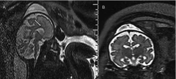
Prenatal brain scan diagnosis of mild, isolated VM carries a 12–13% rate of false negative results; this must be taken into consideration when counselling parents-to-be.32 Fe-tuses with mild, isolated VM have good prognosis, although Fig. 1 T2-weighted MRI performed in a fetus at 31.3 weeks of gestation showing a thin corpus callosum (white arrow) (A) associated with mild macrocrania (B).
Fig. 2 T2-weighted MRI performed in a fetus at 28.4 weeks of gestation: brachycephaly (A), subcuticular frontal edema (B) and hypertelorism (C) were detected.
an overall 11% risk of neurodevelopmental delay in fetuses with isolated mild and moderate VMs is seen.32
A follow-up by brain scan between 28 and 34 weeks of gestation should be indicated in all cases of prenatally ultrasound diagnosis of VM,33as the risk of progression of VM (defined as an increase in the ventricular measurement of more than 3 mm) is of16%.32In addition, infants with progression of VM are at a higher risk of subsequent neuro-developmental delay than those with non-progression.4 Ultrasound and MRI are substantially in agreement in defi n-ing the degree of VM (either isolated or with associated anomalies), although a low correlation between ultrasound and MRI in the evaluation of VM associated either with CNS or non-CNS anomalies has been seen. Magnetic Resonance Imaging may be a useful complementary diagnostic investi-gation in the detection of hemorrhagic foci, porencephaly, cortical and subependymal tubers, midline anomalies and callosal dysgenesis, as well as posterior fossa abnormalities. From 25 weeks onwards, the MRI may add additional infor-mation about cortical development and maturation, and is more accurate in detecting white matter pathology com-pared with the brain scan.34 Nonetheless, fetal MRI has a limited role over ultrasound in assessing the size of the cerebral ventricles, except for cases where fetal position and calvarial ossification cause reverberation artifacts and shadowing.35
In our series, the fetal MRI was performed at a mean gestational age of 31 weeks, 4 days, and allowed to detect, compared with the prenatal brain scan, the following CNS pathologies: microcephaly and microlissencephaly; sus-pected interventricolar septi; brachycephaly; impaired cor-tical gyration; cyst of the thalamocaudate sulcus; abnormal periventricular neuronal migration; increased signal of the temporal white matter and increased apparent diffusion coefficient (ADC); abnormal white matter migration; septum in the left occipital horn; and abnormal hippocampus
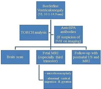
gyra-tion. Following the analysis of the medical records and results from our series, we developed a clinical-diagnostic flowchart to be used in cases of prenatally detected isolated mild and moderate VMs (►Fig. 5). This clinicalflowchart may aid antenatal care management. Specifically, fetal karyotype, TORCH analysis, search for anti-human platelet antigen (anti-HPA) antibodies (justified only if there is a suspicion of intracranial hemorrhage on imaging) and a thorough fetal ultrasound examination should be recommended; the fetal MRI may integrate the brain scan, especially when per-formed in the third trimester of pregnancy, enhancing prenatal counselling and antenatal care management by
the multispecialist team. In addition, the postnatal MRI may also allow the diagnosis of associated cerebral abnormalities undiscovered antenatally. The clinical limitations of the MRI are represented by elevated costs, availability and decreased sensitivity at a gestational age<25 weeks of gestation.
Note
Study performed in the Prenatal Diagnostic Unit, Depart-ment of Obstetrics & Gynecology, Guastalla Civil Hospital, AUSL Reggio Emilia, Italy.
References
1 Parazzini C, Righini A, Doneda C, et al. Is fetal magnetic resonance imaging indicated when ultrasound isolated mild ventriculome-galy is present in pregnancies with no risk factors? Prenat Diagn 2012;32(8):752–757
2 Pilu G, Hobbins JC. Sonography of fetal cerebrospinal anomalies. Prenat Diagn 2002;22(4):321–330
3 Vergani P, Locatelli A, Strobelt N, et al. Clinical outcome of mild fetal ventriculomegaly. Am J Obstet Gynecol 1998;178(2):218–222
4 Ouahba J, Luton D, Vuillard E, et al. Prenatal isolated mild ventri-culomegaly: outcome in 167 cases. BJOG 2006;113(9):1072–1079
5 Levene MI, Chervenak FA, Whittle M. Fetal and neonatal neurolo-gy and neurosurgery. 3rd ed. London: Churchill Livingstone; 2001
6 Signorelli M, Tiberti A, Valseriati D, et al. Width of the fetal lateral ventricular atrium between 10 and 12 mm: a simple variation of the norm? Ultrasound Obstet Gynecol 2004;23(1):14–18
7 Cardoza JD, Goldstein RB, Filly RA. Exclusion of fetal ventriculo-megaly with a single measurement: the width of the lateral ventricular atrium. Radiology 1988;169(3):711–714
8 Kinzler WL, Smulian JC, McLean DA, Guzman ER, Vintzileos AM. Outcome of prenatally diagnosed mild unilateral cerebral ven-triculomegaly. J Ultrasound Med 2001;20(3):257–262
9 Nejat F, Kazmi SS, Ardakani SB. Congenital brain tumors in a series of seven patients. Pediatr Neurosurg 2008;44(1):1–8
10 D’Addario V, Pinto V, Di Cagno L, Pintucci A. Sonographic diagno-sis of fetal cerebral ventriculomegaly: an update. J Matern Fetal Neonatal Med 2007;20(1):7–14
11 Whitelaw A. Neonatal hydrocephalus - clinical assessment and non surgical treatment. In: Levene MI, Chervenak FA, Whittle M, editors. Fetal and neonatal neurology and surgery. 3rd ed. London: Churchill Livingstone; 2001. p. 739–61.
12 Sherer DM, Onyeije CI. Prenatal ultrasonographic diagnosis of fetal intracranial tumors: a review. Am J Perinatol 1998;15(5): 319–328
13 Fong KW, Ghai S, Toi A, Blaser S, Winsor EJ, Chitayat D. Prenatal ultrasoundfindings of lissencephaly associated with Miller-Dieker syndrome and comparison with pre- and postnatal magnetic reso-nance imaging. Ultrasound Obstet Gynecol 2004;24(7):716–723
14 Malinger G, Kidron D, Schreiber L, et al. Prenatal diagnosis of malformations of cortical development by dedicated neuroso-nography. Ultrasound Obstet Gynecol 2007;29(2):178–191
15 Pilu G, Falco P, Gabrielli S, Perolo A, Sandri F, Bovicelli L. The clinical significance of fetal isolated cerebral borderline ventri-culomegaly: report of 31 cases and review of the literature. Ultrasound Obstet Gynecol 1999;14(5):320–326
16 Gaglioti P, Danelon D, Bontempo S, Mombrò M, Cardaropoli S, Todros T. Fetal cerebral ventriculomegaly: outcome in 176 cases. Ultrasound Obstet Gynecol 2005;25(4):372–377
17 Graham E, Duhl A, Ural S, Allen M, Blakemore K, Witter F. The degree of antenatal ventriculomegaly is related to pediatric neurological morbidity. J Matern Fetal Med 2001;10(4): 258–263
18 Pastorino D, Prefumo F, Rossi A, et al. Apparently isolated border-line ventriculomegaly and lissencephaly. Prenat Diagn 2007; 27(5):483–484
19 Nicolaides KH, Berry S, Snijders RJ, Thorpe-Beeston JG, Gosden C. Fetal lateral cerebral ventriculomegaly: associated malformations and chromosomal defects. Fetal Diagn Ther 1990;5(1):5–14
20 Mercier A, Eurin D, Mercier PY, Verspyck E, Marpeau L, Marret S. Isolated mild fetal cerebral ventriculomegaly: a retrospective analysis of 26 cases. Prenat Diagn 2001;21(7):589–595
21 Morris JE, Rickard S, Paley MN, Griffiths PD, Rigby A, Whitby EH. The value of in-utero magnetic resonance imaging in ultrasound diagnosed foetal isolated cerebral ventriculomegaly. Clin Radiol 2007;62(2):140–144
22 Breeze AC, Alexander PM, Murdoch EM, Missfelder-Lobos HH, Hackett GA, Lees CC. Obstetric and neonatal outcomes in severe fetal ventriculomegaly. Prenat Diagn 2007;27(2):124–129
23 Lam SJ, Kumar S. Evolution of fetal ventricular dilatation in relation to severity at first presentation. J Clin Ultrasound 2014;42(4):193–198
24 Gaglioti P, Oberto M, Todros T. The significance of fetal ventricu-lomegaly: etiology, short- and long-term outcomes. Prenat Diagn 2009;29(4):381–388
25 Valsky DV, Ben-Sira L, Porat S, et al. The role of magnetic reso-nance imaging in the evaluation of isolated mild ventriculome-galy. J Ultrasound Med 2004;23(4):519–523, quiz 525–526
26 Benacerraf BR, Shipp TD, Bromley B, Levine D. What does mag-netic resonance imaging add to the prenatal sonographic diagno-sis of ventriculomegaly? J Ultrasound Med 2007;26(11): 1513–1522
27 Levine D, Barnes PD, Robertson RR, Wong G, Mehta TS. Fast MR imaging of fetal central nervous system abnormalities. Radiology 2003;229(1):51–61
28 Salomon LJ, Ouahba J, Delezoide AL, et al. Third-trimester fetal MRI in isolated 10- to 12-mm ventriculomegaly: is it worth it? BJOG 2006;113(8):942–947
29 Manganaro L, Savelli S, Francioso A, et al. Role of fetal MRI in the diagnosis of cerebral ventriculomegaly assessed by ultrasonogra-phy. Radiol Med (Torino) 2009;114(7):1013–1023
30 Parazzini C, Righini A, Doneda C, Rustico M, Lanna M, Triulzi F. Response to“Is fetal magnetic resonance imaging indicated when ultrasound isolated mild ventriculomegaly is present in pregnan-cies with no risk factors?”Prenat Diagn 2014;34(9):919
31 Garel C, Alberti C. Coronal measurement of the fetal lateral ventricles: comparison between ultrasonography and magnetic resonance imaging. Ultrasound Obstet Gynecol 2006;27(1): 23–27
32 Melchiorre K, Bhide A, Gika AD, Pilu G, Papageorghiou AT. Counseling in isolated mild fetal ventriculomegaly. Ultrasound Obstet Gynecol 2009;34(2):212–224
33 Baffero GM, Crovetto F, Fabietti I, et al. Prenatal ultrasound predictors of postnatal major cerebral abnormalities in fetuses with apparently isolated mild ventriculomegaly. Prenat Diagn 2015;35(8):783–788
34 Griffiths PD, Reeves MJ, Morris JE, et al. A prospective study of fetuses with isolated ventriculomegaly investigated by antenatal sonography and in utero MR imaging. AJNR Am J Neuroradiol 2010;31(1):106–111