The efect of quercetin on cerulein-induced acute
pancreatitis
Ahmet KahramanI, Ayhan VurmazI, Halit Buğra KocaI, Hilmi UyarII, Abdulkadir ÇatI, Çiğdem TokyolIII, Coşkun PolatII,
Tülay KökenI
DOI: 10.5935/MedicalExpress.2017.05.02
Afyon Kocatepe University, Faculty of Medicine, I Department of Medical Biochemistry,II Department of General Surgery and III Department of Medical Patology Afyonkarahisar-TURKEY
OBJECTIVE: The aim of this study was to evaluate the protective and therapeutic efects of quercetin on pancreatic injury in cerulein-induced acute pancreatitis.
METHOD: Thirty-two rats were randomly divided into four groups, eight per group: (CT): untreated controls, (CER) treated with cerulein, 50 μg/kg body weight; (Q+CER) pre-treatment with quercetin, 100 mg/kg body weight, followed by cerulein, 50 μg/kg; (CER+Q) post-treatment, cerulein followed by quercetin, same doses. Cerulein was divided into four doses, given at 1-hour intervals by intraperitoneal injection. Quercetin was given either 1-hour before (in pre-treatment group) or 1-hour after (in post-treatment group) cerulein. Pancreatic malondialdehyde (MDA), carbonyl, myeloperoxidase (MPO), tumor necrosis factor-alpha (TNF-a), interleukin-6 (IL-6), reduced and oxidized glutathione (GSH and GSSG, respectively) were measured. Histology of the pancreas was studied.
RESULTS: (1) MDA, carbonyl, MPO, TNF-a and IL-6 levels were signiicantly higher in CER vs CT rats. (2) MDA, carbonyl, MPO and TNF-α decreased signiicantly in pre-treated rats vs. CER. (3) MDA, MPO, TNF-α, IL-6 were signiicantly lower in post-treated rats vs. CER. (4) The reduced vs. oxidized glutathione ratio (GSH/GSSG) of was signiicantly lower CER vs. CT rats. (5) Pre- and post-treatment with quercetin signiicantly increased this ratio. (6) Pancreatic histology showed that quercetin had no signiicant efect on the histological image of the pancreas
CONCLUSION: These results suggest that quercetin can attenuate the severity of cerulein-induced acute pancreatitis by acting as an antioxidant and anti-inlammatory agent and combating oxidative stress. Further studies are needed to clearly explain its utility on acute pancreatitis.
KEYWORDS: Acute pancreatitis, cerulein, quercetin, oxidative stress.
Kahraman A, Vurmaz A, Koca HB, Uyar H, Çat A, Tokyol Ç, Polat C, Köken T. The efect of quercetin on cerulein-induced acute pancreatitis. MedicalExpress (São Paulo, online). 2017 Oct;4(5):M170502
Received for Publication on July 27, 2017; First review on August 17, 2017; Accepted for publication on August 29, 2017; Online on September 26, 2017
E-mail: [email protected]
■
INTRODUCTIONAcute pancreatitis (AP) is an inflammatory disease which is characterized by activation of leukocytes, macrophages and digestive proteases, inflammatory cell infiltration, the release of various inflammatory mediators such as tumor necrosis
factor-alpha (TNF-α), interleukin-1 beta (IL-1β), and interleukin-6 (IL-6).1-4 The digestive enzymes
of pancreas lead to auto-digestion of the gland. The
auto-digestion is a key event in the pathogenesis of
AP. After auto-digestion, a massive infiltration of neu -trophils and macrophages lead to a local and systemic
inflammatory response.3,5 This is partially caused by the release of cytokines from acinar cells. Reactive oxygen substances (ROS) may also contribute to the damage of pancreatic acinar cells.4,6-10 Previous studies
have confirmed the participation of ROS at early stages of AP, regardless of the underlying cause.11-13 The
cha-racteristics of the pancreatitis induced by cerulein (a decapeptide and cholecystokinin analogue) resemble
Experimental Procedures
Acute Pancreatitis (AP) was induced by intraperi-toneal injection of cerulein, diluted in physiological saline
(50 μg/kg body weight), four times at one hour intervals.26 Quercetin was dissolved in DMSO (1%) and given intrape
-ritoneally (100 mg/kg body weight).22 Figure 1 illustrates the timeline of the procedures.
There are still no therapies for acute pancreatitis.
Medical treatment remains largely supportive such as the control of symptoms, and prevention of severe
complica-tions. Therefore, prevention of oxidative stress and acinar
cell injury during the early phase of acute pancreatitis may stop the pathologic progression to severe
pancreati-tis.1,2,4,8,15 The role of oxidative stress in the pathogenesis
of AP and the benefits of antioxidants have been the
subject of numerous studies.14,16-19 Recent studies have
focused on antioxidant and anti-inflammatory properties
of phenolic compounds.2,4,6,20 Quercetin (3,5,7,3’,4’ pen
-tahydroxyflavone) is a plant-derived phenolic compound
belonging to a class of substances known as flavonoids.
Flavonoids are widely found in vegetables, such as black
and green tea, red wine, apple, onion, bean etc. Biologi -cal effects of quercetin have been reported as follows: antioxidant, anti-inflammatory, antiviral, anti-ischemic,
anticancer, antithrombotic, and antihistaminic etc.2,21,22
Moreover, quercetin has been shown to inhibit amylase release induced by agonists such as cholecystokinin,
carbachol, phorbol ester tetra decanoylphorbol-13
--acetate.23-25 A recent study has shown that one hour after the last dose of cerulein administration, quercetin
treatment attenuates the development of AP in mice.2 The aim of the present study was to evaluate the protective and therapeutic effects of quercetin on pancreatic
injury in cerulein-induced acute pancreatitis in rats.
■
MATERIALS AND METHODSChemicals and Drugs
Cerulein, quercetin, 3,3’,5,5’- Tetra Methyl Benzydine (TMP), dimethyl sulfoxide (DMSO), formaldehyde, eosin and hematoxylin were purchased from Sigma Chemical Co. (St. Louis, MO, USA). Malondialdehyde (MDA) and glutathione kits were purchased from Chromosystems Instruments & Chemicals (GmbH, Munich, Germany). ELISA kits including Interleukin-6 (IL-6), Tumor Necrosis Factor-alpha (TNF-α) and ketamine were purchased from eBioscience (USA), Invi
-trogen Co. (Camarrillo, CA, USA), Alfason Int. B.V. (Voerden, Holland), respectively.
Animals
Animal procedures were performed according to
the “Guide for the Care and Use of the Laboratory Animals” set by the Ethics Committee of Afyon Kocatepe University. Thirty-two female Sprague-Dawley rats (250-300 g) were housed in four cages at a temperature of 23±2°C with 12 h of light-dark cycle. Animals were fed with a standard rat chow (Aytekinler feed Industry, Konya, Turkey) and allowed to drink water ad libitum, but were deprived of food for 12 h before the experiments.All procedures were performed
in sterilized conditions.
Figure 1. Timeline of the experimental procedure. Time markers shown for 1 and 6 hours. All injections given intraperitoneally. S: isotonic saline D: dimethyl sulfoxide (DMSO); C: Cerulein; Q: quercetin. A+EU: rats anesthetized and euthanized.
The thirty-two rats were randomly divided into four groups (eight in each group): rats in the CT group
received intraperitoneally (i.p.) physiological saline four times and DMSO twice at 1-h intervals. Rats in the cerulein group (CER) received i.p. cerulein (50 μg/kg body weight in physiological saline), divided into four hourly doses
and DMSO twice at 1-h intervals. Quercetin pre-treatment group (Q+CER) received i.p. quercetin as a single dose one
hour before cerulein treatment applied as described and
DMSO once, 6-h after cerulein treatment. Quercetin post
--treatment group (CER+Q) received i.p. DMSO, once, one
hour before cerulein treatment and quercetin, as a single
dose, six hours after cerulein treatment.
The rats were anesthetized by an intramuscular
injection of ketamine (50 mg/kg body wt.) 6-h after the last administration of DMSO or quercetin. Blood samples
were drawn with a heparinized syringe by cardiac puncture
and collected in heparinized tubes. Rats were euthanized by exsanguination with blood retained for serum harvest.
Their pancreas tissues were taken for biochemical and his-tological analysis and rinsed with ice-cold saline and frozen
at -20 °C until assay. A portion of pancreatic tissue from each rat was reserved for histological analysis. Plasma was obtained by centrifugation at 3000 rpm for 10 minutes at 4°C and stored at -20 °C until the analyses were performed. The pancreas tissues were homogenized in 0.1 M phosphate buffer (pH 7.4) with an Ultra Turrax homogenizer (T25, Janke and Kunkel). Homogenates were centrifuged at 5000 rpm, 4 0C for 10 minutes. Supernatants were removed and
used for further analysis.
Plasma amylase and lipase activities were
Diagnostic kits (GmbH, Mannheim, Germany). Results are expressed as U/L.
Tissue malondialdehyde (MDA), reduced glutathione
(GSH) and oxide glutathione (GSSG) concentrations were determined by high-pressure liquid chromatography (HPLC) in the isocratic phase in the Agilent 1100 series instru
-ment with fluorescent detection (Ex:515, Em: 553 nm for MDA; Ex: 385, Em: 515 for GSH and GSSG) using a kit from Chromsystems Instruments & Chemicals GmbH (Munich, Germany). The results were evaluated as nmol/g protein for
MDA and μmol/g protein for GSH and GSSG. Tumor Necro
-sis Factor-alpha (TNF-α) and Interleukin-6 (IL-6) levels as inflammatory cytokines were measured using commercial colorimetric kits. TNF-α and IL-6 levels were expressed as ng/g protein. Myeloperoxidase (MPO) activities as an indica -tor of polymorphonuclear leukocytes27 and Carbonyl content as an indicator of protein oxidation8 in pancreas tissue were
measured according to previously described methods.
Histopathological examination
Pancreas tissue samples were fixed in 10% formal
-dehyde solution and embedded in paraffin using standard methods. Tissues were sectioned at 3-μm and stained with hematoxylin-eosin (H&E). Then, the stained sections
were assessed under light microscopy and examined by a pathologist blinded to group division for grading of the
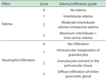
histopathological alterations. Edema and neutrophil infil -tration in pancreatic tissue were assessed using a scoring
system from 0 to 3 as described by Schoenberg et al29 and
shown in Table 1.
Statistical Analysis
The data were analyzed using the SPSS Statistical Package (Version 20.0, Chicago, USA for Windows). The
differences between groups were determined by the
Kruskal-Wallis test. The Conover-Iman test was used to
Table 1. Histopathological grading system in experimental acute
pancreatitis
Efect Score Edema/iniltration grade
Edema
0 No edema
1 İnterlobular edema
2 Moderate interlobular edema+intraacinar edema
3 Maximum interlobular + intra-acinar edema
Neutrophil iniltration
0 No iniltration
1 Intravascular margination of granulocytes
2 Granulocytes present in the perivascular tissue
3 Difuse iniltration of entire pancreatic gland
perform multiple comparisons between different treatment
groups. The results are expressed as the Mean±SEM. The p<0.05 value was accepted as statistically significant.
■
RESULTSPlasma amylase and lipase activities are shown in
Table 2. Plasma amylase and lipase activities significantly
increased in the cerulein-induced AP compared to the
control group (p<0.001). Quercetin treatment (pre- and post-) significantly decreased amylase and lipase activities compared to the CER group (p<0.001) but the enzyme ac
-tivities were higher than those of the controls.
The pancreatic levels of MDA, Carbonyl, MPO, TNF-α, and IL-6 levels in CER group increased significantly compared to the control group (p<0.001, p<0.001, p<0.001, p<0.01, and p<0.05; respectively). Quercetin treatment before cerulein administration (Q+CER) decreased significantly MDA, Car
-bonyl, MPO, and TNF-α levels compared to the CER group (p<0.05, p<0.05, p<0.001, and p<0.05, respectively). As also
shown in Table 2, Quercetin treatment after cerulein
admi-nistration (CER+Q) also significantly decreased MDA, MPO, TNF-α, and IL-6 levels compared to the CER group (p<0.001, p<0.001, p<0.05, and p<0.05, respectively).
The pancreatic levels of reduced and oxidized
gluthatio-ne (GSH and GSSG) are shown in Figure 2. Reduced glutathiogluthatio-ne levels decreased significantly in the CER group compared to the control group (p<0.01); in contrast, oxidized gluthatione in the CER group increased significantly compared to the control group (p<0.01). In sharp contrast, pre- or post-quercetin treat
-ment prevented the cerulein induced decrease of GSH and the increase of GSSG, both of which were not significantly different from their respective control levels. The GSH/GSSG ratio after cerulein induced pancreatitis decreased very sharply vs. con
-trols (p<0.001); quercetin pre-treatment partially impeded the fall in of the GSH/GSSG ratio, whereas post-treatment impeded the fall of the GSH/GSSG ratio, which was significantly higher than that in the CER group and not significantly different from the controls (p<0.05 and p<0.01, respectively).
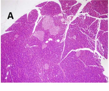
Table 3 shows that Cerulein induced intense
pan-creatic edema and inflammation, neither of which was countered by pre- or post-treatment with quercetin.
Figure 3 illustrates these findings. Panel A, collected
from a control rat shows a typically normal pancreatic
struc-ture; in contrast, panels B and C, collected from cerulein
administered rats revealed extensive tissue damage
cha-racterized by significant interlobular edema and neutrophil infiltration, respectively.
■
DISCUSSIONTable 2. Amylase, Lipase, MDA, Carbonyl, MPO, TNF-α and IL-6 levels in pancreatic tissue (Mean±SEM)
Parameters Control CER Q+CER CER+Q
Amylase (U/L) 848±34 3956±245a 2383±287a,b 1920±176a,b
Lipase (U/L) 8.51±0.17 136.40±15.59a 33.48±7.05a,b 23,37±12.01a,c
MDA (nmol/g protein) 58.91±9.27 195.33±27.24a 115.62±15.43c 94.34±13.16b
Carbonyl (μmol/g protein) 2.82±0.37 6.83±0.63a 4.08±0.66c 5.05±0.82
MPO (U/g protein) 0.38±0.07 1.15±0.12a 0.47±0.07b 0.59±0.13b
TNF-α (ng/g protein) 2.24±0.31 3.49±0.63d 2.03±0.40c 1.88±0.32c
IL-6 (ng/g protein) 62.94±1.86 84.10±5.31e 68.57±9.47 61.11±4.47c
a p < 0.001 vs. Control group; b p <0.05 vs. CER group; c p < 0.001 vs. CER group; d p < 0.01 vs. Control group; e p < 0.05 vs. Control group.
Figure 2. Pancreatic GSSG, GSH levels and GSH/GSSG ratios in control rats and rats with cerulein induced pancreatitis, untreated or treated with quercetin. a p < 0.01 vs.
Control; bp < 0.001 vs. Control; cp < 0.05 vs group; dp < 0.05 vs. CER; ep < 0.01 vs. CER.
Table 3. Pancreas tissue edema and inlammation scores (Mean
± SEM)
Groups Edema Inlammation
Control group 0.12±0.12 0.12±0.12 CER group 1.00±0.00a 1.77±0.22a
Q+CER group 0.87±0.12a 1.75±0.25a
CER+Q group 0.87±0.12a 1.50±0.32a
a p < 0.001 vs Control group
(AP) in rats through anti-inflammatory and antioxidant mechanisms by reducing the inflammatory cytokines, lipid peroxidation and protein oxidation.
Pathophysiology of acute pancreatitis is a multi-factorial process which involved the complex interaction
of pro-inflammatory and anti-inflammatory pathways. Oxidative stress and cytokines have an important role in the formation of the pathways.2 Cerulein-induced AP is one of the best–characterized rat models of experimental
pancreatitis. There is much similarity between experimen -tal and human pancreatitis with regard to morphological,
biochemical and pathophysiological features.2,30
Cerulein, an analogue of cholecystokinin (CCK),
overstimulates the acinar cells of pancreas by leading to pre-maturation to trypsin of trypsinogen; this leads to lysosomal degradation of intracellular organelles within
au-tophagic vacuoles in acinar cells; this followed by interstitial
edema and inflammation in the acinar cells. Inflammatory cells generate reactive oxygen species (ROS) which disrupt
membranes via lipid peroxidation, protein oxidation, and
trigger the inflammatory processes.4,21 Pro-inflammatory
cytokines such as TNF-α and IL-6 released by damaged
pancreas cells play an important role in the pathogenesis
of AP.3,31
There are still no specific therapies for AP. Treatment remains substantially supportive for symptoms. At present, all attention is focused on antioxidant and anti-inflam
-matory therapies.2,3,4,6,18,20 Quercetin has been reported to
exert multiple biological effects such as antioxidant,
anti--inflammatory, and antihistaminic effects etc.2,21,22
The increased serum amylase activity which is at least three times higher than the upper limit of normal and
lipase activity which remains high for up to 8 and 14 days;
this fact allows its comparison to normal serum amylase activity and supports the diagnosis of acute pancreatitis;32 however Bulut et al. report thatthis comparison does not
always match the severity of AP.33 In this study, plasma amylase (approximately fourfold above control) and lipase
(approximately fifteen fold above control) activities in the CER group were significantly higher than the control group; quercetin pre- and post-treatment significantly decreased amylase and lipase activities compared to the CER group.
In the present study, the pancreas tissue MDA, Car
-bonyl, MPO, TNF-α and IL-6 levels in the CER group signifi
-cantly increased compared to the control group. Quercetin pre-treatment significantly prevented increases of MDA, Carbonyl, MPO and TNF-α, but not of IL-6. Quercetin post
--treatment significantly prevented increases of MDA, MPO, TNF-α and IL-6, not of carbonyl. Our previous studies21,22
showed that quercetin was a more effective antioxidant and
anti-inflammatory molecule and that the (3,5,7,4’) positions of hydroxyl groups were associated with ROS inhibition.
Therefore, the protective and therapeutic effect of quercetin
could be attributed to its antioxidant and anti-inflammatory properties by various mechanisms.
and activation of the oxidant-sensitive transcription factor
kB (NF-kB).19 ROS can also activate the transcription of NF
--kB.19 NF-kB increases the transcription of inflammatory
cytokines such as IL-6 and TNF-α which can initiate the pancreatic inflammatory process.19,22 In the present study,
quercetin potently suppressed neutrophil mediated MPO, TNF-α and IL-6, well established markers of inflammation, thereby inhibiting NF-kB activation.2,34-36 Furthermore, it has
been reported that quercetin decreased gene expression
and production of the proinflammatory cytokines as TNF
--alpha, interleukin (IL)-1β, IL-6, and IL-8 in mast cells.22 Our results are compatible with such reports.2,8,11
Glutathione is the most important non-enzymatic
antioxidant molecule in cells. It exists in a reduced (GSH) and in an oxidized form (GSSG): GSH is in equilibrium in GSSG, and the GSH/GSSG ratio is a measure of the redox --status of the cells: the ratio is a reliable indicator of
oxi-dative stress and reflects the balance between antioxidant and oxidant status in cells.10,14 However, GSH depletion and
GSSG status in AP have not been well-established. Pancre
-atic GSH depletion has been shown in the initial phase of acute pancreatitis.8,11,37,38 However, while some reports have
shown increased pancreatic GSSG in AP,13,14,37-39 others have
found that pancreatic GSSG levels and the GSSG/GSH ratio
remains essentially unchanged during the early course of
AP.10,12,14,39 Rau et al.12 demonstrated a significant decrease
of GSH levels parallel with an increased ratio of GSSG/Total glutathione. Gomez-Cambronero et al.14 reported that the
dose of cerulein affects GSSG levels and the ratio of GSH/ GSSG in a dose dependent manner. The pancreatic ratio of GSH/GSSG did not change significantly in rats treated with low or moderate doses of cerulein (8-40 μg/kg), but it
increased after a higher dose of cerulein (80 μg/kg). In this study, given alone, cerulein significantly decreased pancre
-atic GSH and increased GSSG, a sure sign of cellular oxidant stress. Quercetin treatment after and before cerulein partly reversed GSH decrease and GSSG increase: as an obvious consequence, the GSH/GSSG ratio significantly decreased
after untreated pancreatitis induction, but this was effec-tively countered both by pre- and by post-quercetin
treat-ment. ROS is the most probable inducer of this conversion of GSH to GSSG and we believe that quercetin reversed this process, mainly by scavenging ROS.
Our histopathological results showed that cerulein treatment caused edema and leukocyte infiltration of pancreatic acinar cells. Our previous study showed simi
-lar results.9 Pre- and post- treatment of quercetin partly
decreased edema and inflammation, but it did not prevent histologically detected pancreatic edema and inflammation induced by cerulein. However, cerulein-induced MPO acti
-vity as an indicator of leukocyte infiltration and the TNF-α and IL-6 levels as the indicators of inflammation significan
-tly decreased by quercetin administration (Table 3). This confirmed the participation of ROS at early stage of acute Figure 3. Histological features of pancreas tissue (HE x400): (A) normal architecture
pancreatitis, independently of the underlying etiology. It
is supposed that an imbalance between the production of free radicals and antioxidant systems of the organism would
predispose to acute pancreatitis. Thereafter, premature activation of pancreatic enzymes, leukocyte infiltration,
and cytokine production could aggravate local pancreatic
injury and produce the spread of the inflammation to the rest of the organism.11 For this reason, histological reflec
-tions of damage occur after biochemical changes and it also takes a certain period of time for the amelioration of
the damage. The administration of quercetin at different
points in time, to pre- and post-treatment groups can shed
light on this issue.
■
CONCLUSIONQuercetin pre- and post-treatment attenuated the development of cerulein-induced AP and partly attenuated acute pancreatitis through antioxidant and
anti-inflammatory mechanisms by minimizing inflammation, lipid peroxidation, and protein oxidation. In this regard, we
believe that more studies are needed to exactly clarify the molecular mechanisms involved in this interaction, both in
human and experimental models.
■
ACKNOWLEDGEMENTSThis study was supported by the Scientific Investiga
-tion Fund of Afyon Kocatepe University (10.Tip.17).
■
CONFLICT OF INTERESTAuthors declare no conflict of interests regarding
this study
■
AUTHOR PARTICIPATIONKahraman A participated in the planning of the
research, conduction of experiments, the acquisition of
data, and the writing of the manuscript. Vurmaz A, Koca HB, and Çat K participated in the experimental procedure,
and biochemical analysis. Tokyol Ç performed the histopa
-thological examination. Polat C and Köken T participated
in experimental procedure and conduction of biochemical
analysis, respectively.
EFEITO DA QUERCETINA SOBRE A PANCREATITE INDUZIDA POR CERULEÍNA
OBJETIVO: O objetivo deste estudo foi avaliar os
efeitos protetores e terapêuticos da quercetina na lesão
pancreática da pancreatite aguda induzida por ceruleína.
MÉTODO: Trinta e dois ratos foram divididos aleato-riamente em quatro grupos, oito por grupo: (CT): controles
não tratados (CER) tratados com ceruleína, 50 μg/kg de peso corporal; (Q+CER) pré-tratamento com quercetina, 100 mg / kg de peso corporal, seguido de ceruleína, 50 μg/ kg; (CER+Q) pós-tratamento, ceruleína seguida de querceti
-na, mesmas doses. A ceruleína foi dividida em quatro doses, administradas a intervalos de 1 hora por injeção intraperi
-toneal. A quercetina foi administrada 1 hora antes (no grupo de pré-tratamento) ou 1 hora após (no pós-tratamento) a administração de ceruleína. Foram medidos o malondialde
-ído pancreático (MDA), carbonilo, mieloperoxidase (MPO), fator de necrose tumoral alfa (TNF-a), interleucina-6 (IL-6), glutationa reduzida e oxidada (GSH e GSSG, respetivamen
-te). Foi estudada a histologia do pâncreas.
RESULTADOS: Os níveis de MDA, carbonila, MPO,
TNF-a e IL-6 foram significativamente maiores nos ratos CER vs. CT. MDA, carbonila, MPO e TNF-α diminuíram signi
-ficativamente em ratos pré-tratados versus CER. MDA, MPO, TNF-α, IL-6 também foram significativamente menores em ratos pós-tratados versus CER. A proporção reduzida de glutationa oxidada (GSH/GSSG) foi significativamente menor ratos CER vs. CT; pré e pós-tratamento com quer
-cetina aumentaram significativamente esta proporção. A
histologia pancreática mostrou que a quercetina não teve
efeito morfológico significativo.
CONCLUSÃO: Estes resultados sugerem que a
quercetina pode atenuar a gravidade da pancreatite aguda induzida por ceruleína, atuando como agente antioxidante
e anti-inflamatório e combater o estresse oxidativo. Mais
estudos são necessários para explicar claramente suas
utilidades na pancreatite aguda.
PALAVRAS-CHAVE: pancreatite aguda, ceruleína,
quercetina, estresse oxidativo.
■
REFERENCES1. Kim MJ, Bae GS, Choi SB, Jo IJ, Kim DG, Shin JY, et al. Lupeol Protects Against Cerulein-Induced Acute Pancreatitis in Mice. Phytother Res. 2015;29(10):1634-9. DOI:10.1002/ptr.5423
2. Carvalho K, Morais TC, de Melo TS, de Castro Brito GA, de Andrade GM, Rao VS, Santos FA. The natural flavonoid quercetin ameliora -tes cerulein-induced acute pancreatitis in mice. Biol Pharm Bull. 2010;33(9):1534-9. DOI:10.1248/bpb.33.1534
3. Buyukberber M, Savaş MC, Bagci C, Koruk M, Gulsen MT, Tutar E, et al. Therapeutic effect of caffeic acid phenethyl ester on cerulein-induced acute pancreatitis. World J Gastroenterol. 2009;15(41):5181-5. DOI:10.3748/wjg.15.5181
4. Polito F, Bitto A, Irrera N, Squadrito F, Fazzari C, Minutoli L, et al. Flavocoxid, a dual inhibitor of cyclooxygenase-2 and 5-lipoxygenase, reduces pancreatic damage in an experimental model of acute pan-creatitis. Br J Pharmacol. 2010;161(5):1002-11. DOI:10.1111/j.1476-5381.2010.00933.x
6. Babu BI. Malleo G, Genovese T, Mazzon E, Di Paola R, Crisafulli C, et al. Green tea polyphenols ameliorate pancreatic injury in cerulein--induced murine acute pancreatitis. Pancreas 2009;38(8):954-67. DOI:10.1097/MPA.0b013e3181b28d11
7. Çöl C, Dinler K, Hasdemir O, Buyukasik O, Bugdayci G. Oxidative stress and lipid peroxidation products: effect of pinealectomy or exogenous melatonin injections on biomarkers of tissue damage during acute pancreatitis. Hepatobiliary Pancreat Dis Int. 2010;9(1):78-82. 8. Özkan E, Akyüz C, Dulundu E, Topaloğlu U, Sehirli AÖ, Ercan F, et al..
Protective effects of lycopene on cerulein-induced experimental acute pancreatitis in rats. J Surg Res. 2012;176(1):232-8. DOI:10.1016/j. jss.2011.09.005
9. Polat C, Uyar H, Başsorgun I, Kahraman A, Ciftcioglu A, Arikan Y. Lapa -roscopy can aggravate the severity of pancreatitis in an experimental rat model. J Laparoendosc Adv Surg Tech A. 2012;22(10):978-83. DOI:10.1089/lap.2012.0328
10. Pérez S, Pereda J, Sabater L, Sastre J. Redox signaling in acute pancre -atitis. Redox Biol. 2015;5:1-14. DOI: 10.1016/j.redox.2015.01.014 11. Carrasco C, Holguín-Arévalo MS, Martín-Partido G, Rodríguez AB,
Pariente JA. Chemopreventive effects of resveratrol in a rat model of cerulein-induced acute pancreatitis. Mol Cell Biochem. 2014; 387(1-2):217-25. DOI:10.1007/s11010-013-1887-0
12. Rau B, Poch B, Gansauge F, Bauer A, Nüssler AK, Nevalainen T, et al. Pa -thophysiologic role of oxygen free radicals in acute pancreatitis: initia-ting event or mediator of tissue damage? Ann Surg. 2000;231(3):352-60. DOI:10.1097/00000658-200003000-00008
13. Schoenberg MH, Büchler M, Gaspar M, Stinner A, Younes M, Melzner I, Bültmann B, et al. Oxygen free-radicals in acute pancreatitis of the rats. Gut. 1990;31(10):1138-43. DOI:10.1136/gut.31.10.1138 14. Gómez-Cambronero L, Camps B, de La Asunción JG, Cerdá M, Pellín
A, Pallardó, FV, et al. Pentoxifylline ameliorates cerulein-induced pancreatitis in rats: role of glutathione and nitric oxide. J Pharmacol Exp Ther. 2000;293(2):670-6.
15. Choi SB, Bae GS, Park KC, Jo IJ, Seo SH, Song K, et al. Opuntia hu -mifusa ameliorated cerulein-induced acute pancreatitis. Pancreas. 2014;43(1):118-27. DOI:10.1097/MPA.0b013e318296f903 16. Wang G, Sun B, Zhu H, Gao Y, Li X, Xue D, et al. Protective effects of
emodin combined with danshensu on experimental severe acute pancreatitis. Inflamm Res. 2010;59:479-88. DOI:10.1007/s00011-009-0152-1
17. Alhan E, Usta A, Türkyılmaz S, Kural BV, Erçin C. Effects of glutami -ne alo-ne on the acute -necrotizing pancreatitis in rats. J Surg Res. 2015;193(1):161-7. DOI: 10.1016/j.jss.2014.07.029
18. Turkyilmaz S, Alhan E, Ercin C, Vanizor KB, Kaklikkaya N, Ates B, et al. Effects of caffeic acid phenethyl ester on pancreatitis in rats. J Surg Res. 2008;145(1):19-24. DOI:10.1016/j.jss.2007.04.019
19. Lima PR, Melo TS, Carvalho KM, Oliveira IB, Arruda BR, Castro Brito GA, et al. 1,8-cineole (eucalyptol) ameliorates cerulein-induced acute pancreatitis via modulation of cytokines, oxidative stress and NF-κB activity in mice. Life Sci. 2013;92(24-26),1195-1201. DOI:10.1016/j. lfs.2013.05.009
20. Lampropoulos P, Lambropoulou M, Papalois A, Basios N, Manousi M, Simopoulos C, et al. The role of apigenin in an experimental model of acute pancreatitis. J Surg Res. 2013;183(1):129-37. DOI:10.1016/j. jss.2012.11.053
21. Kahraman A, Erkasap N, Köken T, Serteser M, Aktepe F, Erkasap S. The antioxidative and antihistaminic properties of quercetin in ethanol-induced gastric lesions. Toxicology. 2003;183(1-3):133-42. DOI:10.1016/S0300-483X(02)00514-0
22. Kahraman A, Çakar H, Köken T. The protective effect of quercetin on long-term alcohol consumption-induced oxidative stress. Mol Biol Rep. 2012;39(3):2789-94. DOI:10.1007/s11033-011-1037-2
23. Lee PC, Shimizu K, Rossi TM, Cumella JC. Selectivity of quercetin inhi -bition on stimulated amylase release in rat pancreatic acini. Pancreas. 1988;3(3):317-23. DOI:10.1097/00006676-198805000-00013 24. Feick P, Gerloff A, Singer MV. Effect of non-alcoholic compounds of
alcoholic drinks on the pancreas. Pancreatology. 2007;7(2-3):124-30. DOI:10.1159/000104237
25. Jedinák A, Maliar T, Grancai D, Nagy M. Inhibition activities of natural products on serine proteases. Phytother Res. 2006;20(3):214-47. DOI:10.1002/ptr.1836
26. Gultekin FA, Kerem M, TatlıcıoĞlu E, Arıcıoglu A, Unsal C, Bukan N. Leptin treatment ameliorates acute lung injury in rats with cerulein --induced acute pancreatitis. World J Gastroenterol. 2007;13(21):2932-8.
27. Suzuki K, Ota H, Sasagawa S, Sakatani T, Fujikura T. Assay method for myeloperoxidase in human polymorphonuclear leukocytes. Anal Biochem. 1983;132(2):345-52. DOI:10.1016/0003-2697(83)90019-2 28. Levine RL, Garland D, Oliver CN. Determination of carbonyl content in oxidatively modified proteins. Method Enzymol. 1990;186:464-78. 29. Schoenberg MH, Büchler M, Pietrzyk C, Uhl W, Birk D, Eisele S, et al.
Lipid peroxidation and glutathione metabolism in chronic pancreatitis. Pancreas. 1995;10(1):36-43. DOI:10.1097/00006676-199501000-00005
30. Yu JH, Lim JW, Kim H. Altered gene expression in cerulein-stimulated pancreatic acinar cells: pathologic mechanism of acute pancreatitis. Korean J Physiol Pharmacol. 2009;13(6):409-16. DOI:10.4196/ kjpp.2009.13.6.40
31. Ueno N, Kashiwamura S, Ueda H, Okamura H, Tsuji NM, Hosohara K, et al. Role of interleukin 18 in nitric oxide production and pancre -atic damage during acute pancreatitis. Shock. 2005;24(6):564-70. DOI:10.1097/01.shk.0000184285.57375.bc
32. Matull WR, Pereira SP, O’Donohue JW. Biochemical markers of acute pancreatitis. J Clin Pathol 2006; 59(4):340-4. DOI:10.1136/ jcp.2002.002923
33. Bulut NE, Özkan E, Ekinci O, Dulundu E, Topaloğlu Ü, Şehirli AÖ, et al. Beneficial effects of alpha lipoic acid on cerulein-induced expe -rimental acute pancreatitis in rats. Ulus Travma Acil Cerrahi Derg. 2011;17(5):383-9. DOI: 10.5505/tjtes.2011.99835
34. Min YD, Choi CH, Bark H, Son HY, Park HH, Lee S, et al. Quercetin inhibits expression of inflammatory cytokines through attenuation of NF-kappa B and p38 MAPK in HMC-1 human mast cell line. Inflamm Res. 2007;56(5):210-25. DOI:10.1007/s00011-007-6172-9 35. Ruiz PA, Braune A, Hölzlwimmer G, Bauer A, Nüssler AK, Nevalainen T,
et al. Quercetin inhibits TNF-induced NF-kappa B transcription factor recruitment to proinflammatory gene promoters in murine intestinal epithelial cells. J Nutr 2007;137(5):1208-15.
36. Lee YK, Park SY, Kim YM, Lee WS, Park OJ. AMP kinase/cyclooxyge -nase-2 pathway regulates proliferation and apoptosis of cancer cells treated with quercetin. Exp Mol Med. 2009;41(3):201-7. DOI:10.3858/ emm.2009.41.3.023
37. Palmieri VO, Grattagliano I, Palasciano G. Ethanol induces secretion of oxidized proteins by pancreatic acinar cells. Cell Biol Toxicol. 2007;23(6):459-64. DOI:10.1007/s10565-007-9007-0
38. Lutgendorff F, Trulsson LM, van Minnen LP, Rijkers GT, Timmerman HM, Franzén LE, et al. Probiotics enhance pancreatic glutathione biosyn -thesis and reduce oxidative stress in experimental acute pancreatitis. Am J Physiol Gastrointest Liver Physiol. 2008;295(5):G1111-21. DOI: 10.1152/ajpgi.00603.2007