224 Radiol Bras. 2017 Jul/Ago;50(4):224–230
Accuracy of contrast-enhanced spectral mammography for
estimating residual tumor size after neoadjuvant chemotherapy
in patients with breast cancer: a feasibility study
Acurácia da mamograia espectral com contraste para seguimento de tumor residual pós-quimioterapia neoadjuvante em pacientes com câncer de mama: um estudo de viabilidade
Filipe Ramos Barra1, Fernanda Freire de Souza1, Rosimara Eva Ferreira Almeida Camelo1, Andrea Campos de Oliveira
Ribeiro1, Luciano Farage2
Barra FR, Souza FF, Camelo REFA, Ribeiro ACO, Farage L. Accuracy of contrast-enhanced spectral mammography for estimating residual tumor size after neoadjuvant chemotherapy in patients with breast cancer: a feasibility study. Radiol Bras. 2017 Jul/Ago;50(4):224–230.
Abstract
Resumo
Objective: To assess the feasibility of contrast-enhanced spectral mammography (CESM) of the breast for assessing the size of residual tumors after neoadjuvant chemotherapy (NAC).
Materials and Methods: In breast cancer patients who underwent NAC between 2011 and 2013, we evaluated residual tumor mea-surements obtained with CESM and full-ield digital mammography (FFDM). We determined the concordance between the methods, as well as their level of agreement with the pathology. Three radiologists analyzed eight CESM and FFDM measurements separately, considering the size of the residual tumor at its largest diameter and correlating it with that determined in the pathological analysis. Interobserver agreement was also evaluated.
Results: The sensitivity, speciicity, positive predictive value, and negative predictive value were higher for CESM than for FFDM (83.33%, 100%, 100%, and 66% vs. 50%, 50%, 50%, and 25%, respectively). The CESM measurements showed a strong, consistent correlation with the pathological indings (correlation coeficient = 0.76–0.92; intraclass correlation coeficient = 0.692–0.886). The correlation between the FFDM measurements and the pathological indings was not statistically signiicant, with questionable consistency (intraclass correlation coeficient = 0.488–0.598). Agreement with the pathological indings was narrower for CESM measurements than for FFDM measurements. Interobserver agreement was higher for CESM than for FFDM (0.94 vs. 0.88). Conclusion: CESM is a feasible means of evaluating residual tumor size after NAC, showing a good correlation and good agreement with pathological indings. For CESM measurements, the interobserver agreement was excellent.
Keywords: Mammography/methods; Breast neoplasms/diagnosis; Magnetic resonance imaging; Neoadjuvant therapy/methods.
Objetivo: Avaliar a viabilidade da utilização da mamograia espectral com meio de contraste (CESM) na avaliação do tumor residual em mulheres com câncer de mama submetidas a quimioterapia neoadjuvante.
Materiais e Métodos: Foi avaliada a concordância entre a mensuração do tumor residual na CESM e na mamograia digital (FFDM) com os dados histopatológicos de mulheres submetidas a quimioterapia neoadjuvante entre 2011 e 2013. Após as exclusões, três radiologistas analisaram oito CESMs e FFDMs separadamente. A maior dimensão do tumor residual foi considerada para comparação com os resultados histopatológicos. Concordância e correlação da CESM e FFDM com resultados histopatológicos e a concordância interobservador foram avaliadas.
Resultados: A CESM teve sensibilidade, especiicidade e valores preditivos positivos e negativos maiores que a FFDM – 83,33%, 100%, 100% e 66% versus 50%, 50%, 50% e 25%, respectivamente. A CESM teve correlação boa e consistente com os achados histopatológicos (coeiciente de correlação = 0,76–0,92; coeiciente de correlação intraclasse = 0,692–0,886). A correlação entre FFDM e os achados histopatológicos não foi estatisticamente signiicante, com consistência questionável (coeiciente de correlação intraclasse = 0,488–0,598). A concordância entre as dimensões do estudo histopatológico foi mais estreita com a CESM do que com a FFDM. A concordância interobservador foi maior na CESM (0,94) do que na FFDM (0,88).
Conclusão: A CESM é viável e pode ser utilizada para avaliação de tumor residual após quimioterapia neoadjuvante. A CESM tem boa correlação e concordância com o estudo histopatológico e excelente concordância interobservador.
Unitermos: Mamograia/métodos; Neoplasias de mama/diagnóstico; Ressonância magnética; Terapia neoadjuvante/métodos.
Study conducted in the Department of Breast Imaging, Imagens Médicas de Bra-sília – IMEB, BraBra-sília, DF, Brazil.
1. MD, Radiologist in the Department of Breast Imaging, Imagens Médicas de Brasília – IMEB, Brasília, DF, Brazil.
2. MD, Professor at the School of Medical Sciences, Universidade de Brasília (UnB), Brasília, DF, Brazil.
Mailing address: Dr. Filipe Ramos Barra. Imagens Médicas de Brasília – IMEB. SMHN, Quadra 2, Conjunto C, Sobreloja 18. Brasília, DF, Brazil, 70710-100. E-mail:
[email protected]. Received February 14, 2016. Accepted after revision July 29, 2016.
INTRODUCTION
of micrometastatic disease, and in vivo assessment of tu-mor response(1,2). The accurate assessment of residual
tumor extent after NAC is critical for surgical planning. Overestimation of the tumor extent can lead to unneces-sary mastectomy, whereas underestimation can increase the risk of positive surgical margins.
Although a complete pathological response is not prognostic for disease-free survival in all breast cancer sub-types, the post-NAC extent of residual disease in the breast and lymph nodes is associated with patient survival(3).
Patients with a complete pathological response have a lower risk of locoregional relapse and are candidates for less extensive locoregional treatment(4).
Physical examination, ultrasound, and mammography have been used in order to assess residual tumor size in breast cancer patients after NAC, although the accuracy of these techniques is not satisfactory(5–7). Magnetic reso-nance imaging (MRI) of the breast is currently the best modality for monitoring tumor response and for assess-ing residual disease after NAC because it is more accu-rate than are mammography, ultrasound, and clinical ex-amination(8,9). However, MRI is a time-consuming exam,
usually lasting 30–45 min, and requires a dedicated coil, as well as trained readers.
Contrast-enhanced spectral mammography (CESM) is an imaging modality that combines contrast enhance-ment with digital mammography. Nonionic iodinated contrast, which is administered intravenously, allows le-sions to be characterized based on their enhancement. Each CESM exposure is composed of a low-energy image, similar to that obtained with full-ield digital mammog -raphy (FFDM), and a high-energy image with an X-ray spectrum above the k-edge of iodine (33.2 keV). The two images are recombined, and a subtraction image of the lesions is produced(10,11). Initial studies comparing
CESM with mammography, ultrasound, and MRI show that CESM is better at detecting suspicious lesions than are mammography and mammography plus ultrasound, as well as having an accuracy in lesion size measurement similar to that of MRI(12–16).
The purpose of this study was to evaluate the feasibil-ity of using CESM to assess residual tumor extent after NAC in breast cancer patients. Speciic objectives were to evaluate the accuracy of CESM in determining residual tumor size, using pathology results as the gold standard, to compare the performance of CESM with that of FFDM (low-energy images only), in terms of their performance, and to analyze interobserver agreement.
MATERIALS AND METHODS Patients and treatment
This was a retrospective study. The study protocol was approved by the local research ethics committee, and in-formed consent was waived. The patients enrolled in this study were selected from among all patients undergoing
CESM at our institution between October 2011 and March 2013. The inclusion criteria were as follows: being fe-male; being ≥ 18 years of age; having histologically proven primary breast cancer; and having received NAC as part of the treatment. Patients who had undergone surgical treatment other than lumpectomy or mastectomy were excluded, as were those for whom there were no results from the histological analysis of the surgical specimen. The precise regimen of NAC varied and was at the discre-tion of medical oncologist in charge.
CESM examination
All CESM examinations were performed with a com-mercially available FFDM system (SenoDS/SenoBright; GE Healthcare, Buc, France). The dual-energy technique was applied under the supervision of a radiologist.
Dual-energy CESM exams were performed by acquir-ing a pair of low- and high-energy images duracquir-ing a sacquir-ingle breast compression. Low-energy images were obtained with a molybdenum or rhodium target and ilter, whereas high-energy images were acquired with a molybdenum or rho-dium target and a copper ilter. Both images were acquired with automatic optimization of parameters.
A 1–2 mL/kg dose of a nonionic contrast agent (iohe-xol, 300 mg/mL) was injected intravenously with an auto-mated injector at a low rate of 3 mL/s. Imaging was initi -ated 1.5–2 min after the injection and continued for 3–5 minutes. Bilateral craniocaudal and mediolateral oblique views were acquired. The complete examination protocol has been described and explained in detail elsewhere(17).
It has been demonstrated that low-energy images are equivalent to FFDM, even in the presence of intravenous iodinated contrast(18). In this study, we use the terms FFDM and CESM to refer to low-energy images and recombined images, respectively.
Image analysis
Before the readings, a radiologist with three years of experience in CESM (reader 1) conducted a training ses-sion. Training cases were provided in order to familiarize the other radiologists with CESM and with the reading protocol. The same radiologist selected the cases for this study, split the low-energy and recombined images, ano-nymized them, and loaded them (separately and together) into a workstation.
A breast radiologist with over ten years of practice (reader 2) and a breast imaging fellow (reader 3) reviewed all studies using the same radiology workstation (Seno Ad-vantage 2.2; GE Medical Systems, Milwaukee, WI, USA). Tumor laterality was the only background information available.
included in the second review session. During the second session, the images were reviewed separately or together. Data from readers 2 and 3 were comparable to those in the original report submitted by reader 1.
Pathology
Specimen processing was performed at a hospital, ac-cording to the protocols of the local institution. All patho-logical data were extracted from the pathology reports.
Tumor size measurement
Suspicious indings were measured on low-energy and recombined images, in the craniocaudal and medio-lateral oblique views. Some tumors presented as multiple enhancing spots, irregular masses, or ill-deined asym -metric masses. In those cases, the measurement included the largest tumor diameter
For analysis purposes, the largest diameters of the residual tumor documented on low-energy (FFDM) and recombined (CESM) images were compared with that de-termined for the pathological specimen.
Statistical analysis
The size of the residual tumor determined by pathol-ogy was set as the “gold standard” and was compared to the size determined from the analysis of the low-energy and recombined images. The agreement between the size determined by pathology and that determined from the low-energy (FFDM) and recombined (CESM) images was assessed with the Bland-Altman 95% limits of agreement and intraclass correlation coeficient (ICC)(19). Tumor size
based on the FFDM and CESM images was also catego-rized as in agreement, underestimated, or overestimat-ed, in relation to the size determined by pathology. We used scatter plots and Pearson’s correlation coeficients to explore whether the size of the residual tumor deter-mined by pathology correlated with that deterdeter-mined from the CESM and FFDM images. Values of p < 0.05 were considered signiicant. The interobserver agreement for each imaging technique was calculated using the limits of agreement and ICC. Statistical analysis was performed
using MedCalc for Windows, version 14.8.1 (MedCalc Software, Mariakerke, Belgium).
RESULTS Study population
We identiied 12 lesions in 11 patients who met the inclusion criteria. Three patients (with a collective total of four lesions) were excluded because two died before surgery and the pathology result was not available for one. Therefore, the inal sample comprised eight lesions in eight patients. The mean patient age was 46.41 ± 15.19 years (range, 22–76 years). The mean time from CESM and surgery was 32.6 ± 22.4 days (range, 5–66 days).
Residual tumor size
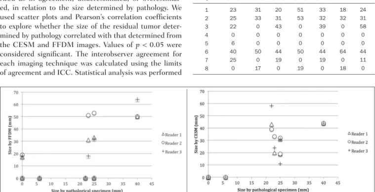
Residual tumor size ranged from microscopic (not measurable) to 40 mm, with a mean size of 17 mm. The size of the residual tumor determined from analysis of the pa-thology specimen is shown in Table 1, as are the sizes determined by all readers from the FFDM and CESM im-ages. Scatter plots of those measurements are shown in Figure 1.
The pathological analysis revealed residual tumors in six (75%) of the eight patients evaluated. Three residual tumors were not visible on FFDM. Among those three tumors, CESM missed one, overestimated one, and un-derestimated one (Figure 2). CESM was true negative in
Figure 1. Scatter plot of the largest diameter of post-NAC residual breast tumors, as determined by pathology, from FFDM images, and from CESM images.
Table 1—Residual breast tumor size determined by pathology, from CESM im-ages, and from FFDM images.
both of the patients in whom the pathological analysis failed to identify a residual tumor, CESM also revealed no residual tumor at histopathology, whereas, in one patient, a focal asymmetry on FFDM was interpreted as a residual tumor by all readers.
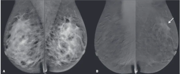
One patient also had a ibroadenoma (Figure 3). On the basis of the FFDM images, all of the readers incor-rectly held it as suspicious and concluded that the index tumor, which was located in the same breast, had been overrun. On CESM, the index tumor showed mild en-hancement and the ibroadenoma showed none.
The sensitivity and speciicity of CESM for detect -ing residual tumors were 83.33% and 100%, respectively,
compared with only 50% (for both) for FFDM. A positive CESM examination was predictive of a residual tumor in 100% of the cases, twice as many as did a positive FFDM examination, whereas a negative CESM result predicted the absence of a residual tumor in 66%, compared with 25% for a negative FFDM result.
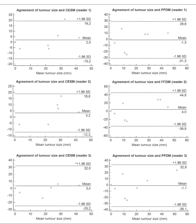
Among all readers, the ICC between the size of the re-sidual tumor determined by imaging and that determined by pathology was higher for CESM than for FFDM. The LoA was also better for CESM than for FFDM, as shown in Table 2 and Figure 4.
Residual tumor size was underestimated by FFDM and CESM in 50% and 37.5% of the cases, respectively, Figure 2. Residual (2.2-cm) tumor in a 23-year-old woman. FFDM (A) was negative, whereas CESM (B) overestimated the tumor size.
B A
Figure 3. Residual (2.2-cm) tumor in a 45-year-old woman. On FFDM (A), all readers mistook a ibroadenoma for the tumor (arrow). On CESM (B), there was het-erogeneous enhancement consistent with a residual tumor (asterisk).
overestimated by both in 37.5%, and correctly assessed in 12.5% and 25%, respectively.
There was a perfect agreement among readers re-garding the presence or absence of residual tumor based on both FFDM and CESM (Table 1). The interobserver agreement was very good for both methods, although it was higher for CESM (Table 2).
DISCUSSION
of MRI. In this feasibility study, we assessed the diagnos-tic performance of CESM in the detection and size deter-mination of residual tumors after NAC in breast cancer patients.
Our indings make it clear that CESM is a feasible means of detecting residual tumors after NAC. In com-parison with FFDM, CESM increased the sensitivity and speciicity of residual tumor detection from 50% to 83.33% and from 50% to 100%, respectively. On post-NAC FFDM images, residual tumors were missed in three patients (37.5%), compared with only one (12.5%) on post-NAC CESM images. Our data also suggest that a positive CESM indicates the presence of residual tumor after NAC (positive predictive value, 100%).
Previous studies have demonstrated the greater ac-curacy of CESM in breast tumor measurement in com-parison with MRI(13,15), ultrasound(16), and FFDM(12). In
the present study, we were able to show that the accuracy of CESM in residual tumor size determination was better than was that of FFDM, and both methods showed good agreement with the pathological indings (ICC = 0.692– 0.886 and 0.488–0.598, respectively), although the cor-relation was signiicant only for CESM. The residual tu -mor size was overestimated less often by CESM than by FFDM. That might be especially meaningful in order to avoid unnecessary mastectomies and reduce the extent of breast conservation surgery.
Although interobserver agreement was very good for CESM and FFDM, it was slightly better for CESM. There was perfect agreement among readers regarding the presence or absence of residual tumors as determined by FFDM and CESM. The use of CESM increased the di-agnostic performance of all readers, as was also reported by Dromain et al.(28) in a study with six readers. Cheung
et al.(29) found that, among four blinded readers
(radiolo-gists) who scored lesions in terms of the probability of ma-lignancy, interobserver agreement was signiicantly higher for CESM than for FFDM (0.62 vs. 0.38), whereas the difference between the two methods was smaller in the present study (0.94 vs. 0.88). Although this discrepancy is most likely attributable to a difference in sample size, it could also be because, in our study, the correlation was made on the basis of measurements, rather than scores, which are less subjective.
The present study has certain limitations, chief among which is the small size of the sample. The small sample size precluded an analysis of diverse cancer types and the collection of data regarding the inluence of molecular subtypes. In addition, because CESM was not performed before or during NAC, we were unable to predict the re-sponse or estimate tumor size reduction. Readers were not totally blinded, because the laterality of the tumor was known to them. We were also unable to perform an analy-sis of the impact of CESM on surgical decision-making.
CONCLUSION
Our results conirm that CESM is a feasible, easily performed method for evaluating residual tumor size after NAC. CESM correlates well and shows good agreement with the pathology, as well as showing good interobserver agreement. Therefore, our indings might be used as refer -ence data for future prospective studies designed to eval-uate the impact of CESM on surgical decision-making.
REFERENCES
1. Tardivon AA, Ollivier L, El Khoury C, et al. Monitoring therapeutic eficacy in breast carcinomas. Eur Radiol. 2006;16:2549–58. 2. van der Hage JA, van de Velde CJ, Julien JP, et al. Preoperative
chemotherapy in primary operable breast cancer: results from the European Organization for Research and Treatment of Cancer trial 10902. J Clin Oncol. 2001;19:4224–37.
3. von Minckwitz G, Untch M, Blohmer JU, et al. Deinition and im-pact of pathologic complete response on prognosis after neoadju-vant chemotherapy in various intrinsic breast cancer subtypes. J Clin Oncol. 2012;30:1796–804.
4. von Minckwitz G. Neoadjuvant chemotherapy in breast cancer—in-sights from the German experience. Breast Cancer. 2012;19:282–8. 5. Chagpar AB, Middleton LP, Sahin AA, et al. Accuracy of physical
examination, ultrasonography, and mammography in predicting re-sidual pathologic tumor size in patients treated with neoadjuvant chemotherapy. Ann Surg. 2006;243:257–64.
6. Croshaw R, Shapiro-Wright H, Svensson E, et al. Accuracy of clini-cal examination, digital mammogram, ultrasound, and MRI in de-termining postneoadjuvant pathologic tumor response in operable breast cancer patients. Ann Surg Oncol. 2011;18:3160–3.
7. Marinovich ML, Houssami N, Macaskill P, et al. Meta-analysis of magnetic resonance imaging in detecting residual breast cancer af-ter neoadjuvant therapy. J Natl Cancer Inst. 2013;105:321–33. 8. Ramirez SI, Scholle M, Buckmaster J, et al. Breast cancer tumor
size assessment with mammography, ultrasonography, and magnetic resonance imaging at a community based multidisciplinary breast center. Am Surg. 2012;78:440–6.
9. Lobbes MBI, Prevos R, Smidt M, et al. The role of magnetic
reso-Table 2—Comparison among the three readers in terms of the mean residual breast tumor size as determined from CESM and FFDM images, as well as the limits
of agreement, intraclass correlation coeficient, and correlation coeficient in comparison with the size determined by pathology.
Measure Mean ± SD (mm) LoA (mm) ICC (95% CI) CC (p-value)
FFDM
−1.3 ± 15.33 −31.3 to 28.8
0.598 (0.11–0.90) 0.63 (0.094)
CESM 2 ± 8.8
−15.2 to 19.2
0.859 (0.45–0.97) 0.89 (0.003)
FFDM 4 ± 20.8
−36.6 to 44.9
0.488 (0.26–0.87) 0.57 (0.138)
CESM 3.25 ± 7.94
−12.3 to 18.8
0.886 (0.53–0.97) 0.92 (0.001)
FFDM
−1.1 ± 17.35 −35.1 to 32.9
0.579 (0.14–0.90) 0.64 (0.086)
FFDM
−1.1 ± 17.35 −35.1 to 32.9
0.579 (0.14–0.90) 0.64 (0.086)
Reader 1 Reader 2 Reader 3
nance imaging in assessing residual disease and pathologic com-plete response in breast cancer patients receiving neoadjuvant che-motherapy: a systematic review. Insights Imaging. 2013;4:163–75. 10. Dromain C, Balleyguier C, Adler G, et al. Contrast-enhanced digital
mammography. Eur J Radiol. 2009;69:34–42.
11. Barra FR, Barra RR, Barra Sobrinho A. Novel functional methods in the evaluation of breast lesions. Radiol Bras. 2012;45:340–4. 12. Lobbes MBI, Lalji U, Houwers J, et al. Contrast-enhanced spectral
mammography in patients referred from the breast cancer screen-ing programme. Eur Radiol. 2014;24:1668–76.
13. Jochelson MS, Dershaw DD, Sung JS, et al. Bilateral contrast-en-hanced dual-energy digital mammography: feasibility and comparison with conventional digital mammography and MR imaging in wom-en with known breast carcinoma. Radiology. 2013;266:743–51. 14. Dromain C, Thibault F, Muller S, et al. Dual-energy
contrast-en-hanced digital mammography: initial clinical results. Eur Radiol. 2011;21:565–74.
15. Fallenberg EM, Dromain C, Diekmann F, et al. Contrast-enhanced spectral mammography versus MRI: initial results in the detec-tion of breast cancer and assessment of tumour size. Eur Radiol. 2014;24:256–64.
16. Blum KS, Rubbert C, Mathys B, et al. Use of contrast-enhanced spectral mammography for intramammary cancer staging: prelimi-nary results. Acad Radiol. 2014;21:1363–9.
17. Barra FR, Ribeiro AC, Mathieu OD, et al. Dual-energy contrast-en-hanced digital mammography: examination protocol. Diagn Interv Imaging. 2014;95:351–2.
18. Francescone MA, Jochelson MS, Dershaw DD, et al. Low energy mammogram obtained in contrast-enhanced digital mammography (CEDM) is comparable to routine full-ield digital mammography (FFDM). Eur J Radiol. 2014;83:1350–5.
19. Bland JM, Altman DG. Comparing methods of measurement: why
plotting difference against standard method is misleading. Lancet. 1995;346:1085–7.
20. Villar VCFL, De Seta MH, Andrade CLT, et al. Evolution of mam-mographic image quality in the state of Rio de Janeiro. Radiol Bras. 2015;48:86–92.
21. Avelar MS, Almeida O, Alvares BR. Mammographic artifact leading to false-positive result. Radiol Bras. 2015;48:198–9.
22. Badan GM, Roveda Júnior D, Ferreira CAP, et al. Complete internal audit of a mammography service in a reference institution for breast imaging. Radiol Bras. 2014;47:74–8.
23. Correia PD, Granzotti CRF, Santos YS, et al. Characterization of a lead breast shielding for dose reduction in computed tomography. Radiol Bras. 2014;47:223–7.
24. Valentim MH, Monteiro V, Marques JC. Primary neuroendocrine breast carcinoma: a case report and literature review. Radiol Bras. 2014;47:125–7.
25. Bitencourt AGV, Lima ENP, Chojniak R, et al. Correlation between PET/CT results and histological and immunohistochemical ind-ings in breast carcinomas. Radiol Bras. 2014;47:67–73.
26. Pinheiro DJPC, Elias S, Nazário ACP. Axillary lymph nodes in breast cancer patients: sonographic evaluation. Radiol Bras. 2014;47:240– 4.
27. Campos GCP, Castro MVK, Mattos VFE, et al. Lymphocytic mas-topathy mimicking breast malignancy: a case report. Radiol Bras. 2014;47:256–8.
28. Dromain C, Thibault F, Diekmann F, et al. Dual-energy contrast-enhanced digital mammography: initial clinical results of a multi-reader, multicase study. Breast Cancer Res. 2012;14:R94.