Breast-conserving surgery in locally advanced breast
cancer submitted to neoadjuvant chemotherapy. Safety
and effectiveness based on ipsilateral breast tumor
recurrence and long-term follow-up
Guilherme Freire Angotti Carrara,ICristovam Scapulatempo-Neto,IILucas Faria Abraha˜o-Machado,IIMaria Mitzi Brentani,IIIJoa˜o Soares Nunes,IVMaria Aparecida Azevedo Koike Folgueira,IIIRene´ Aloisio da Costa VieiraI,V,*
IHospital de Caˆncer de Barretos, Programa de Po´s-Graduac¸a˜o em Oncologia, Barretos/SP, Brazil.IIHospital de Caˆncer de Barretos, Departamento de
Patologia, Barretos/SP, Brazil.IIIFaculdade de Medicina da Universidade de Sa˜o Paulo, Disciplina de Oncologia, Departamento de Radiologia, Sa˜o Paulo/SP, Brazil.IVHospital de Caˆncer de Barretos, Departamento de Oncologica Clı´nica, Barretos/SP, Brazil.VHospital de Caˆncer de Barretos, Programa de Po´s-Graduac¸a˜o em Oncologia, Departamento de Mastologia e Reconstruc¸a˜o Mama´ria, Barretos/SP, Brazil.
OBJECTIVE:To evaluate ipsilateral breast tumor recurrence after breast-conserving surgery for locally advanced breast cancer.
METHODS:A retrospective observational cohort study was performed in patients with locally advanced breast cancer submitted to breast-conserving surgery after neoadjuvant chemotherapy based on an adriamycin-cyclophosphamide-paclitaxel regimen. We evaluated the clinical, pathologic, immunohistochemistry, and surgical factors that contribute to ipsilateral breast tumor recurrence and locoregional recurrence. A Kaplan-Meier analysis and Cox model were used to evaluate the main factors related to disease-free survival.
RESULTS: Of the 449 patients who received neoadjuvant chemotherapy, 98 underwent breast-conserving
surgery. The average diameter of the tumors was 5.3 cm, and 87.2% reached a size of up to 3 cm. Moreover, 86.7% were classified as clinical stage III, 74.5% had T3-T4 tumors, 80.5% had N1-N2 axilla, and 89.8% had invasive ductal carcinoma. A pathologic complete response was observed in 27.6% of the tumors, and 100.0% of samples had free margins. The 5-year actuarial overall survival rate was 81.2%, and the mean follow-up was 72.8 months. The rates of ipsilateral breast tumor recurrence and locoregional recurrence were 11.2% and 15.3%, respectively. Multifocal morphology response was the only factor related to ipsilateral breast tumor recurrence disease-free survival (p=0.04). A multivariate analysis showed that the pathologic response
evaluation criteria in solid tumors (RECIST)-breast cutoff was the only factor related to locoregional recurrence disease-free survival (p=0.01).
CONCLUSIONS:Breast-conserving surgery is a safe and effective therapy for selected locally advanced breast tumors.
KEYWORDS: Breast Neoplasms; Neoadjuvant Therapy; Drug Therapy Combination; Breast-Conserving Surgery; Recurrence; Disease-Free Survival.
Carrara GF, Scapulatempo-Neto C, Abraha˜o-Machado LF, Brentani MM, Nunes JS, Folgueira MA, et al. Breast-conserving surgery in locally advanced breast cancer submitted to neoadjuvant chemotherapy. Safety and effectiveness based on ipsilateral breast tumor recurrence and long-term follow-up. Clinics. 2017;72(3):134-142
Received for publication onAugust 20, 2016;First review completed onOctober 9, 2016;Accepted for publication onDecember 8, 2016 *Corresponding author. E-mail: [email protected]
’ INTRODUCTION
Breast cancer is the most common cancer among women. Approximately 1.7 million new cases are estimated to occur
worldwide, and mortality is increasing in developing coun-tries, primarily because the disease is not diagnosed until it is in an advanced stage (1). In the past decade, advanced stage III and IV carcinomas represented 8.5% of tumors in United States and 44.7% of tumors in Brazil, making advanced carci-noma a public health problem in the latter country (2).
From clinical, biological and pathologic perspectives, locally advanced breast cancer (LABC) represents a relatively heterogeneous group of tumors. Although neoadjuvant che-motherapy (NC) does not increase the survival rates, it is used to improve tumor resection, increase the rates of breast-conservative surgery (BCS) (3, 4), and identify patients with DOI:10.6061/clinics/2017(03)02
Copyright&2017CLINICS–This is an Open Access article distributed under the terms of the Creative Commons License (http://creativecommons.org/licenses/by/ 4.0/) which permits unrestricted use, distribution, and reproduction in any medium or format, provided the original work is properly cited.
better prognoses, that is, patients who exhibit a pathologic complete response (pCR) (5).
The rate of conservative surgery after NC varies from 37% to 82% (6, 7); however, only 1.7% to 28% of these cases are classified as LABC (7, 8). The role of conservative surgery in the treatment of breast cancer is well established if the sur-gery is combined with radiotherapy (5, 9). Studies of large cohorts of patients with LABC who underwent NC and conservative surgery are limited (10). The safety of BCS may be assessed based on the rates of local and locoregional recurrence (LRR). The selection of patients for BCS depends on the tumor characteristics, pre- and post-chemotherapy clinical-radiologic correlation, chemotherapy type, the mark-ing and resection of the tumor bed, excision margins, the type of response to chemotherapy, and the molecular sub-type (11-14). Local recurrence and LRR are influenced by the tumor characteristics, the size of the initial or residual tumor, the rate of initial or residual lymph node metastatic disease, the type of response to NC, the duration of follow-up, the expression of markers measured by immunohistochemistry, and the molecular subtype (11-15). However, studies of large cohorts are necessary to assess the safety of BCS in patients with LABC subjected to the same chemotherapy regimen. These studies should evaluate the clinical, pathologic, and molecular factors associated with ipsilateral breast tumor recurrence (IBTR) and LRR.
’ MATERIALS AND METHODS
This study examined a retrospective observational cohort of sequential patients with a clinical diagnosis of non-metastatic LABC who had not undergone previous treatment but recei-ved NC and BCS at an oncology tertiary hospital between October 2005 and December 2012. LABC was defined as patients with clinical stage III disease, i.e., advanced tumors at diagnosis with tumors larger than 5 cm, clinical N2 metastasis, clinical skin infiltration, or clinical ‘‘peau d’orange’’ at dia-gnosis. Of the 449 patients subjected to NC, 98 received BCS and were included in the study. Patients who underwent a mastectomy were excluded from the analysis. Patients were selected for BCS based on tumor size, breast-tumor relation, clinical aspects, response to NC, radiological and post-NC
evaluation and surgeon expertise. T3 tumor selection was based breast-tumor relation, associated with free surgical margins. Only localized clinical T4 tumors were selected for BCS. Inflammatory breast cancer was not selected for BCS. Two patients who initially underwent BCS had intraoperative positive margins and received a mastectomy. Thus, they were excluded from the analysis. Table 1 presents the differences between the groups of surgeries performed.
At the time of the observation, the standard NC regi-men consisted of 4 cycles of AC (doxorubicin [adriamycin] 60 mg/m2+ cyclophosphamide 600 mg/m2), followed by 4 cycles or 12 cycles of T (Taxols
[paclitaxel] 175 mg/m2or 80 mg/m2, respectively). Standard Brazilian surgical treatment consisted of quadrantectomy combined with level III axillary lymph node dissection, and all patients were subjected to whole-breast adjuvant radiotherapy (5040 cGy) combined with a boost to the surgical bed (1000 cGy). They eventually also received radiotherapy to the supraclavicular fossa. Axillary radiotherapy was not performed. Five-year adjuvant hormonal therapy was indicated for patients with estrogen/progesterone-receptor (ER/PR)-positive tumors. No additional chemother-apy was administered after surgery and was only indicated as a palliative treatment in cases of recurrence. Neoadjuvant trastuzumab was not administered, and adjuvant trastuzumab was provided to some patients.
Clinical, surgical, and histological data as well as tumor marker expression were assessed using immunohistochem-istry; all data were standardized. For bilateral tumors, the tumors exhibiting the most advanced stage were considered. The tumor slides were assessed by pathologists (CSN and LFAM), and pathologic characteristics were based on open biopsy specimen at the time of diagnosis. The tumor, node, metastasis (TNM) clinical staging system (7th edition, 2010) was used. Prior to chemotherapy, clinical and radiologic staging and systematic breast assessment were performed for all patients. These clinical criteria were used to identify LABC. If radiologic evaluation resulted in down-staging, the patients were considered for analysis, but clinical-breast radiologic evaluation defined the staging. The classification of the morphologic response formulated by Chen et al. (11) from MD Anderson Cancer Center (MDA) was used to clas-sify the pathologic response to chemotherapy in the breast,
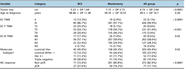
Table 1-Main characteristics of the patients submitted to NC.
Variable Category BCS Mastectomy All group p
Tumor size cm 5.23±DP 1.64 7.15±DP 2.73 6.73±DP 2.64 o0.0001
Age at diagnosis years 48.48±DP 11.38 49.76±DP 10.55 49.5±DP 10.7 0.30
EC TNM II 13 (13.3%) 10 (2.9%) 23 (5.1%) o0.0001
III 85 (86.7%) 341 (97.1%) 426 (94.9%)
EC-T TNM T2 25 (25.5%) 18 (5.1%) 43 (9.6%)
T3 53 (54.1%) 178 (50.7%) 231 (51.4%) o0.001
T4 20 (20.4%) 155 (44.2%) 175 (3.9%)
EC-N TNM N0 17 (17.3%) 26 (7.4%) 43 (9.6%) 0.02
N1 56 (57.1%) 207 (59.0%) 263 (58.6%)
N2 22 (22.4%) 105 (29.9%) 127 (28.3%)
N3 3 (3.1%) 13 (3.7%) 16 (3.6%)
Molecular Luminal/ Her- 45 (45.9%) 158 (45.3%) 203 (45.4%) 0.02
Subtype* Luminal B/Her+ 12 (12.2%) 93 (26.6%) 105 (23.5%)
Her 2+ 11 (11.2%) 41 (11.7%) 52 (11.6%)
Triple negative 30 (30.6%) 57 (16.3%) 87 (19.5%)
NC response Non pCR 71 (72.4%) 301 (85.8%) 372 (82.9%) o0.0001
pCR 27 (27.6%) 50 (14.2%) 77 (17.1%)
which is based on the response of the solid mass, residual multifocal disease, and no residual tumor (absence of tumor and in situ carcinoma). Stable disease was added to this clas-sification, and we considered it a morphologic classification. The response evaluation criteria in solid tumors (RECIST) radiologic classification cutoff values were adapted for 1-dimensional pathologic breast assessment [(RECIST-breast (RECIST-B)], with the cutoff points for 1-dimensional invasive breast disease established as 30% for partial response and 10% for progressive disease (16). A pCR was defined as the absence of invasive disease in the breast and axilla.
Follow-up
The patients were selected based on clinical and radiologic aspects before and after NC. The selected patients were sub-jected to surgery, which was performed by a team compris-ing 5 surgical oncologists and 1 breast cancer specialist. The team’s preferential approach was the resection of the entire disease area prior to NC (17). The planning was based on tumor marking performed prior to NC, clinical assessment, and radiologic assessment before and after NC. The frozen section analyses were available during the surgeries. All patients were assessed by multidisciplinary staff during the postoperative period.
The patients were assessed every 6 months for 5 years, and then once per year for an additional 5 years. The total follow-up period was counted from the first visit to the last available day. Breast clinical and radiological annual evaluations were performed. In the presence of clinical signs, symptoms, or proven recurrence, the patient was submitted for new radio-logical staging. The time between the first evaluation to the surgery was evaluated. Disease-free survival (DFS) was defi-ned as the time elapsed from the performance of quad-rantectomy to either the recurrence of disease or the last follow-up visit. All patients who missed 2 scheduled visits were considered lost to follow-up.
Endpoints
The response to chemotherapy, IBTR, and LRR were asses-sed. IBTR was defined as a local relapse of breast cancer, even in the case of secondary breast local invasion. LRR was defi-ned as local relapse associated with ipsilateral regional lymph node disease. Metachronous contralateral breast tumors were not considered to be recurrences. In the presence of recurrence, the patients were submitted to thoracic and abdominal tomog-raphy associated with bone scintigtomog-raphy.
Molecular subtypes
Systematic assessment of samples in 10% neutral buffe-red formalin blocks was performed using a tissue microarray (TMA). The approximate molecular subtypes were assessed by immunohistochemistry. The tumor markers ER SP1, anti-PR 1E2, anti-HER2/neu (clone 4B5), and anti-Ki-67 (clone 30-9) were used (Roche Diagnostics). Tumors were rated ER/PR-positive when nuclear labeling was evident in more than 1% of tumor cells. A semi-quantitative score was used for HER2 staining. A score of 0, 1+, or 2+ was considered negative staining, and a score of 3+ was considered positive staining. Fluorescence in situ hybridization (FISH) was performed for samples with a 2+score. The approximate molecular subtypes were clustered as a function of the immunohistochemical results as follows: luminal A (ER/PR-positive, HER2-negative, and Ki-67o14), luminal B/HER-negative (ER/PR-positive,
HER2-negative, and Ki-67X14), luminal B/HER-positive (ER/
PR-positive and HER2-positive), HER2 (ER/PR-negative and positive), and triple negative (ER/PR-negative and HER2-negative). In the absence of TMA information, the primary immunohistochemical lamina was reviewed. Discrepancies were discussed until reaching consensus. To evaluate the analysis, luminal A and luminal B/HER-negative samples were grouped into the luminal/HER-negative group.
Statistical analysis
The data were collected, standardized, tabulated, and analyzed using the SPSS 20.0 software for Macs
(Armonk, New York, NY). Univariate analyses of the categorical var-iables related to local and locoregional DFS were performed using the Kaplan-Meier method. The difference between the curves was assessed with the log-rank method. A Cox model was used to identify variables independently associated with local and locoregional DFS. We evaluated continuous vari-ables without dichotomization. For Cox modeling, we evalua-ted categorical interest variables and variables exhibiting po0.10 in the univariate analysis. An exploratory model was used for Cox multivariate analyses. The significance level was set topo0.05.
Ethics
The study was approved by the Research Ethics Commit-tee, no. 135/2008.
STROBE statement
This study adhered to the STROBE guidelines for cohort studies.
’ RESULTS
Ninety-eight patients with LABC who had undergone NC and BCS were evaluated. Tumor size, clinical TNM stage, pT-TNM, and pN-TNM were lower, whereas the incidence of triple-negative tumors was higher in the BCS group than the mastectomy group; age did not significantly differ between these groups (Table 1). The average age of patients who had undergone BCS was 48.5 years old, and the average duration of complaint was 8 months. Bilateral tumors were identified in 2% of samples. The average diameter of the tumors was 5.3 cm (2 to 8.5 cm). Clinical-radiologic staging was performed for all patients, but 4 patients (4.2%) were classified as stage IIa (T2N0) after initial radiologic examination and were further analyzed. Bone scintigraphy was performed for all patients. In 58.2% of patients, staging was based on chest radiographs and abdominal ultrasound, whereas it was based on thoracic and abdominal computed tomography in the remaining patients. All patients underwent mammography with breast ultrasound (64.3%) or ultrasound and magnetic resonance imaging (MRI) (23.5%). Lesions were preoperatively marked on the skin in 23.5% patients. The main characteristics related to tumor staging and treatment are described in Table 2.
Chemotherapy treatment included the 4AC+4T (81.6%) and 4AC+12T (11.2%) regimens, and the standard regimen was modified in 7.1% of patients because of toxicity (4.1%) or disease progression (3.1%).
Surgery
quadrantectomy, and oncoplastic surgery was performed in 26.5% of patients, which was distributed as follows: cen-tral quadrantectomy (8.1%), rotation flap (7.1%), periareolar (5.1%), inferior pedicle (4.1%), and superior pedicle (2.0%). Level III axillary lymph node dissection was performed in 97.0% of patients, the sentinel lymph node was investigated in 2.0% of patients, and no axillary approach was employed in 1.0% of patients.
The margins were tumor-free in all patients, and 81.6% of patients harbored tumors measuring 12.3 mm (1 to 40 mm) on average. Moreover, 13.3% of patients had pCR, and the margins were not evaluated. In 5.1% of patients (5), the margins were considered free, and the distance measure-ment was not evaluated. The average weight of the surgi-cal specimens was 233 g (41.5 to 980 g). The average number of dissected lymph nodes reported during pathologic evaluation was 18.5 (4 to 42). Table 3 presents the response to NC.
Adjuvant treatment
With regard to adjuvant therapy, 98% of patients received radiotherapy to the chest wall (5040 cGy) and a boost to the breast (1000 cGy) near the incision. Radiotherapy to the sup-raclavicular fossa was performed in 89.4% of patients. Two patients did not receive radiotherapy, 1 because of rapid disease progression and the other refused because of clau-strophobia. Hormonal therapy was administered to 57.1% of patients and consisted of tamoxifen alone, anastrozole alone, or a combined regimen in 35.7%, 3%, and 18.4% of patients, respectively. Adjuvant trastuzumab was administered to only 2.0% of patients.
Follow-up
The average duration of follow-up was 64.1 months (13.4 to 105.7 months), with the follow-up period decreasing to 55.8 months (3.6 to 95.7 months) after surgery. After the exclusion of deaths due to disease, the average total duration
Table 2-Univariate analysis of factors related to local and locoregional recurrence -free survival.
Category Variable n (%) Local p Locoregional p
60 months DFS 60 months DFS Pre-operative
EC TNM II 13 (13.3) 77.1 0.54 67.5 0.37
III 85 (86.7) 89.5 82.8
ECT - TNM T2 25 (25.5) 78.6 0.53 66.8 0.23
T3 53 (54.1) 89.2 85.6
T4 20 (21.4) 95.0 95.0
ECN - TNM N0 17 (17.3) 100.0 0.71 100.0 0.40
N1 56 (57.1) 85.2 81.0
N2-3 25 (25.5) 87.2 77.8
Tumor marking Absent 75 (76.5) 86.0 0.68 79.9 0.37
Present 23 (23.5) 95.0 95.0
Histologic type CDI 90 (89.8) 89.5 0.13 84.1 0.31
CLI+other 8 (8.2) 70.0 70.0
Nottinghan grade* G1+2 56 (56.4) 85.1 0.29 76.4 0.07
G3 40 (41.7) 91.2 91.2
Necrosis* Absent 60 (61.2) 81.9 0.039 73.7 0.008
Present 36 (37.5) 97.1 97.1
Peritumoral Slight 56 (57.2) 89.9 0.80 85.2 0.68
Infiltration* Moderate/intense 10 (40.8) 85.0 79.4
Lymphatic Absent 85 (88.5) 86.6 0.29 82.1 0.75
embolization* Present 11 (11.5) 100.0 90.9
ER Negative 45 (45.9) 86.9 0.50 82.7 0.90
Positive 53 (54.1) 88.9 83.4
PR Negative 52 (53.1) 88.6 0.80 86.7 0.49
Positive 46 (46.9) 86.9 78.8
Her2 Positive 23 (23.5) 86.1 0.79 80.0 0.41
Negative 75 (76.5) 88.8 89.0
Molecular Luminal / Her - 45 (45.9) 85.5 0.63 79.0 0.40
Subtype Luminal B Her+ 12 (12.2) 91.7 91.7
Her2 11 (11.2) 78.8 79.0
Triple negative 30 (30.6) 93.2 89.8
Postoperative
Oncoplastic Absent 72 (73.5) 85.0 0.50 79.7 0.52
surgery Present 26 (26.5) 96.0 92.0
RECIST-B Complete response 31 (31.6) 92.6 0.16 92.7 0.003
Partial response 64 (64.3) 86.9 80.6
Stable disease 3 (3.1) 66.7 33.3
Morphology Solid mass 50 (51.0) 89.9 0.04 82.7 0.03
MDA (11) Multifocal disease 13 (13.3) 77.3 69.5
Without disease 31 (31.6) 92.0 92.1
Stable disease 4 (4.1) 75.0 50.0
pCR/ NSABP Absent 71 (72.4) 87.3 0.58 80.6 0.28
Present 27 (27.6) 90.4 90.4
of follow-up was 72.8 months (34.4 to 105.7 months) and 64.1 months (26.3 to 95.9 months) after surgery. Moreover, 6.1% of patients (6) were considered lost of follow-up, and 5/6 patients exhibited DFS with a mean time of 39.4 months (26.3-48.6 months).
By the end of the follow-up period, 19.5% of the patients had died from breast cancer, 5.1% had died from other causes, 7.1% were living with cancer, and 68.4% were alive and free of disease. The main sites of metastases were in the bones (16.3%), lungs (13.3%), liver (8.2%), and brain (4.1%). The overall actuarial survival (OS) rates at 36, 60, and 96 months were 87.7%, 81.2%, and 71.4%, respectively (Figure 1).
Recurrence
The average months after surgery for recurrence vary from 1.8 to 81.3 months and was 26.4, 26.8 and 27.1 months in general, IBTR and LRR, respectively. At 36 months 72.4% of the general recurrence occurred, 83.3% of the IBTR and 76.5% of the LRR. The overall DFS rates at 36, 60 and 96 months were 77.9%, 68.9% and 67.0%, respectively (Figure 1).
The IBTR rate was 11.2%. IBTR was classified as local recurrence associated with systemic disease (3.1%), local plus LRR (3.1%), breast recurrence alone (3.1%), and secondary breast invasion from sternal recurrence (2.0%). When asses-sing disease progression, some patients exhibited rapidly progressive disease with early local recurrence plus LRR (2.0%), sternal recurrence extending to the breast (2.0%), breast recurrence alone (3.1%), and multiple local recurrence plus LRR (4.1%). Excluding the instance of local sternal recurrence that infiltrated the breast, the primary recurrence rate was 9.3%.
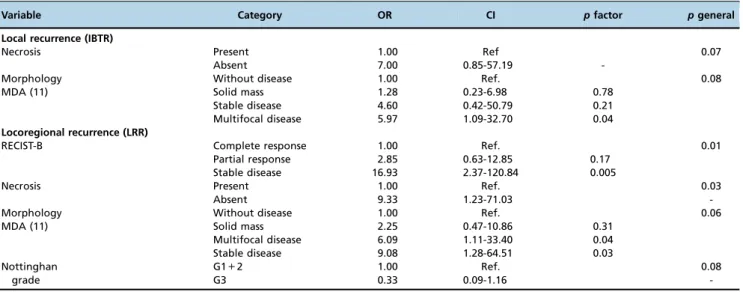
A univariate analysis of local DFS relative to the categorical variables (Table 1) showed that the absence of necrosis (p=0.04) and the morphologic response to che-motherapy characterized by multifocal disease and stable disease were associated with poorer survival (p=0.04). Neither age [p=0.33, risk ratio 1.01, confidence interval (CI) 0.99-1.03] nor initial tumor size (p=0.35, risk ratio 1.06, CI 0.93-1.20) influenced IBTR DFS. The Cox univariate analysis, which was used to explore factors possibly related to IBTR
DFS (Table 2), showed that multifocal morphology was the only factor associated with IBTR because it increased the IBTR 5.97-fold (p=0.04). Figure 1 shows the overall and morphology factor risk curves related to the hazard risk of local DFS.
The LRR rate was 15.3% and was distributed as follows: ipsilateral supraclavicular fossa (4.0%), ipsilateral axilla (3.1%), local and systemic recurrence (3.1%), breast rence alone (3.1%), and breast associated with sternal recur-rence (2.0%).
A univariate analysis of the categorical variables relative to locoregional DFS (Table 1) showed that the pathologic RECIST-B response (p=0.003), necrosis (p=0.008), and mor-phology were related to locoregional DFS. Neither age (p=0.41, risk ratio 1.00, CI 0.98-1.03) nor initial tumor size (p=0.27, risk ratio 1.08, CI 0.94-1.23) influenced LRR DFS. The Cox univariate analysis showed that the absence of tumor necrosis at diagnosis increased the LRR 9.33-fold (p=0.03), multifocal morphology increased the risk of LRR 6.09-fold (p=0.04), and stable disease increased the risk of LRR 9.08-fold (p=0.03). However, the RECIST-B pathologic response was the main factor related to locoregional DFS (p=0.01) because stable RECIST-B disease increased the risk of LRR 16.93-fold (p=0.005). An exploratory Cox multi-variate analysis model showed that RECIST-B pathologic response was the only factor related to locoregional DFS. Figure 2 shows the curves related to the hazard risk of locoregional DFS.
’ DISCUSSION
NC provides global survival similar to adjuvant che-motherapy, with the added advantage of identifying patients with better prognoses, that is, patients who exhibit pCR in addition to increasing the rates of BTC (18). The primary indication for NC is larger tumors or tumors with higher rates of lymph node involvement. When comparing patients treated with NC and BCS with patients subjected to mas-tectomy, the former have a lower T-TNM stage at diagnosis; higher rates of pCR; and higher rates of ER-negative, PR-negative, and triple-negative tumors, indicating bias in the
Table 3-Cox analysis of factors related to local and locoregional recurrence-free survival.
Variable Category OR CI pfactor pgeneral
Local recurrence (IBTR)
Necrosis Present 1.00 Ref 0.07
Absent 7.00 0.85-57.19
-Morphology Without disease 1.00 Ref. 0.08
MDA (11) Solid mass 1.28 0.23-6.98 0.78
Stable disease 4.60 0.42-50.79 0.21
Multifocal disease 5.97 1.09-32.70 0.04
Locoregional recurrence (LRR)
RECIST-B Complete response 1.00 Ref. 0.01
Partial response 2.85 0.63-12.85 0.17
Stable disease 16.93 2.37-120.84 0.005
Necrosis Present 1.00 Ref. 0.03
Absent 9.33 1.23-71.03
-Morphology Without disease 1.00 Ref. 0.06
MDA (11) Solid mass 2.25 0.47-10.86 0.31
Multifocal disease 6.09 1.11-33.40 0.04
Stable disease 9.08 1.28-64.51 0.03
Nottinghan G1+2 1.00 Ref. 0.08
grade G3 0.33 0.09-1.16
analysis of this subgroup. This bias may influence the rates of recurrence and survival (19). In our group of patients, patients who underwent BCS exhibited better survival than the mastectomy group (p=0.002), which corroborated pre-vious reports. Bias selection likely occurred based on tumor size, breast-tumor relation, response to NC, and molecular subtype (Table 1) because BCS was performed in patients with smaller tumors, lower clinical TNM stage, and a better response to NC. Because the characteristics differed between groups, we only evaluated patients who underwent BCS. Bleicher et al. evaluated Surveillance, Epidemiology, and End Results (SEER) data from a cohort of 5,685 patients aged466
years old with tumors45 cm. Of these patients, 887 (15.6%)
underwent BCS, and only 205 (3.6%) received NC. BCS was associated with a lower clinical stage and more NC, but only 101 patients received both NC and BCS, and these patients were not evaluated separately (20). Our study represents one of the largest institutional retrospective cohort studies of LABC treated with NC and BCS (11).
BCS is safe provided that the excision margins are free of disease and this treatment is combined with adjuvant radio-therapy to the breast (21, 22). BCS was initially used to treat tumors smaller than 3 cm associated with a 1-cm free margin.
These criteria are changing, and smaller margins are currently accepted (23). A meta-analysis of randomized controlled trials showed that BCS is a safe treatment for patients with clinical stage I and II disease and tumors smaller than 5 cm (24). SEER data evaluated for tumors45 cm indicated that breast
cancer-specific survival did not differ between patients who received BCS and patients who underwent a mastectomy, but the women in this study were older, the IBTR and molecular subtype were not evaluated, and few patients received NC (20). The rate of conservative surgery after NC varies from 37% to 82% (6, 7); however, only 1.7% to 28% of patients exhibit LABC (7, 8). The LABC candidates who were initially selected were patients without skin or chest wall involve-ment and who were free of multicentric disease or extensive microcalcifications. They harbored tumors smaller than 5 cm, exhibited favorable tumor localization, had no contraindica-tions for radiotherapy, and had negative margins. Primary inflammatory carcinoma is a contraindication for BCS (25). Patients with N2-3 lymph nodes, residual tumors 42 cm,
residual multifocal components, and the presence of lym-phovascular embolization should be cautiously assessed because of the higher risk of IBTR (11, 26). Therefore, the cutaneous infiltration criteria have become more flexible
for localized cutaneous infiltration and the breast/tumor volume ratio, and the initial indications for oncoplastic surgery have been expanded (17). Although the average size of the initial tumors was 5.3 cm (varying from 2 to 8.5 cm) in the present cohort, the margins were disease-free in 100% of cases, with a distance to the tumor of 12.3 mm. In addition, oncoplastic techniques were used in 26.5% of patients, which supports the use of BCS in selected cases of LABC.
The preoperative planning was based on clinical-radiolo-gic data and operative freezing. Two patients were excluded from the cohort because of positive surgical margins, which resulted in conversion to mastectomy.
Diagnostic imaging tests are essential to pretreatment therapeutic planning (3) and were performed before and after the administration of NC in 100% and 87.7% of patients, respectively. Although not shown numerically, a tendency toward the resection of the entire tumor bed before NC was observed in this study. Not all patients who exhibited a com-plete clinical response (21) reached pCR, and the anatomic-pathologic assessment is not always uniform. In this regard, the‘‘Residual Cancer Burden’’method renders the resection of the full area necessary prior to NC (27), but it is used in
prospective studies. Pathologic sampling interferes with the pathologic results. In the present study, the average number of blocks per surgical specimen was 20, but a consensus for pathologic evaluation was obtained in 2015 (28).
Upon the assessment of patients subjected to BCS and radiotherapy, we should consider studies of patients who did not receive NC that demonstrate the long-term safety of BCS. For example, Veronesi (9) assessed tumors smaller than 2 cm and identified a recurrence rate of 8% at 20 years, whereas Fisher (NSAPB-B06), who assessed tumors smaller than 4 cm, reported recurrence rates at 20 years of 14.3% for patients who underwent lumpectomy and breast radiation and 39.2% for patients who did not receive radiation (21). In patients subjected to NC and BCS, this rate was reported to be 14% at 5.8 years (29), 19% at 4.6 years (15), and 21.5% at 20 years (30); however, the assessed tumors differed diagnostically and in their initial staging (19). Therefore, the possibility of new surgical margins remains open for discussion, but case-control studies assessing locally advanced tumors are lacking. NSABP B-27, which assessed patients with T1c-3N0 or T1-3N1M0 disease, was designed to evaluate the addition of taxanes to anthracyclines and reported an average tumor size of 4.4 cm and a 6% IBTR rate at 102 months; however,
only 30% of cases exhibited lymph node involvement. In the present cohort, the average tumor size was 5.3 cm, and 87.2% of tumors were larger than 3 cm; 88.9% of patients were diagnosed with stage III disease, 74.5% of patients harbored stage T3-4 disease, and 82.6% of patients had stage N1-3 disease. The IBTR rate was 11.2% at 64.1 months. Although this rate is high, it is lower than the rate reported in a study by Fisher of patients subjected exclusively to lumpectomy without radiotherapy (21). These findings demonstrate the effectiveness of BCS in patients with LABC subjected to NC and adjuvant radiotherapy.
In the assessment of IBTR, we must discriminate true recurrence at the surgical site, ipsilateral second primary tumors, and ipsilateral thoracic wall tumors (31). Although ipsilateral thoracic wall events involving the sternal bone were defined as a distant event in 2014 (31), previous studies with long follow-up period did not specify this form of recurrence (32). In the present study, we observed 2 patients with simultaneous IBTR and sternal infiltration, but 1 patient underwent local full-thickness chest wall resection. We opted to consider this case as local recurrence to better compare our results to those of other studies with long follow-up periods. No pattern is associated with the type of local recurrence, but many recurrences are defined as multiple recurrence. Alternatively, recurring tumors may indicate resistance to treatment and subsequent multiple recurrences. In the present study, the LRR rate was 15.3% and consisted of all patients with local recur-rence and the 4 patients with locoregional lymph node involve-ment. This finding corroborates the analysis of the DFS results. The chi-squared test may be used to calculate recurrence, but we also assessed DFS because recurrence depends on time. Several factors are associated with IBTR and LRR. Better results were observed in patients who showed an early response to treatment (33) and were positive for hor-monal receptors (12); poorer outcomes were reported for patients with lymphovascular invasion(11), residual tumors larger than 2 cm (11), multifocal disease after chemotherapy (11, 34), no expression of hormonal receptors, stage III and N2-3 axillary nodal status (15), age p40 years old, excision marginsp2 mm, and S-phase fraction44% (30).
Few studies have examined a sufficient number of cases to assess the rates of recurrence in patients subjected to NC and BTC (11, 33, 34). In the present study, which evaluated a large sample over a long follow-up period, the morphologic res-ponse, although valid, was not significantly associated with recurrence, whereas the RECIST-B response was shown to have prognostic value in a multivariate analysis. The response to NC can be classified into several categories (35). Chen et al. suggested a morphologic classification of the response, show-ing that the response correlates with the occurrence of IBTR (11). In the present study, morphologic assessment showed an association, albeit a non-significant one, between the presence of multifocal disease/stable disease and higher rates of IBTR and LRR. The RECIST-B pathologic response (p=0.02) was the only variable retained in the multivariate model related to LRR: the risk was 2.85 times higher among patients with a partial response (p=0.17) and 16.93 times higher among patients with stable disease (p=0005).
In the present cohort, the absence of necrosis in the pretreatment biopsy sample was the only histological factor that was associated with IBTR and LRR. This finding is corroborated by other studies, which detected an association between complete response and the presence of necrosis (36). The absence of necrosis was associated with a 7.00-fold higher
rate of IBTR, but this increase was not significant (p=0.07). It was also associated with a 9.33-fold higher rate of LRR (p=0.03) in univariate analysis. The only significant factor related do LRR in multivariate analysis was the RECIST-B response.
In the assessment of IBTR, other variables, such as the type of tumor fragmentation and the presence of surgical margins, should also be considered. Thus, the rates of IBTR were reported to be 12.7% and 20.3% in the presence and absence of tumor-free margins, respectively (30). In the present study, all patients exhibited tumor-free margins. In a previous study, the presence of multifocal disease increased the risk of IBTR 3.3-fold (12), which is corroborated by this study: multifocal disease increased the rate of IBTR 5.97-fold (p=0.04). In addition, the molecular subtypes are related to the rate of recurrence in an adjuvant (37) and neoadjuvant setting (38). Specifically, the rates of recurrence were 0.8% for luminal tumors (ER/PR-positive and HER2-negative), 1.5% for luminal B tumors (ER/PR-positive and HER2-positive), 8.4% for HER2 tumors (ER/PR-negative and HER2 positive), and 7.1% for triple-negative tumors.
A possible limitation of the present study is the retro-spective and nonrandomized design in which cases were selected in a continuous manner, that is, based on the feasibility of BCS. Thus, multiple elements influenced the selection of patients, including age, comorbidities, breast-volume ratio, and response to NC. Another limitation of this study is the absence of NC association with trastuzumab, which may affect the pCR, OS, and DFS (39). Because HER2 tumors represent 23.5% (29) of patients, we observed only 3 incidences of local recurrence/LRR in this group and a 0.487-fold reduction in the recurrence rate (39). The addition of trastuzumab would slightly decrease the overall recurrence rate, but this drug was not used by the Brazilian Public Health System at the time of NC treatment.
The present study corroborates the fact that in cases selected by clinical and radiologic findings with a satisfac-tory response to NC, BCS is feasible and safe for the treat-ment of locally advanced tumors, provided that the tumor is completely resected, surgical margins are clear, and patients are subjected to complementary multimodal treatment. This finding was corroborated by the occurrence of acceptable rates of local recurrence and LRR.
’ ACKNOWLEDGMENTS
We are grateful to Fundac¸ão de Amparo a Pesquisa do Estado de São
Paulo (FAPESP), which provided grants for this study. Project number 2012/19642-0.
’ AUTHOR CONTRIBUTIONS
Carrara GF participated in the data review, data analysis, and writing of the manuscript. Scapulatempo-Neto C and Abrahão-Machado LF performed the pathological review and reviewed thefinal version of the manuscript. Brentani MM and Folgueira MA participated in the study design, data analysis, and review of thefinal version of the manuscript. Nunes JS participated in the case selection, chemotherapy, submission to the Ethics Committee, and review of thefinal version of the manuscript. Vieira RA participated in the study design, submission to the Ethics Committee, case selection, surgery, data analysis, and writing of the manuscript.
’ REFERENCES
2. Vieira RA, Uemura G, Zucca-Matthes G, Costa AM, Micheli RA, Oliveira CZ. Evaluating Breast Cancer Health System Between Countries: The Use of USA/SEER and Brazilian Women as a Cohort Sample. Breast J. 2015; 21(3):322-3, http://dx.doi.org/10.1111/tbj.12410.
3. Caudle AS, Kuerer HM. Breast conservation therapy after neoadjuvant chemotherapy: optimization of a multimodality approach. J Surg Oncol. 2014;110(1):32-6, http://dx.doi.org/10.1002/jso.23595.
4. Mauri D, Pavlidis N, Ioannidis JP. Neoadjuvant versus adjuvant systemic treatment in breast cancer: a meta-analysis. J Natl Cancer Inst. 2005; 97(3):188-94, http://dx.doi.org/10.1093/jnci/dji021.
5. Fisher B, Brown A, Mamounas E, Wieand S, Robidoux A, Margolese RG, et al. Effect of preoperative chemotherapy on local-regional disease in women with operable breast cancer: findings from National Surgical Adjuvant Breast and Bowel Project B-18. J Clin Oncol. 1997;15(7):2483-93, http://dx.doi.org/10.1200/JCO.1997.15.7.2483.
6. van der Hage JA, van de Velde CJ, Julien JP, Tubiana-Hulin M, Vander-velden C, Duchateau L. Preoperative chemotherapy in primary operable breast cancer: results from the European Organization for Research and Treatment of Cancer trial 10902. J Clin Oncol. 2001;19(22):4224-37, http://dx.doi.org/10.1200/JCO.2001.19.22.4224.
7. Scholl SM, Asselain B, Palangie T, Dorval T, Jouve M, Garcia Giralt E, et al. Neoadjuvant chemotherapy in operable breast cancer. Eur J Cancer. 1991;27(12):1668-71, http://dx.doi.org/10.1016/0277-5379(91)90442-G. 8. Wolmark N, Wang J, Mamounas E, Bryant J, Fisher B. Preoperative
che-motherapy in patients with operable breast cancer: nine-year results from National Surgical Adjuvant Breast and Bowel Project B-18. J Natl Cancer Inst Monogr. 2001(30):96-102, http://dx.doi.org/10.1093/oxfordjournals. jncimonographs.a003469.
9. Veronesi U, Cascinelli N, Mariani L, Greco M, Saccozzi R, Luini A, et al. Twenty-year follow-up of a randomized study comparing breast-conser-ving surgery with radical mastectomy for early breast cancer. N Engl J Med. 2002;347(16):1227-32, http://dx.doi.org/10.1056/NEJMoa020989. 10. Gianni L, Baselga J, Eiermann W, Guillem Porta V, Semiglazov V, Lluch A,
et al. Feasibility and tolerability of sequential doxorubicin/paclitaxel fol-lowed by cyclophosphamide, methotrexate, and fluorouracil and its effects on tumor response as preoperative therapy. Clin Cancer Res. 2005;11(24 Pt 1):8715-21, http://dx.doi.org/10.1158/1078-0432.CCR-05-0539.
11. Chen AM, Meric-Bernstam F, Hunt KK, Thames HD, Oswald MJ, Outlaw ED, et al. Breast conservation after neoadjuvant chemotherapy: the MD Anderson cancer center experience. J Clin Oncol. 2004;22(12):2303-12, http://dx.doi.org/10.1200/JCO.2004.09.062.
12. Ishitobi M, Ohsumi S, Inaji H, Ohno S, Shigematsu H, Akiyama F, et al. Ipsilateral breast tumor recurrence (IBTR) in patients with operable breast cancer who undergo breast-conserving treatment after receiving neoad-juvant chemotherapy: risk factors of IBTR and validation of the MD Anderson Prognostic Index. Cancer. 2012;118(18):4395-93, http://dx.doi. org/10.1002/cncr.27377.
13. James T, McCahill L, Ratliff J, Ashikaga T, Single R, Sheehey-Jones J, et al. Quality assessment of neoadjuvant therapy use in breast conservation: barriers to implementation. Breast J. 2009;15(5):524-6, http://dx.doi.org/ 10.1111/j.1524-4741.2009.00771.x.
14. Morrow M. Margins in breast-conserving therapy: have we lost sight of the big picture? Expert Rev Anticancer Ther. 2008;8(8):1193-6, http://dx. doi.org/10.1586/14737140.8.8.1193.
15. Min SY, Lee SJ, Shin KH, Park IH, Jung SY, Lee KS, et al. Locoregional recurrence of breast cancer in patients treated with breast conservation surgery and radiotherapy following neoadjuvant chemotherapy. Int J Radiat Oncol Biol Phys. 2011;81(5):e697-705, http://dx.doi.org/10.1016/ j.ijrobp.2010.10.014.
16. Khokher S, Qureshi MU, Chaudhry NA. Comparison of WHO and RECIST criteria for evaluation of clinical response to chemotherapy in patients with advanced breast cancer. Asian Pac J Cancer Prev. 2012; 13(7):3213-8, http://dx.doi.org/10.7314/APJCP.2012.13.7.3213. 17. Zucca Matthes AG, Uemura G, Kerr L, Matthes AC, Michelli RA,
Folgueira MA, et al. Feasibility of oncoplastic techniques in the surgical management of locally advanced breast cancer. Int J Surg. 2012;10(9): 500-5, http://dx.doi.org/10.1016/j.ijsu.2012.07.009.
18. Hennequin C, Espie M, Misset JL, Maylin C. [Does primary chemotherapy really increase the rate of breast conserving treatments?]. Cancer Radio-ther. 2004;8(1):48-53.
19. Cho JH, Park JM, Park HS, Park S, Kim SI, Park BW. Oncologic safety of breast-conserving surgery compared to mastectomy in patients receiving neoadjuvant chemotherapy for locally advanced breast cancer. J Surg Oncol. 2013;108(8):531-6, http://dx.doi.org/10.1002/jso.23439. 20. Bleicher RJ, Ruth K, Sigurdson ER, Daly JM, Boraas M, Anderson PR,
et al. Breast conservation versus mastectomy for patients with T3 primary tumors (45 cm): A review of 5685 medicare patients. Cancer. 2016; 122(1):42-9, http://dx.doi.org/10.1002/cncr.29726.
21. Fisher B, Anderson S, Bryant J, Margolese RG, Deutsch M, Fisher ER, et al. Twenty-year follow-up of a randomized trial comparing total mas-tectomy, lumpectomy, and lumpectomy plus irradiation for the treatment of invasive breast cancer. N Engl J Med. 2002;347(16):1233-41, http://dx. doi.org/10.1056/NEJMoa022152.
22. Veronesi U, Saccozzi R, Del Vecchio M, Banfi A, Clemente C, De Lena M, et al. Comparing radical mastectomy with quadrantectomy, axillary dis-section, and radiotherapy in patients with small cancers of the breast. N Engl J Med. 1981;305(1):6-11, http://dx.doi.org/10.1056/NEJM198107 023050102.
23. Houssami N, Macaskill P, Marinovich ML, Morrow M. The association of surgical margins and local recurrence in women with early-stage invasive breast cancer treated with breast-conserving therapy: a meta-analysis. Ann Surg Oncol. 2014;21(3):717-30, http://dx.doi.org/10.1245/s10434-014-3480-5.
24. Yang SH, Yang KH, Li YP, Zhang YC, He XD, Song AL, et al. Breast conservation therapy for stage I or stage II breast cancer: a meta-analysis of randomized controlled trials. Ann Oncol. 2008;19(6):1039-44, http://dx.doi.org/10.1093/annonc/mdm573.
25. Buchholz TA, Hunt KK, Whitman GJ, Sahin AA, Hortobagyi GN. Neoadjuvant chemotherapy for breast carcinoma: multidisciplinary con-siderations of benefits and risks. Cancer. 2003;98(6):1150-60, http://dx. doi.org/10.1002/cncr.11603.
26. Singletary SE, McNeese MD, Hortobagyi GN. Feasibility of breast-con-servation surgery after induction chemotherapy for locally advanced breast carcinoma. Cancer. 1992;69(11):2849-52, http://dx.doi.org/10.1002/1097-0142(19920601)69:11o2849::AID-CNCR282069113443.0.CO;2-P. 27. Symmans WF, Peintinger F, Hatzis C, Rajan R, Kuerer H, Valero V, et al.
Measurement of residual breast cancer burden to predict survival after neoadjuvant chemotherapy. J Clin Oncol. 2007;25(28):4414-22, http://dx. doi.org/10.1200/JCO.2007.10.6823.
28. Provenzano E, Bossuyt V, Viale G, Cameron D, Badve S, Denkert C, et al. Standardization of pathologic evaluation and reporting of post-neoadjuvant specimens in clinical trials of breast cancer: recommenda-tions from an international working group. Mod Pathol. 2015;28(9):1185-201, http://dx.doi.org/10.1038/modpathol.2015.74.
29. Cance WG, Carey LA, Calvo BF, Sartor C, Sawyer L, Moore DT, et al. Long-term outcome of neoadjuvant therapy for locally advanced breast carcinoma: effective clinical downstaging allows breast preservation and predicts outstanding local control and survival. Ann Surg. 2002;236(3): 295-302, http://dx.doi.org/10.1097/00000658-200209000-00006. 30. Rouzier R, Extra JM, Carton M, Falcou MC, Vincent-Salomon A, Fourquet
A, et al. Primary chemotherapy for operable breast cancer: incidence and prognostic significance of ipsilateral breast tumor recurrence after breast-conserving surgery. J Clin Oncol. 2001;19(18):3828-35, http://dx.doi.org/ 10.1200/JCO.2001.19.18.3828.
31. Moossdorff M, van Roozendaal LM, Strobbe LJ, Aebi S, Cameron DA, Dixon JM, et al. Maastricht Delphi consensus on event definitions for classification of recurrence in breast cancer research. J Natl Cancer Inst. 2014;106(12)., doi: http://dx.doi.org/10.1093/jnci/dju288.
32. Cebrecos I, Cordoba O, Deu J, Xercavins J, Rubio IT. Can we predict local recurrence in breast conserving surgery after neoadjuvant chemotherapy? Eur J Surg Oncol. 2010;36(6):528-34, http://dx.doi.org/10.1016/j.ejso. 2010.04.004.
33. Ishitobi M, Komoike Y, Motomura K, Koyama H, Inaji H. Early response to neo-adjuvant chemotherapy in carcinoma of the breast predicts both successful breast-conserving surgery and decreased risk of ipsilateral breast tumor recurrence. Breast J. 2010;16(1):9-13, http://dx.doi.org/ 10.1111/j.1524-4741.2009.00864.x.
34. Ishitobi M, Ohsumi S, Inaji H, Ohno S, Shigematsu H, Akiyama F, et al. Ipsilateral breast tumor recurrence (IBTR) in patients with operable breast cancer who undergo breast-conserving treatment after receiving neoad-juvant chemotherapy: Risk factors of IBTR and validation of the M. D. Anderson Prognostic Index. Cancer. 2012;118(18):4385-93, http://dx.doi. org/10.1002/cncr.27377.
35. Bear HD, Anderson S, Smith RE, Geyer CE Jr, Mamounas EP, Fisher B, et al. Sequential preoperative or postoperative docetaxel added to preoperative doxorubicin plus cyclophosphamide for operable breast cancer:National Surgical Adjuvant Breast and Bowel Project Protocol B-27. J Clin Oncol. 2006;24(13):2019-27, http://dx.doi.org/10.1200/JCO.2005.04.1665. 36. Bhargava R, Beriwal S, Dabbs DJ, Ozbek U, Soran A, Johnson RR, et al.
Immunohistochemical surrogate markers of breast cancer molecular classes predicts response to neoadjuvant chemotherapy: a single institu-tional experience with 359 cases. Cancer. 2010;116(6):1431-9, http://dx. doi.org/10.1002/cncr.24876.
37. Voduc KD, Cheang MC, Tyldesley S, Gelmon K, Nielsen TO, Kennecke H. Breast cancer subtypes and the risk of local and regional relapse. J Clin Oncol. 2010;28(10):1684-91, http://dx.doi.org/10.1200/JCO.2009.24.9284. 38. Meyers MO, Klauber-Demore N, Ollila DW, Amos KD, Moore DT, Drobish AA, et al. Impact of breast cancer molecular subtypes on locor-egional recurrence in patients treated with neoadjuvant chemotherapy for locally advanced breast cancer. Ann Surg Oncol. 2011;18(10):2851-7, http://dx.doi.org/10.1245/s10434-011-1665-8.