Joana Christina Carvalho(a) Heliana Dantas Mestrinho(b)
(a) School of Medicine and Dentistry, Catholic
University of Louvain, Belgium.
(b) Department of Odontology, Faculty of
Health Sciences, Universidade de Brasília - UnB, Brasília, DF, Brazil.
Corresponding Author:
Joana Christina Carvalho
E-mail: [email protected]
Diagnosing non-cavitated lesions in
epidemiological studies: practical and
scientific considerations
*Abstract: Over the last decade, there has been growing interest in di-agnosing non-cavitated lesions in epidemiological studies involving large numbers of preschool children, schoolchildren and young adults. In this context, assessment of lesions characteristics indicating whether or not there is ongoing mineral loss is also considered relevant. The reasoning sustained by these studies is that diagnosis of the caries process limited to the cavitated level is no longer in accordance with current state-of-the-art knowledge in cariology. This paper highlights one topic of the lecture en-titled “Caries Process: Evolving Evidence and Understanding,” presented at the 18th Congress of the Brazilian Association for Oral Health
Pro-motion (Associação Brasileira de Odontologia de Promoção de Saúde
- ABOPREV) in April 2013. In the framework of epidemiological
stud-ies, the interest in diagnosing active and inactive non-cavitated lesions was elucidated. However, relevant questions associated with the diagno-sis of non-cavitated lesions that might raise concerns among researchers and health administrators were not addressed. The present paper aims to bring these questions into discussion. The contribution of this discussion in terms of developing the understanding of caries decline is analyzed by using data from a caries trends study of Brazilian preschool children residing in the Federal District of Brazil as an example. The inclusion of active and inactive non-cavitated lesions in the diagnosis of the car-ies process allowed us to demonstrate that, in Brazilian 1- to 5-year-old children, caries prevalence decreased signiicantly from 1996 to 2006, simultaneously with a reduction in the rate of caries progression.
Descriptors: Dental Caries; Diagnosis; Epidemiology.
Introduction
Over the last decade, there has been growing interest in diagnosing non-cavitated lesions in epidemiological studies involving large numbers of preschool and school-age children.1-7 In this context, the assessment of
lesion characteristics indicating whether or not there is ongoing mineral loss is also considered relevant. The reasoning sustained by these studies is that diagnosis of the caries process limited to the cavitated level is no longer in accordance with current state-of-the-art knowledge in cariolo-gy. In particular, it contrasts with the current understanding of the devel-opment and arrest of the caries process at subclinical and clinical levels. One may argue that the threshold for caries diagnosis should be
tai-Declaration of Interests: The authors certify that they have no commercial or associative interest that represents a conflict of interest in connection with the manuscript.
Submitted: Jul 22, 2013
Accepted for publication: Sep 01, 2013 Last revision: Oct 18, 2013
* Paper presented at the “Equity, Social Inclusion and Oral Health Promotion: Major Challenges” International Symposium, Held at the 18th Congress of the Brazilian
Association for Oral Health Promotion (Associação Brasileira de Odontologia de Promoção de Saúde - ABOPREV), April 2013, Bauru, SP, Brazil.
lored to the aim of the epidemiological study, since arguments can be made both ways.8 This is indeed
a relevant aspect to consider because objectives may in some cases be achieved with the estimate mea-sured only at a more severe stage of caries develop-ment. The need for operative treatment at the pop-ulation or individual levels may well be measured by the diagnosis of the caries process into dentin or even into dental pulp. While this information is in itself important, when evaluated alone, it perpetu-ates the outdated concept that the caries process should be treated by restorations and extractions only. Thus, the contemporary needs for planning, delivery and monitoring of oral health care servic-es at the population level are not fulilled.9 This is
true for populations of children, adolescents, young adults and, to some extent, adults up to 45 years of age.2,3,10,11
Currently, the major challenge in the manage-ment of the caries process is to control caries pro-gression, mainly by delivering non-operative treat-ments and by limiting the number of individuals in a population subject to operative treatment.12 In this
context, it is important to bear in mind that not all non-cavitated lesions progress to dentinal lesions requiring operative treatment,8 but many of them
certainly require non-operative treatment to control further caries progression.
This paper highlights one topic of the lecture entitled “Caries Process: Evolving Evidence and Understanding” presented at the 18th Congress of
the Brazilian Association for Oral Health Promo-tion (Associação Brasileira de Odontologia de Pro-moção de Saúde - ABOPREV) in April 2013. In the framework of epidemiological studies, the interest in diagnosing active and inactive non-cavitated lesions was elucidated. However, relevant questions associ-ated with the diagnosis of non-cavitassoci-ated lesions that might raise concerns among researchers and health administrators were not addressed. The present pa-per aims to bring these questions into the discus-sion. The contribution of this discussion in terms of developing the available understanding of caries de-cline is analyzed by using data from a caries trends study in Brazilian preschool children residing in the Federal District of Brazil13 as an example.
Are the field conditions suitable for the diagnosis of non-cavitated lesions?
A major challenge in carrying out epidemiologi-cal studies is to develop the conditions required to perform a thorough clinical examination for caries and other oral health conditions as these studies are carried out at nurseries, schools, nursing homes and in-home settings. This includes training and calibration of the examiners and positioning of the participant for clinical examination, in addition to obtaining good lighting, and clean and dry teeth. The ield equipment should include at least material and instruments such as toothbrushes, dental loss, portable lights, mouth mirrors, periodontal probes, dental probes and gauze bandages. Additional mate-rial and equipment may be required according to the subject of study and the resources available to the researcher, institution or government responsible for carrying out the epidemiological survey.
Thorough clinical examination may be per-formed under ield conditions, including the diag-nosis of non-cavitated lesions, by having calibrated dentists assess caries activity and severity at the non-cavitated level, by having participants lie down on tables and having their teeth professionally brushed, lossed and dried with gauze bandage and by working with portable lights. Good lighting may be obtained by means of frontal lamps. It is recom-mended that the professional perform the brushing and lossing while the participant lies down and subsequently dry the teeth with a gauze bandage.13
Thus, ield conditions may become suitable for the diagnosis of non-cavitated lesions.
Is the diagnosis of non-cavitated lesions reliable under field conditions?
Several epidemiological studies recording non-cavitated lesions have shown good reliability of the clinical assessments.3-7,10-12 Moreover, lesion activity
assessment indicating whether or not there is ongo-ing mineral loss14,15 provides information of major
importance for treatment decisions and implementa-tion of strategies to control caries progression.
In our study,13 two calibrated examiners
Is the extra time required to record non-cavitated lesions in the field work a barrier?
Time is an important issue when it comes to epi-demiological surveys at a national level or any oth-er survey including a high numboth-er of participants. There is no doubt that more time is required to car-ry out a clinical examination that includes the re-cording of non-cavitated lesions than one limited to cavitated lesions. Depending on the system chosen, the time required to record non-cavitated lesions may be double that of the system recommended by the World Health Organisation.17
Practitioners who are dealing with the record-ing of non-cavitated lesions for the irst time spend more time than those who became familiar with it during their university training. In today’s scenario, these aspects may be seen in the light of the increas-ing number of general practitioners whose university training in diagnosis of the caries process has been updated. In Brazil, this training seems to be spread across the country. In the near future, the recording of non-cavitated lesions will become very natural as a continuation of university training and this issue will lose its importance in due time. The extra time required to record non-cavitated lesions should not be considered a barrier, but should be taken into ac-count when planning the ield work.
What is the impact of non-cavitated
lesions on caries prevalence and severity?
Our data showed signiicant differences in car-ies prevalence and severity when non-cavitated le-sions were considered in the individual’s caries ex-perience.13 In this context, it is worth mentioning
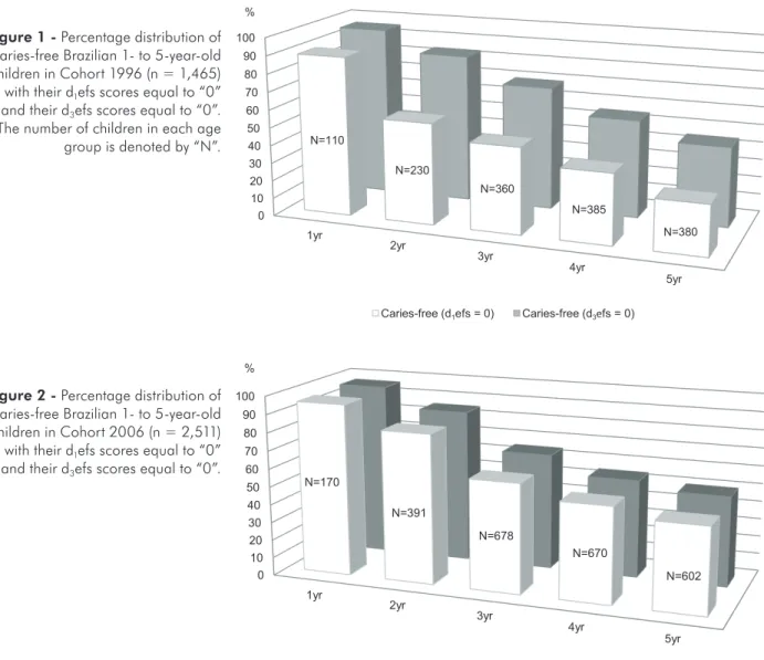
that in our studies very few children presented in-active non-cavitated lesions only. These cases were included in the group of participants classiied as caries-free. Figures 1 and 2 illustrate the percentage distribution of caries-free Brazilian 1- to 5-year-old children in Cohort 1996 (n = 1,465) and in Cohort 2006 (n = 2,511) according to two levels of exami-nation. The children were classiied as caries-free at two levels of examination. Free from any form of treated or untreated active as well as inactive car-ies lesions, i.e. d1efs = 0; and free from cavitated
Under ield conditions, the overall intra-examiner agreement showed non-weighted kappa values of 0.88 (95% CI 0.86–0.90) for examiner 1 and 0.83 (95% CI 0.80–0.86) for examiner 2. The inter-ex-aminer value of 0.82 (95% CI 0.81–0.83) demon-strated almost perfect agreement.16
We went one step further in our analysis in order to independently verify the inter-examiner reliability in the diagnosis of active and inactive non-cavitated lesions. We also showed the non-weighted Kappa values obtained for active and inactive cavitated le-sions since these values might be of interest.
The inter-examiner reliability in the diagnosis of active non-cavitated lesions was 0.75 (95% CI 0.71–0.79), whereas for cavitated lesions the corre-sponding value was 0.81 (95% CI 0.79–0.84). These indings indicated substantial agreement for both non-cavitated and cavitated active lesions. Exam-iner 1 had participated in data collection in previous epidemiological surveys and scored higher numbers of active non-cavitated lesions than Examiner 2 who was dealing for the irst time with an epidemiologi-cal survey. However, the difference between them was not signiicant (p > 0.06).
Additionally, the inter-examiner reliability in the diagnosis of inactive lesions was also measured at the non-cavitated and cavitated levels. The inter-ex-aminer reliability for inactive non-cavitated lesions was 0.47 (95% CI 0.40–0.55), whereas that for cavi-tated lesions was 0.88 (95% CI 0.81–0.94). These results showed that the reliability of the diagnosis scores for inactive non-cavitated lesions was moder-ate, in contrast to that of cavitated lesions, which was 0.88 (95% CI 0.82–0.94), almost substantial agreement.16
A more detailed analysis of our data looking at the intra-examiner reliability showed that Examiner 1 had a kappa value of 0.64 (95% CI 0.44–0.85) and the corresponding value for Examiner 2 was
0.57 (95% CI 0.32–082). Whereas Examiner 1
lesions, i llings and extracted teeth, i.e. d3efs = 0
(d1 = decayed at non-cavitated level for both active
and inactive lesions, d3 = decayed at cavitated level
for both active and inactive lesions, e = extracted, f = i lled, t = tooth, s = surface). When comparing the percentages of caries-free children according to age and level of examination, signii cant differences, from the age of 2 years, were observed in Cohort 1996 (p < 0.001). In Cohort 2006, differences were still present, but they were signii cant at the i rst lev-el of examination only (p < 0.05). Concerning caries severity, signii cant differences were observed be-tween the mean number of d1eft/s and d3eft/s scores
in Cohort 1996 (p < 0.001), while in Cohort 2006
this difference was important, but no longer signii -cant (p = 0.10; for details see Carvalho et al.13). The
inclusion of active and inactive non-cavitated lesions gives a more realistic estimate of caries prevalence; at the same time, it gives further information about possible changes in caries activity and severity.
What is the impact of non-cavitated lesions on caries decline?
In our study, caries prevalence in preschool chil-dren decreased almost by half from 1996 to 2006. The number of children free of any form of treated and untreated caries increased signii cantly in all age groups, as shown in Figures 1 and 2 (p < 0.05).
0 10 20 30 40 50 60 70 80 90 100
1yr
2yr
3yr
4yr
5yr
Caries-free (d1efs=0) Caries-free (d3efs = 0) N=110
N=230
N=360
N=385
N=380 %
Cohort 1996
0 10 20 30 40 50 60 70 80 90 100
1yr
2yr
3yr
4yr
5yr
Caries-free (d1efs= 0) Caries-free (d3efs = 0) %
N=170
N=391
N=678
N=670
N=602
Cohort 2006
Figure 2 - Percentage distribution of caries-free Brazilian 1- to 5-year-old children in Cohort 2006 (n = 2,511) with their d1efs scores equal to “0” and their d3efs scores equal to “0”.
A marked reduction in terms of severity was reg-istered for d1eft/s scores from the age of 2 years
(p < 0.0005). However, a signiicant reduction at the d3eft/s level was recorded at the tooth level only and
limited to the 5-year-old age group (p < 0.0005). The reduction in the rate of caries progression dur-ing early childhood manifested itself as a signiicant reduction in the number of cavitated lesions at 5 years of age. The recording of non-cavitated lesions had a major impact on the results of the caries trends study,13 as it clearly showed that caries decline was
still signiicant in the population studied. The ob-served caries decline was related to both active and inactive non-cavitated lesions.
Discussion
The traditional view that the diagnosis of the caries process is equivalent to that of cavitated le-sions and that its treatment should be based on ill-ings and extractions is questioned in the present pa-per.
The main indings of the present paper were that the diagnosis of non-cavitated lesions was suitable and reliable when performed under ield conditions and that the diagnosis of active and inactive non-cavitated lesions signiicantly inluenced caries out-comes.
There is general agreement that obtaining good conditions to perform a thorough clinical exami-nation in the context of an epidemiological study is a major challenge. The present study described a methodology that circumvents this challenge, since it allows data collection with suficient lexibility to be used in a variety of ield settings and situations. It is therefore recommended for ield work in countries with limited economic resources. This methodology was irst implemented in the mid 1990s1 and since
then it has been used by our group for large epide-miological surveys.2,3,13 Concerning the reliability of
the diagnosis of non-cavitated lesions in ield work, most of the values obtained were compatible, either with almost perfect agreement or with substantial agreement.16 However, more efforts should be made
to improve the diagnosis of inactive non-cavitated lesions, since it showed moderate agreement. In ad-dition, experienced examiners seemed to score
bet-ter than those who were dealing for the irst time with an epidemiological survey.
Any system that records non-cavitated lesions re-quires more time for the clinical examination than the system recommended by the World Health Or-ganization. This statement is in accordance with the literature, since the time required to record non-cav-itated lesions may be doubled depending on the sys-tem chosen.17 However, it should be appreciated that
practitioners who are dealing with the recording of non-cavitated lesions for the irst time will spend more time than those who are familiar with it. The information obtained by recording active and inac-tive non-cavitated lesions should, in one way or an-other, compensate for the extra time employed dur-ing ield work.
It is important to bear in mind that, tradition-ally, the caries process has been diagnosed at cavi-tated level for both prevalence and time-trend stud-ies.18-26 Since the 1980s, a leveling out of the decline
of caries in the primary dentition has been reported in the literature.18,20,23,24 In some countries, however,
recent studies still documented a decline in mean defs scores.2,3,19,21 In agreement with our results,
studies in the literature showed signiicant differ-ences in caries prevalence and severity in children, adolescents and young adults when non-cavitated lesions were considered in the individual’s caries ex-perience.1-4,7,10,11,17 The recording of active and
inac-tive non-cavitated lesions had a major impact on the results of our caries trends study, as it clearly dem-onstrated that caries decline was still signiicant in the population studied.13
Finally, one may speculate about the reasons for the observed decline during the study period. The authors have no clear explanation, but they con-sider the following to be important. The children who participated in both cohort studies were from the same geographical area of the Federal District of Brazil, and were from low socio-economic back-grounds based on the family’s monthly income and mother’s educational level.
addi-tion, daily access to luoride toothpaste was provid-ed by the nursery schools for children from 3 years of age in Cohort 1996 and from 1 year of age in Co-hort 2006. Fluoride reduces the rate of caries pro-gression and this may partially explain our results. Other factors may have contributed to the caries de-cline, but they were not investigated in our study.
Conclusion
The interest in carrying out clinical examina-tions for caries diagnosis in epidemiological stud-ies, which include diagnosis of active and inactive non-cavitated lesions, is highlighted in the present paper. By doing so, it was possible to demonstrate that caries prevalence and caries activity decreased signiicantly from 1996 to 2006 among Brazilian 1- to 5-year-old children, simultaneously with a
reduc-tion in the rate of caries progression.
The reduction in the rate of caries progression manifested itself irst by a signiicant increase in the percentage of children who were free from any form of clinically treated or untreated caries, including active and inactive non-cavitated lesions, and, sec-ond, by a signiicant reduction in the mean d1eft/s
scores in which non-cavitated lesions were included.
Acknowledgment
The author thanks the ABOPREV for the op-portunity to lecture at the “Equity, Social Inclusion and Oral Health Promotion: Major Challenges” In-ternational Symposium, held at the 18th Congress of
the Brazilian Association for Oral Health Promo-tion (Associação Brasileira de Odontologia de Pro-moção de Saúde - ABOPREV).
References
1. Carvalho JC, Mestrinho HD, Bezerra AC, Maltz M. Onset, development and arrest of dental caries in Brazilian pre-school children. Clin Oral Investig. 1998 Jun;2(2):96-100.
2. Carvalho JC, Van Nieuwenhuysen JP, D’Hoore W. The decline in dental caries among Belgian children between 1983 and 1998. Community Dent Oral Epidemiol. 2001 Feb;29(1):55-61.
3. Carvalho JC, D’Hoore W, Van Nieuwenhuysen JP. Caries decline in the primary dentition of Belgian children over 15 years. Community Dent Oral Epidemiol. 2004 Aug;32(4):277-82.
4. Machiulskiene V, Nyvad B, Baelum V. Prevalence and severity of dental caries in 12-year-old children in Kaunas, Lithuania 1995. Caries Res. 1998 May-Jun;32(3):175-180.
5. Silva BB, Maltz M. Prevalence of dental caries, gingivitis and fluorosis in 12-year-old students from Porto Alegre-RS, Brazil, 1998/1999. Pesqui Odontol Bras. 2001 Jul-Sep;15(3):208-214. Portuguese.
6. Alm A, Wendt LK, Koch G, Birkhed D. Prevalence of approxi-mal caries in posterior teeth in 15-year-old Swedish teenagers in relation to their caries experience at 3 years of age. Caries Res. 2007 Aug;41(5):392-8.
7. Ismail AI, Lim S, Sohn W, Willem JM. Predictors of den-tal caries progression in primary teeth. J Dent Res. 2009 Mar;88(3):270-5.
8. Fejerskov O, Kidd E. Dental Caries. The Disease and its Clini-cal Management. 2nd ed. Oxford: Blackwell Munksgaard; 2008. 126 p.
9. Pitts NB. Modern concepts of caries measurement. J Dent Res. 2004 Jul;83 Spec No C:C43-7. Review.
10. Fontana M, Jackson R, Eckert G, Zandona AF, Ando M, Stookey GK, et al. Identification of caries risk factors in tod-dlers. J Dent Res. 2011 Feb;90(2):209-14.
11. Ismail AI, Sohn W, Tellez M, Willem JM, Betz J, Lepkowski J. Risk indicators for dental caries using the International Caries Detection and Assessment System (ICDAS). Community Dent Oral Epidemiol. 2008 Feb;36(1):55-68.
12. Carvalho JC, Thylstrup A, Ekstrand KR. Results after 3 years of non-operative occlusal caries treatment of erupting perma-nent first molars. Community Dent Oral Epidemiol. 1992 Aug;20(4):187-92.
13. Carvalho JC, Figueiredo MJ, Vieira EO, Mestrinho HD. Car-ies trends in Brazilian non privileged preschool children in 1996 and 2006. Caries Res. 2009 Mar;43(1):2-9.
14. Thylstrup A, Bruun C, Holmen L. In vivo caries models-mechanisms for caries initiation and arrestment. Adv Dent Res. 1994 Jul;8(2):144-57. Review.
15. Nyvad B. Diagnosis versus detection of caries. Caries Res. 2004 May-Jun;38(3):192-8. Review.
16. Landis JR, Koch GG. The measurement of observer agree-ment for categorical data. Biometrics. 1977 Mar;33(1):159-74. 17. Braga MM, Oliveira LB, Bonini GA, Bönecker M, Mendes
FM. Feasibility of the International Caries Detection and Assessment System (ICDAS-II) in epidemiological surveys and comparability with standard World Health Organization criteria. Caries Res. 2009 Jul;43(4):245-249.
18. Marthaler TM. Changes in dental caries 1953-2003. Caries Res. 2004 May-Jun;38(3):173-81. Review.
in a North-East Italian Health district. Eur J Paediatr Dent. 2007 Dec;8(4):199-204.
20. Truin GJ, van Rijkom HM, Mulder J, van’t Hof MA. Caries trends 1966-2002 among 6- and 12-year-old children and erosive wear prevalence among 12-year-old children in The Hague. Caries Res. 2005 Jan-Feb;39(1):2-8.
21. Martins RJ, Garben CA, Garbin AJ, Moimaz SA, Saliba O. Declining caries rate in a municipality in northwestern São Paulo State, Brazil, 1998-2004. Cad Saude Publica. 2006 May;22(5):1035-41. Portuguese.
22. Ferreira SH, Béria JU, Kramer PF, Feldens EG, Feldens CA. Dental caries in 0- to 5-year-old Brazilian children: preva-lence, severity, and associated factors. Int J Paed Dent. 2007 Jul;17(4):289-96.
23. Pitts NB, Boyles J, Nugent ZJ, Thomas N, Pine CM. The dental caries experience of 5-year-old children in Great Britain
(2005/6). Surveys co-ordinated by the British Association for the study of community dentistry. Community Dent Health. 2007 Mar;24(1):59-63.
24. Bönecker M, Ardenghi TM, Oliveira LB, Sheiham A, Marce-nes W. Trends in dental caries in 1- to 4-year-old children in a Brazilian city between 1997 and 2008. Int J Paediatr Dent. 2010 Mar;20(2):125-31.