Mariel Soeiro MAAS(a) Yvette ALANIA(a)
Livia Camargo NATALE(a)
Marcela Charantola RODRIGUES(a) David Christopher WATTS(b) Roberto Ruggiero BRAGA(a)
(a) Universidade de São Paulo – USP, School
of Dentistry,Department of Biomaterials and Oral Biology, São Paulo, SP, Brazil.
(b) University of Manchester School of Medical
Sciences, Division of Dentistry, Manchester, United Kingdom.
Trends in restorative composites
research: what is in the future?
Abstract: Clinical trials have identiied secondary caries and bulk fracture as the main causes for composite restoration failure. As a measure to avoid frequent reinterventions for restoration replacement, composites with some sort of defense mechanism against bioilm formation and demineralization, as well as materials with lower susceptibility to crack propagation are necessary. Also, the restorative procedure with composites are very time-consuming and technically demanding, particularly concerning the application of the adhesive system. Therefore, together with bulk-ill composites, self-adhesive restorative composites could reduce operator error and chairside time. This literature review describes the current stage of development of remineralizing, antibacterial and self-healing composites. Also, an overview of the research on iber-reinforced composites and self-adhesive composites, both introduced for clinical use in recent years, is presented.
Keywords: Composite Resins; Calcium Phosphates; Anti-Bacterial Agents; Adhesives
Introduction
As resin composites approach a half century of clinical use, it is possible to identify their “developmental cycles” motivated by deiciencies observed in the clinic. In the irst two decades (1980’s and 1990’s), the focus was on iller systems that would allow for materials with superior mechanical properties, wear resistance and good polishing, resulting in the development of microhybrid composites.1 From the mid-1990’s to mid-2000’s, efforts were directed towards reducing polymerization shrinkage as an strategy to reduce post-operative sensitivity, cuspal delection, and interfacial gap formation.2 In this decade, bulk-ill composites are becoming increasingly popular due to the clinical appeal of reducing the time necessary to insert the composite into the cavity preparation.3
While the use of restorative resin composites becomes more and more ubiquitous and indication boundaries are extended, their service time is usually abbreviated by the development of new caries lesions at the tooth-restoration interface (“secondary caries”) or by fracture of the material.4,5 Such occurrences are not necessarily related to a material deiciency. The skill level of the professional and the patient’s awareness regarding good dietary and oral hygiene habits seem to be determinative for restoration success.6,7 Still, the accumulated clinical experience suggests
Declaration of Interests: The authors certify that they have no commercial or associative interest that represents a conflict of interest in connection with the manuscript.
Corresponding Author:
Roberto Ruggiero Braga E-mail: [email protected]
https://doi.org/10.1590/1807-3107BOR-2017.vol31.0055
Submitted: May 11, 2017
that improvements in composite fracture toughness (i.e., resistance to crack propagation), as well as the
incorporation of protective mechanisms to reduce the risk of caries development are necessary to increase the restoration’s longevity.
The aim of this review is to present to the reader some of the technologies recently made available for clinical use and to describe some of the current research efforts that, if translated to the clinical practice, may allow for composite restorations with extended service life. The topics discussed can be divided into three main strategy groups: 1) simpliication of the restorative procedure (self-adhesive composites); 2) strategies to reduce the risk of composite bulk fracture (iber-reinforced and self-healing composites); 3) defense mechanisms against new caries lesions at the tooth-restoration interface (remineralizing and antibacterial agents).
Strategy 1: Self-adhesive restorative composites
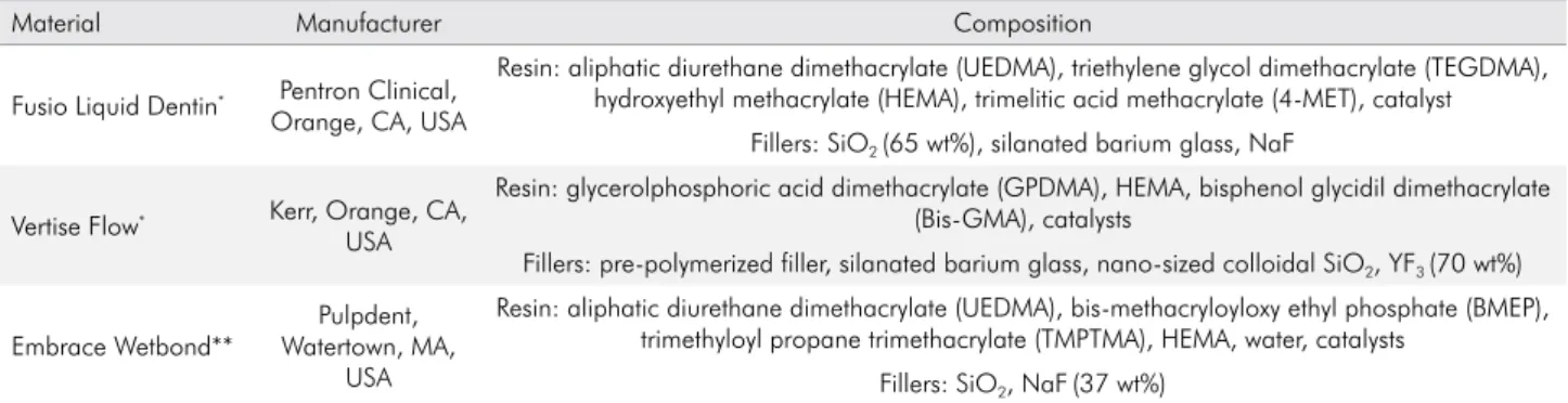
Self-adhesive restorative composites (SACs) were introduced in the dental market in 2009 and, currently, there are three examples available for clinical use (Table 1). These low-viscosity materials are indicated for small class I cavities and non-carious cervical lesions.8 Unfortunately, reports of in vitro evaluations of these materials are scarce and clinical studies are almost non-existent.
A key difference between SACs and self-adhesive resin cements (SARCs) is that SACs do not undergo acid-base neutralization reactions nor contain luoride-releasing glass illers.10 In fact, SACs are more
akin to self-etch adhesive systems due to the presence of acidic monomers such as glycerol phosphate dimethacrylates (GPDM), carboxylic methacrylates (for example, 4-MET) or phosphate ethyl methacrylates (BMEP). These monomers vary in acidity from mild (for example, GPDM with a pH = 1.9) to ultra-mild (4-MET, pH=3-4)11 and are responsible for partially etching the tooth substrate and penetrating through the smear layer, forming a submicron-thick hybrid layer.12 Hydroxyethyl methacrylate (HEMA) is added to increase the wettability of the material on the dentin surface.13
Similar to self-adhesive resin cements, though, is the fact that SAC interaction with dentin is limited by the extent of decalciication produced by the acidic monomers. Furthermore, their relatively high viscosity as the result of iller incorporation makes wetting of the bonding substrate even more dificult,12,14,15 limiting the diffusion of monomers into the collagen ibers network.16 Notwithstanding, some chemical interaction with calcium from hydroxyapatite has been veriied, suggesting that the retention relies not only on micro-mechanical interlocking.17,18
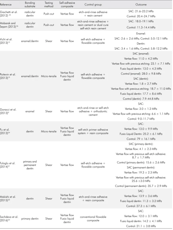
Overall, studies agree that SACs exhibit lower bond strength values to dental tissues than conventional restorative systems (Table 2),12,19,20,21,22,23,24 with a few exceptions where composites were tested on radicular dentin25,26 Several studies suggest that pre-etching enamel and dentin with phosphoric acid signiicantly increases bond strength, as phosphoric acid removes the smear layer and enhances surface area.8,9,19,27,28,29 Using a self-etch adhesive prior to the application of Vertise Flow signiicantly increased
Table 1. Composition of commercially available self-adhesive composites.
Material Manufacturer Composition
Fusio Liquid Dentin* Pentron Clinical,
Orange, CA, USA
Resin: aliphatic diurethane dimethacrylate (UEDMA), triethylene glycol dimethacrylate (TEGDMA), hydroxyethyl methacrylate (HEMA), trimelitic acid methacrylate (4-MET), catalyst
Fillers: SiO2 (65 wt%), silanated barium glass, NaF
Vertise Flow* Kerr, Orange, CA,
USA
Resin: glycerolphosphoric acid dimethacrylate (GPDMA), HEMA, bisphenol glycidil dimethacrylate (Bis-GMA), catalysts
Fillers: pre-polymerized filler, silanated barium glass, nano-sized colloidal SiO2, YF3 (70 wt%)
Embrace Wetbond**
Pulpdent, Watertown, MA,
USA
Resin: aliphatic diurethane dimethacrylate (UEDMA), bis-methacryloyloxy ethyl phosphate (BMEP), trimethyloyl propane trimethacrylate (TMPTMA), HEMA, water, catalysts
Fillers: SiO2, NaF(37 wt%)
Table 2. Bond strength studies.
Reference Bonding
substrate
Testing method
Self-adhesive
composite Control group Outcome
Giachetti et al. (2012) 25
radicular
dentin Push-out Vertise flow
etch-and-rinse adhesive + resin cement
SAC: 21.6–23.2 MPa
Control: 20.4–24.7 MPa
Mobarak and Seyam (2013) 26
radicular
dentin Push-out Vertise flow
etch-and-rinse adhesive + resin cement or dual cure
self-etch resin cement
SAC: 18.0–19.1 MPa
Control: 11.3–14.4 MPa
Vichi et al.
(2013)12 enamel/dentin Shear Vertise flow
self-etch adhesive + flowable composite
Enamel:
SAC: 2.6 ± 2.6 MPa; Control: 5.0–12.1 MPa
Dentin:
SAC: 3.4 ± 1.6 MPa; Control: 5.8–12.2 MPa
Poitevin et al.
(2013)19 enamel/dentin Micro-tensile
Vertise flow Fusio liquid
dentin
self-etch adhesive + flowable composite
SAC (enamel):
Vertise flow: 11.0 ± 4.2 MPa
Vertise flow with previous etching: 23.1 ± 7.1 MPa Fusio liquid dentin: 13.0 ± 4.3 MPa
Control (enamel): 28.0 ± 9.8 MPa
SAC (dentin):
Vertise flow: 1.8 ± 2.7 MPa
Vertise flow with previous etching: 18.7 ± 11.0 MPa
Fusio liquid dentin: 17.7 ± 8.6 MPa
Control (dentin): 7.9–44.8 MPa
Goracci et al.
(2013)9 enamel Shear Vertise flow
etch-and-rinse or self-etch adhesive + orthodontic
cement
SAC:
Vertise flow: 3.0 ± 1.2 MPa
Vertise flow with previous etching: 6.6 ± 1.1 MPa
Control: 9.0–11.7 MPa
Fu et al.
(2013)24 dentin Micro-tensile
Vertise flow Fusio liquid
dentin
self-etch primer adhesive system + resin composite
SAC:
Vertise flow: 13.0 ± 9.9 MPa Fusio Liquid Dentin: 25.2 ± 6.1 MPa
Control: 79 ± 16.1 MPa
Tuloglu et al. (2014)21
primary and permanent
dentin
Shear Vertise flow self-etch adhesive + flowable composite
SAC (primary dentin):
Vertise flow: 4.1 ± 2.3 MPa
Vertise flow with previous self-etch adhesive: 8.7 ± 1.7 MPa
Control (primary dentin): 15.6 ± 2.6 MPa
SAC (permanent dentin):
Vertise flow: 19.3 ± 2.3 MPa
Vertise flow with previous self-etch adhesive: 25.6 ±3.0 MPa
Control (permanent dentin): 35.7 ± 2.9 MPa
Makishi et al.
(2015)22 dentin Shear
Vertise flow Fusio liquid
dentin
etch-and-rinse adhesive + resin composite
SAC:
Vertise flow: 13.9 ± 3.6 MPa Fusio liquid dentin: 11.3 ± 3.2 MPa
Control: 27.3 ± 6.1 MPa
Sachdeva et al.
(2016)23 primary dentin Shear
Vertise flow Fusio liquid
dentin
conventional flowable composite
SAC:
Vertise flow: 12.0 ± 3.1 MPa
Fusio liquid dentin: 14.2 ± 4.1 MPa
the dentin bond strength and reduced microleakage scores, when compared with the application of the SAC alone.21 Obviously, adding an extra step to the restorative procedure with self-adhesive composites defeats the purpose of simplifying the procedure. Interestingly, despite their low bond strength, Vertise Flow showed better marginal sealing ability in comparison with self-adhesive and etch-and-rinse adhesive systems possibly due to hygroscopic expansion and a relatively low polymerization stress development at the bonded interface.12,14,23 Another evidence of the limited adhesion of these materials to tooth structure was the retention rate of only 33% displayed by Fusio liquid dentin in non-carious cervical lesions after a six-month clinical evaluation, in comparison to the 100% retention of a conventional restorative composite.30
The physical properties of SACs have not been thoroughly evaluated. In one study, Vertise Flow showed lexural strength similar to conventional (i.e., non-self-adhesive) lowable composites.31 After
toothbrushing abrasion, Vertise low had a rougher surface compared to that resulting from the application of Fusio liquid dentin and a conventional restorative composite.32 Results can be related to the type of organic matrix, since urethane-based composites have a better wear resistance than Bis-GMA-based composites, other factors being equal.33 SACs also showed a much poorer performance in terms of gloss retention than a conventional control.32 Self-adhesive composites are more hydrophilic than conventional composites due to the presence of acidic monomers and HEMA. In fact, Vertise Flow showed much higher hygroscopic expansion34 and higher water sorption35 when compared with conventional composites after 150 days in water. A drawback indeed, since SACs hydrophilicity may facilitate network plasticization, enhance bioilm formation and increase degradation.28,34
The idea of bonding the composite directly to the tooth structure is enticing. However, based on the limited amount of information available, it seems that self-adhesive restorative composites are still at their inception and more research is necessary to solve the limitations found with the current materials.
Strategy 2A: Fiber-reinforced composites
Current restorative composites present fracture toughness (KIc) values varying between 0.9 to 1.8 MPa.m0,5,1 which seems insuficient to avoid clinical failures by bulk fracture.4,5 The incorporation of small fractions of short, random glass ibers as part of the iller system is one of the strategies currently in use to create tougher composites.
The use of glass fibers as reinforcement in dental composites is not new. The use of randomly-oriented low aspect ratio (AR: length-to-diameter ratio) glass ibers as reinforcing phase in composites was published in 198936 and in the 1990s composites containing low AR ibers (20–120 µm in length, 6 µm in diameter) associated with iller particles appeared in the market (Restolux, Lee Pharmaceutical, South El Monte, CA, USA, and ALERT, Pentron, Orange, CA, USA). In fact, studies showed higher KIc values for ALERT compared to other packable and regular consistency materials, but no difference in lexural strength was observed.37,38
Recently, a bulk-ill composite containing high AR E-glass ibers (1–2 mm in length, 17 µm in diameter) was released (EverX Posterior, GC Europe, formerly known as Xenius Base). Its total mass iller fraction is 74.2 wt% (53.6 vol%), with 8.6 wt% (7.2 vol%) of glass ibers.39 Its resin matrix is constituted by BisGMA, TEGDMA and PMMA (polymethyl methacrylate), forming a semi-interpenetrating polymer network (semi-IPN).40 As ibers impair polishing, it is indicated as a substructure material and a final layer of particulate composite is mandatory. Overall, its fracture strength does not seem to differ from those of particle-only composites.20,41 Fatigue strength of teeth restored with this material also did not differ from that of a conventional composite.42 The only report on the material’s KIc showed a signiicantly higher value, compared to other commercial composites.43
(“critical length”), estimated from the iber strength and diameter (therefore, related to iber AR) and on the interfacial bond strength between the iber and the matrix.44 For example, the addition of 20 vol% high AR ibers (AR=68) to a commercial lowable composite (iller fraction: 43.6 vol%) had a positive effect on lexural strength compared to low AR ibers (AR=5.2).45
The replacement of up to 7.5 vol% of particles by ibers (AR = 140) did not increase the lexural strength of experimental composites containing 60 vol% of illers. Though randomly oriented ibers are supposed to result in a material with isotropic behavior, it is possible that the insertion of the composite into the mold during specimen preparation results in some iber orientation perpendicular to load application, which reduces reinforcement eficiency.46
On the other hand, studies agree that fiber-containing commercial composites present higher KIc than particle-only composites.37,38,39,43,47 In fact, the replacement of 5 vol% of glass particles by ibers in a composite with 60 vol% of illers resulted in a two-fold increase in KIc (from 1.25 to 2.6 MPa.m1/2).46 The presence of ibers increases “crack bridging”, i.e., when a crack
propagates through the material, ibers pull the crack faces together and, as a result, more energy is necessary for the crack to propagate further.48
Besides increasing KIc, ibers may also interfere with composite polymerization shrinkage. Composites containing continuous, oriented fibers present anisotropic behavior in terms of shrinkage, with lower values being registered in the direction of the ibers in comparison to the perpendicular direction.43,49 On the other hand, composites containing short random ibers are expected to show isotropic behavior. However, depending on the specimen coniguration and testing method, iber-containing composites may show lower shrinkage than particle-only composites. For example, Xenius base showed 43–51% lower post-gel shrinkage in comparison to composites with higher iller fractions when tested by the strain gage method.43 Also using the strain gage method, an inverse relationship between iber content and post-gel shrinkage was observed when 2.5 vol% to 7.5 vol% of the iller particles were replaced by 1.4 mm glass ibers in a 60 vol% iller experimental composite, with shrinkage reductions reaching 70%
at the highest iber content.46 Strain gages record mostly the shrinkage taking place adjacently to the sensor grid and during specimen preparation, some degree of iber orientation may result from pressing the composite against it. Consequently, the material does not behave as totally isotropic. When using the mercury dilatometer, a composite containing 6 vol% of fibers and 54 vol% particles showed a much subtler reduction in total shrinkage (11%) in comparison to the 60 vol% particle-only composite. As polymerization stress development is the result of complex interactions between composite elastic modulus, shrinkage and testing system compliance, such difference in shrinkage was not enough to reduce stress magnitude.46 Other studies showed that EverX Posterior developed either similar or higher polymerization stress and gap formation in vitro
compared to other bulk-ill materials.3,50
Strategy 2B: Self-healing composites
Failure mechanism in polymers can be described as the result of damage accumulation, where microcracks propagate due to thermal or mechanical stress concentration at the crack tip.51 Therefore, bringing crack extension to a halt by sealing the crack faces (“crack healing”) may increase composite life-span. The development of self-healing polymers was a major breakthrough in polymer chemistry. According to Huyang et al.52 “self-healing mechanisms are biomimetic models of autonomic repair systems in living tissues that eficiently handle damage, for example, the healing of a broken bone”. The “proof of concept” for this approach was published by Dry.53 In a series of experiments, she used hollow glass ibers illed with either a two-part epoxy system or a cyanoacrylate adhesive embedded in epoxy specimens to demonstrate the material’s self-healing ability. Besides recovering part of the initial strength after fracture, the self-repair system was shown to arrest microcracks and prevent crack reopening.
ruptured and DCPD was released within the crack plane by capillarity. Polymerization of the healing agent was triggered by contact with a transition metal catalyst (Grubbs’ catalyst) incorporated in the matrix. A healing eficiency of 60% was reported in notched specimens containing 10 wt% of microcapsules and 2.5 wt% of catalyst, monotonically loaded to failure in mode I (i.e., perpendicular to the precrack) and re-tested
after being allowed to heal for 48 hours. In a subsequent development, they tested microcapsules illed with an epoxy-solvent mixture.55 In this case, healing occurs by swelling of the set epoxy and transport of the residual amine to the crack plane. The additional epoxy released from the microcapsule increases the chance of crack healing by crosslinking. After a irst healing event with a 100% eficiency (i.e., full recovery of the initial K1c), a maximum of ive healing events were veriied, with decreasing eficiency due to the depletion of healing agent, as well as the available amine.
Fatigue loading represents a more clinically relevant scenario for testing polymer self-repair than static conditions. It has been veriied that under cyclic loading healing eficiency is related to crack growth rate (deined by stress amplitude and frequency), the polymerization rate of the healing agent and the occurrence of rest periods. For example, in an epoxy resin containing 20 wt% of DCPD-illed UF microcapsules, if stress intensity is such that crack extension occurs at a similar rate of that of healing agent curing, the fatigue life extension ranged from 89% to 213%.56
The irst attempt to formulate a self-healing dental composite used the DCPD-illed UF microcapsules developed by White et al.54 Specimens made of a BisGMA/UDMA/TEGDMA resin containing 55 wt% of silanated silica, 5 wt% microcapsules (average size: 50 µm) and 2 wt% of Grubbs’ catalyst were tested for K1c and re-tested after a 7-day healing period at room temperature, with average healing eficiency of 57%.57 It should be noted that the presence of the capsules within the resin may provide some beneit in terms of mechanical properties, regardless of the self-healing effect. For example, polyurethane (PU) nanocapsules (~500 nm) containing TEGDMA were synthesized and loaded into a commercial two-step, single-bottle adhesive system. Crack healing was not
expected, as no extra initiators were incorporated. Still, a signiicantly higher bond strength to dentin was veriied for the material containing 9 wt% of nanocapsules, possibly due to a toughening effect provided by the lexible PU shells.58
Concerns about the biological safety of DCPD59 prompted the research of different self-repair systems for dental composites, also based on microencapsulated healing agents. Reports on the use of poly(urea formaldehyde) microcapsules (average diameter: 70 µm) filled with TEGDMA and a tertiary amine (N,N-dihydroxyethyl-p-toluidine, DHEPT) as activator in experimental composites were recently published.60,61,62 This self-repair system uses benzoyl peroxide (BPO) added to the resin matrix as initiator to polymerize the healing liquid. In the irst study,62 these microcapsules were added to a BisGMA-TEGDMA resin. For microcapsule mass fractions up to 15%, no adverse effects were observed in lexural properties while K1c increased by 40% at the maximum microcapsule loading. Healing eficiency showed a plateau starting at 10% of microcapsules, of approximately 65%. In a follow-up study, experimental composites containing 35% of reinforcing illers, 20 wt% of ACP nanoparticles (to foster remineralization), 3.75 wt% of an antibacterial agent (dimethylaminohexadecyl methacrylate, DMAHDM), 0.5 wt% of BPO and different mass fractions of TEGDMA-DHEPT microcapsules were tested. The initial K1c was not affected by the presence of microcapsules up to 7.5 wt%. Twenty-four hours after repositioning the fragments in the mold, specimens were re-tested and healing eficiency was found to be linearly related to microcapsule content, ranging from 25% (2.5 wt% of microcapsules) to 81% (10 wt%).61 Healing eficiency of composites with 7.5% of TEGDMA-DHEPT microcapsules was not affected by prolonged water storage of the specimen prior to initial K1c test (up to six months), or when healing took place in water.60
particles. The idea was to form a reparative glass ionomer cement in the crack when the healing liquid is released. Microcapsule silanization was used to improve the interfacial strength between the shell and the resin matrix to favor its rupture rather than interfacial debonding upon meeting with the crack front. Re-testing K1c specimens after four days allowed a maximum healing eficiency of 25%, obtained with a microcapsules content of 10 wt%.
Strategy 3A: Remineralizing composites
Calcium orthophosphate (CaP) particles have been studied as ion-releasing illers in resin-based composites for decades.63,64 Calcium and phosphate ions released from the composite would make the surrounding medium supersaturated, favoring their deposition on the enamel hydroxyapatite (HAP) crystals.65 Several CaP phases have been tested as bioactive illers in restorative composites, for example, amorphous calcium phosphate (ACP),66,67 dicalcium phosphate dihydrate (DCPD),68 dicalcium phosphate anhydrous (DCPA)69 and tetracalcium phosphate (TTCP).70
The efficacy of CaP-containing composites in promoting mineral recovery of dental tissues were demonstrated in several studies. For example, a material containing 40 wt% of ACP in a BisGMA/TEGDMA/HEMA matrix was capable of recovering 71% of the enamel mineral content of non-cavitated bovine enamel lesions after four weeks under a static remineralization model and 38% after two weeks under pH cycling (dynamic model).64 A resin cement containing 20 wt% of DCPA particles (1.1 µm), 60 wt% of TTCP particles (16 µm) and 1.5 % of sodium hexafluorosilicate in contact with demineralized human dentin promoted an increase in mineral in mineral content between 38% and 47% after 5-week immersion in saliva-like solution.71 A resin-based material containing 40 wt% of Zr-modified ACP (55 µm) dispersed in a BisGMA/TEGDMA/HEMA matrix led to a 14% mineral recovery in non-cavitated human enamel lesions after 30 days of pH cycling.72 The irst study associating ACP nanoparticles (116 nm, 40 wt%) and reinforcing glass illers (1.4 µm, 20 wt%) in a BisEMA/TEGDMA/HEMA matrix veriied a 22% remineralization in pH cycling conditions similar to those used in the previous study.63 It is interesting
to point out that these in vitro studies share the
characteristic of formulating their experimental materials with fairly hydrophilic resin matrices, which facilitates luid transit through the material, consequently increasing ion release. Also, notice that experimental models utilizing pH cycling result in lower mineral recovery in comparison to static remineralization models (i.e., immersion in calcium
solutions). The protective effect of CaP-containing composites against enamel demineralization was veriied in an in situ study. Cavity preparations in
human enamel fragments were restored with an experimental composite containing ACP (116 nm, 40 wt%) and glass illers (1.4 µm, 20 wt%) in a less hydrophilic matrix (BisGMA/TEGDMA). After 14 days in the presence of biofilm, mineral loss was 59% lower in fragments restored with the ACP-containing composite in comparison to a control.73
Composites containing CaP particles are considered “smart materials” because ion release increases in more acidic conditions due to an increase in particle erosion.74 Also, in order to maximize ion release, particles with high surface area are preferable.75 Ion release increases exponentially with particle volume fraction.76 Finally, the hydrophilicity of the resin matrix must also be taken into account, as it interferes with the water access to the particles and, consequently, with ion release.77 As it could be expected, ion release does not occur indeinitely and ine-tuning all the variables to come up with a material able to provide a long-lasting protection against demineralization is challenging. Recently, it was demonstrated that a composite containing 20 wt% ACP and 50 wt% silanated glass was capable of recharging after complete exhausting its ion release (after 70-days immersion at pH = 4) by immersion in calcium and phosphatesolutions (one minute, 3x/day, for three days).78
of some of the glass fillers with DCPD. However, after 28 days of storage in water, a 35% reduction was observed in the DCPD-containing composite, while no signiicant change in occurred in the control.68
The main cause of the negative effect of CaP particles on the mechanical properties is the lack of a strong chemical bonding between them and the resin matrix.70,80 To eliminate or at least reduce this limitation, functionalization of the CaP particles was attempted.81,82 The use of silane to functionalize DCPA particles caused a signiicant increase in composite fracture strength, compared to a control containing non-silanized DCPA; however, ion release was compromised due to the silane hydrophobic character, which hinders the access of water to the particles.75 Good mechanical results were also obtained when HAP particles were treated with acrylic and methacrylic acids. Unfortunately, ion release was not tested.82 Recently, the synthesis of TEGDMA-functionalized DCPD particles was described.83 TEGDMA is capable of bonding to Ca2+ via ion-dipole interactions with the oxygen atoms from the ethylene glycol groups. The incorporation of 20 vol% of these particles in a BisGMA/TEGDMA resin resulted in a 32% increase in lexural strength in comparison to the material containing non-functionalized particles.84 A similar improvement was observed in a subsequent study where an experimental composite containing 40 vol% of barium glass (0.5 µm) and 20 vol% of TEGDMA-functionalized DCPD (19 µm) was compared to a similar formulation containing non-functionalized DCPD.85
Composites containing CaP particles may undergo more severe degradation after prolonged water immersion than their conventional counterparts, possibly due to the higher water sorption allowed by the transit of luids along CaP-matrix interfaces.86 For example, composites containing DCPD particles showed up to 33% reduction in flexural strength after 28 days in water versus a 16% reduction for composites without DCPD.68 But no difference in strength was observed after two years of immersion in water for composites containing 10–20 wt% of ACP nanoparticles (112 nm) and 65–50 wt% of barium glass (1.4 µm) and the control with 75 wt% of glass illers.87
Despite all the research activity involving CaP composites, it seems that the concept has not yet been embraced by dental materials manufacturers. The sole
exception is a restorative composite containing 38 wt% ACP, released in 2012 (Aegis V, Bosworth, Skokie, USA). Literature reports and manufacturer information are scarce. Its lexural strength is reduced, comparable to that of a microhybrid composite, which explains its indication only for restoring class V cavities. From the same manufacturer, ACP-containing sealant and orthodontic cement showed remineralizing potential similar to luoride-containing materials in vitro.88,89
Strategy 3B: Antibacterial composites
While adhesive systems containing antibacterial agents have been on the market for several years, restorative composites with antibacterial activity are still under development. Ideally, antibacterial composites must meet a critical set of requirements, including: 1) non-toxicity,90 2) antibacterial action against a broad spectrum of microorganisms91 and 3) maintain a long-lasting effect.92,93 Also, it is very important that incorporation of antibacterial agents does not compromise the mechanical and optical properties of the restorative material.93,94
The association between quaternary ammonium (QAM) and methacrylate terminal groups results in an antibacterial monomer with low lixiviation (i.e., leaching) levels.90,91,92,95 The first attempt of
incorporating a copolymerizable antibacterial monomer in experimental composites was reported more than 20 years ago. When MDPB (12-methacryloyloxydodecylpyridinium bromide was incorporated into a BisGMA/TEGDMA composite (iller fraction: 83 wt%) in 0.1 wt% and 0.2 wt% fractions, the growth of S.mutans on the composite surface was
inhibited for up to 90 days, without signiicant effects on the composite lexural properties.96 The proposed antibacterial mechanism for these monomers is that quaternary ammonium would damage the bacterial cell membrane, leading to cell death.90
reduce their initial lexural properties andinhibited the growth of Smutans and A viscous for up to four weeks.98
Another example of an antibacterial monomer with methacrylate functionality is dimethylaminohexadecyl methacrylate (DMAHDM). It has been tested in association with a protein-repellent biopolymer, 2-methacryloyloxyethyl phosphorylcholine (MPC), which reduces bacterial adsorption on the composite surface. Composites containing 70 wt% of glass illers and 1.5 wt% of DMAHDM, associated with 3 wt% of MPC showed higher antibacterial activity against cariogenic and periopathogenic bacteria than either components alone. Flexural properties were not affected by the presence of either compound.99 However, MPC is hydrophilic and its presence signiicantly increases composite water sorption, which may accelerate matrix degradation.91
Bioactive glass (BAG) particles were also investigated as antibacterial agents in dental composites. An experimental BisGMA/TEGDMA composite with 57 wt% of reinforcing glass and 15 wt% BAG particles (0.04–3 µm, SiO2, CaO and P2O5) were shown to reduce bacterial penetration into the tooth-restoration interface and tooth demineralization in comparison to a control. The possible hypothesis for bacterial inhibition is a local increase in pH and/or some of the ions directly affecting bacteria.100 Silver-doped BAG particles (25 µm,
SiO2-CaO-P2O5-Al2O3-Na2O-K2O-Ag2O) incorporated into a commercial lowable composite at mass fractions of 5% and 15% resulted in signiicant reduction in
S mutans activity. As pH remained stable throughout
the study, antibacterial activity was ascribed to Ag+ ion release from the material.101
Though composites containing silver compounds or metallic silver (Ag0) nanoparticles have shown great eficacy against bacteria without compromising their mechanical properties,102 their presence even in small concentrations causes signiicant darkening of the composite.103 Zinc oxide particles have a more tooth-like color and were shown to possess antibacterial activity probably due to the release of Zn2+ ions, which inhibit the metabolism of sugars and interfere with bacterial enzymatic activity by displacing Mg2+ ions.104 Their eficacy, however, is much lower than that of silver, as 10 wt% of ZnO nanoparticles (40–100 nm) added to a commercial composite showed signiicant lower
Streptococci inhibition compared to 1 wt% of silver.104
In another study, the incorporation of up to 5 wt% of ZnO nanoparticles (20 nm) to a commercial lowable composite signiicantly inhibited S. mutans growth
in non-aged and 48-h aged specimens. However, specimens aged for one week and four weeks did not show any inhibitory effect.94
Chlorhexidine (CHX) was also investigated as an antimicrobial agent in composites. Because CHX salts are not soluble in the resin matrix, they tend to form relatively large agglomerates, which negatively affects composite mechanical properties.105 To overcome these problems, CHX was loaded into mesoporous silica nanoparticles (MSNs), with signiicant improvements in mechanical properties compared to materials containing directly added CHX. Furthermore, composites with MSNs sustained a lower surface roughness and allowed for a more controlled CHX release over time.93
Final Remarks: Can All These
Strategies Co-Exist?
Some of the above strategies may be mutually exclusive, because there are practical limits to intelligent addition of multiple chemicals - especially to a unitary (light cured) material formulation. Also, with self-adhesive materials there may be limited shelf life due to the incorporation of reactive chemicals. However the strategies 2A and 2B to produce stronger composites might be compatible by incorporation of both ibers and self-healing capsules into the resin matrix. Despite the great potential of bulk ill composites, it may be desirable to have separate base-layer composites with anti-microbial and/or remineralizing functionality. It seems inevitable that some kind of trade off will still be required between ease/simplicity of application and the number of material functions desired and achievable. It is important that some of these strategies be advanced from in vitro to in situ studies, and then
to controlled clinical trials.
1. Ferracane JL. Resin composite: state of the art. Dent Mater. 2011;27(1):29-38. https://doi.org/10.1016/j.dental.2010.10.020 2. Braga RR, Ferracane JL. Alternatives in
polymerization contraction stress management. Crit Rev Oral Biol Med. 2004;15(3):176-84. https://doi.org/10.1177/154411130401500306 3. Fronza BM, Rueggeberg FA, Braga RR,
Mogilevych B, Soares LE, Martin AA et al. Monomer conversion, microhardness, internal marginal adaptation, and shrinkage stress of bulk-fill resin composites. Dent Mater. 2015;31(12):1542-51. https://doi.org/10.1016/j.dental.2015.10.001 4. Rasines Alcaraz MG, Veitz-Keenan A,
Sahrmann P, Schmidlin PR, Davis D, Iheozor-Ejiofor Z. Direct composite resin fillings versus amalgam fillings for permanent or adult posterior teeth. Cochrane Database Syst Rev. 2014(3):CD005620.
https://doi.org/10.1002/14651858.CD005620.pub2 5. Nedeljkovic I, Teughels W, De Munck J, Van Meerbeek B,
Van Landuyt KL. Is secondary caries with composites a material-based problem? Dent Mater. 2015;31(11):e247-77. https://doi.org/10.1016/j.dental.2015.09.001
6. Beck F, Lettner S, Graf A, Bitriol B, Dumitrescu N,
Bauer P et al. Survival of direct resin restorations in posterior teeth within a 19-year period (1996-2015): A meta-analysis of prospective studies. Dent Mater. 2015;31(8):958-85. https://doi.org/10.1016/j.dental.2015.05.004
7. Opdam NJ, van de Sande FH, Bronkhorst E, Cenci MS, Bottenberg P, Pallesen U et al. Longevity of posterior composite restorations: a systematic review and meta-analysis. J Dent Res. 2014;93(10):943-9. https://doi.org/10.1177/0022034514544217 8. Altunsoy M, Botsali MS, Sari T, Onat H. Effect of
different surface treatments on the microtensile bond strength of two self-adhesive flowable composites. Lasers Med Sci. 2015;30(6):1667-73. https://doi.org/10.1007/s10103-014-1640-2 9. Goracci C, Margvelashvili M, Giovannetti A, Vichi A,
Ferrari M. Shear bond strength of orthodontic brackets bonded with a new self-adhering flowable resin composite. Clin Oral Investig. 2013;17(2):609-17. https://doi.org/10.1007/s00784-012-0729-x 10. Ferracane JL, Stansbury JW, Burke FJ. Self-adhesive
resin cements - chemistry, properties and clinical considerations. J Oral Rehabil. 2011;38(4):295-314. https://doi.org/10.1111/j.1365-2842.2010.02148.x
11. Van Meerbeek B, Peumans M, Poitevin A, Mine A, Van Ende A, Neves A et al. Relationship between bond-strength tests and clinical outcomes. Dent Mater. 2010;26(2):e100-21. https://doi.org/10.1016/j.dental.2009.11.148
12. Vichi A, Margvelashvili M, Goracci C,
Papacchini F, Ferrari M. Bonding and sealing ability of a new self-adhering flowable composite resin in class I restorations. Clin Oral Investig. 2013;17(6):1497-506. https://doi.org/10.1007/s00784-012-0846-6 13. Van Landuyt KL, Snauwaert J, De Munck J,
Peumans M, Yoshida Y, Poitevin A et al. Systematic review of the chemical composition of contemporary dental adhesives. Biomaterials. 2007;28(26):3757-85. https://doi.org/10.1016/j.biomaterials.2007.04.044 14. Rengo C, Goracci C, Juloski J, Chieffi N, Giovannetti A,
Vichi A et al. Influence of phosphoric acid etching on microleakage of a self-etch adhesive and a self-adhering composite. Aust Dent J. 2012;57(2):220-6.
https://doi.org/10.1111/j.1834-7819.2012.01689.x 15. Bektas OO, Eren D, Akin EG, Akin H. Evaluation
of a self-adhering flowable composite in terms of micro-shear bond strength and microleakage. Acta Odontol Scand. 2013;71(3-4):541-6. https://doi.org/10.3109/00016357.2012.696697 16. Miyazaki M, Ando S, Hinoura K, Onose H, Moore BK.
Influence of filler addition to bonding agents on shear bond strength to bovine dentin. Dent Mater. 1995;11(4):234-8. https://doi.org/10.1016/0109-5641(95)80055-7
17. Yoshida Y, Nagakane K, Fukuda R, Nakayama Y, Okazaki M, Shintani H et al. Comparative study on adhesive performance of functional monomers. J Dent Res. 2004;83(6):454-8. https://doi.org/10.1177/154405910408300604 18. Monticelli F, Osorio R, Mazzitelli C, Ferrari M,
Toledano M. Limited decalcification/diffusion of self-adhesive cements into dentin. J Dent Res. 2008;87(10):974-9. https://doi.org/10.1177/154405910808701012 19. Poitevin A, De Munck J, Van Ende A, Suyama Y, Mine A,
Peumans M et al. Bonding effectiveness of self-adhesive composites to dentin and enamel. Dent Mater.
2013;29(2):221-30. https://doi.org/10.1016/j.dental.2012.10.001 20. Goracci C, Cadenaro M, Fontanive L,
Giangrosso G, Juloski J, Vichi A et al. Polymerization efficiency and flexural strength of low-stress restorative composites. Dent Mater. 2014;30(6):688-94. https://doi.org/10.1016/j.dental.2014.03.006
21. Tuloglu N, Sen Tunc E, Ozer S, Bayrak S. Shear bond strength of self-adhering flowable composite on dentin with and without application of an adhesive system. J Appl Biomater Funct Mater. 2014;12(2):97-101. https://doi.org/10.5301/jabfm.5000166 22. Makishi P, Pacheco RR, Sadr A, Shimada Y,
23. Sachdeva P, Goswami M, Singh D. Comparative evaluation of shear bond strength and nanoleakage of conventional and self-adhering flowable composites to primary teeth dentin. Contemp Clin Dent. 2016;7(3):326-31. https://doi.org/10.4103/0976-237X.188549 24. Fu J, Kakuda S, Pan F, Hoshika S, Ting S,
Fukuoka A et al. Bonding performance of a newly developed step-less all-in-one system on dentin. Dent Mater J. 2013;32(2):203-11. https://doi.org/10.4012/dmj.2012-204 25. Giachetti L, Scaminaci Russo D, Baldini M, Bertini F, Steier L,
Ferrari M. Push-out strength of translucent fibre posts cemented using a dual-curing technique or a light-curing self-adhering material. Int Endod J. 2012;45(3):249-56. https://doi.org/10.1111/j.1365-2591.2011.01969.x 26. Mobarak E, Seyam R. Interfacial nanoleakage and bonding
of self-adhesive systems cured with a modified-layering technique to dentin of weakened roots. Oper Dent. 2013;38(5):E154-65. https://doi.org/10.2341/12-103-L 27. İşman E, Karaarslan ES, Okşayan R, Tunçdemır AR,
Üşümez S, Adanir N et al. Inadequate shear bond
strengths of self-etch, self-adhesive systems for secure orthodontic bonding. Dent Mater J. 2012;31(6):947-53. https://doi.org/10.4012/dmj.2012-103
28. Eliades A, Birpou E, Eliades T, Eliades G. Self-adhesive restoratives as pit and fissure sealants: a comparative laboratory study. Dent Mater. 2013;29(7):752-62. https://doi.org/10.1016/j.dental.2013.04.005
29. Yazici AR, Agarwal I, Campillo-Funollet M, Munoz-Viveros C, Antonson SA, Antonson DE et al. Effect of laser preparation on bond strength of a self-adhesive flowable resin. Lasers Med Sci. 2013;28(1):343-7. https://doi.org/10.1007/s10103-012-1158-4 30. Çelik EU, Aka B, Yilmaz F. Six-month clinical evaluation
of a self-adhesive flowable composite in noncarious cervical lesions. J Adhes Dent. 2015;17(4):361-8. https://doi.org/10.3290/j.jad.a34556
31. Czasch P, Ilie N. In vitro comparison of mechanical properties and degree of cure of a self-adhesive and four novel flowable composites. J Adhes Dent. 2013;15(3):229-36. https://doi.org/10.3290/j.jad.a29530
32. Malavasi CV, Macedo EM, Souza KC, Rego GF, Schneider LF, Cavalcante LM. Surface texture and optical properties of self-adhering composite materials after toothbrush abrasion. J Contemp Dent Pract. 2015;16(10):775-82. https://doi.org/10.5005/jp-journals-10024-1756 33. Söderholm KJ, Lambrechts P, Sarrett D, Abe Y,
Yang MC, Labella R, et al. Clinical wear performance of eight experimental dental composites over three years determined by two measuring methods. Eur J Oral Sci. 2001;109(4):273-81. https://doi.org/10.1034/j.1600-0722.2001.00064.x 34. Wei YJ, Silikas N, Zhang ZT, Watts DC.
Hygroscopic dimensional changes of self-adhering and new resin-matrix composites during water
sorption/desorption cycles. Dent Mater. 2011;27(3):259-66. https://doi.org/10.1016/j.dental.2010.10.015
35. Wei YJ, Silikas N, Zhang ZT, Watts DC. Diffusion and concurrent solubility of self-adhering and new resin-matrix composites during water
sorption/desorption cycles. Dent Mater. 2011;27(2):197-205. https://doi.org/10.1016/j.dental.2010.10.014
36. Krause WR, Park SH, Straup RA. Mechanical properties of BIS-GMA resin short glass fiber composites. J Biomed Mater Res. 1989;23(10):1195-211. https://doi.org/10.1002/jbm.820231008 37. Choi KK, Ferracane JL, Hilton TJ,
Charlton D. Properties of packable dental composites. J Esthet Dent. 2000;12(4):216-26. https://doi.org/10.1111/j.1708-8240.2000.tb00224.x 38. Knobloch LA, Kerby RE, Seghi R, Berlin JS, Clelland N.
Fracture toughness of packable and conventional composite materials. J Prosthet Dent. 2002;88(3):307-13.
https://doi.org/10.1067/mpr.2002.128069
39. Lassila L, Garoushi S, Vallittu PK, Säilynoja E. Mechanical properties of fiber reinforced restorative composite with two distinguished fiber length distribution. J Mech Behav Biomed Mater. 2016;60:331-8. https://doi.org/10.1016/j.jmbbm.2016.01.036 40. Garoushi SK, Hatem M, Lassila LVJ, Vallittu PK. The
effect of short fiber composite base on microleakage and load-bearing capacity of posterior restorations. Acta Biomater Odontol Scand. 2015;1(1):6-12. https://doi.org/10.3109/23337931.2015.1017576. 41. Leprince JG, Palin WM, Vanacker J, Sabbagh J, Devaux J,
Leloup G. Physico-mechanical characteristics of commercially available bulk-fill composites. J Dent. 2014;42(8):993-1000. https://doi.org/10.1016/j.jdent.2014.05.009
42. Barreto BC, Van Ende A, Lise DP, Noritomi PY, Jaecques S, Sloten JV et al. Short fibre-reinforced composite for extensive direct restorations: a laboratory and computational assessment. Clin Oral Investig. 2016;20(5):959-66. https://doi.org/10.1007/s00784-015-1576-3 43. Garoushi S, Säilynoja E, Vallittu PK, Lassila L. Physical
properties and depth of cure of a new short fiber reinforced composite. Dent Mater. 2013;29(8):835-41. https://doi.org/10.1016/j.dental.2013.04.016
44. Petersen RC. Discontinuous fiber-reinforced composites above critical length. J Dent Res. 2005;84(4):365-70. https://doi.org/10.1177/154405910508400414
45. Shouha P, Swain M, Ellakwa A. The effect of fiber aspect ratio and volume loading on the flexural properties of flowable dental composite. Dent Mater. 2014;30(11):1234-44. https://doi.org/10.1016/j.dental.2014.08.363 46. Bocalon AC, Mita D, Narumyia I, Shouha P, Xavier TA,
47. Bijelic-Donova J, Garoushi S, Lassila LV, Keulemans F, Vallittu PK. Mechanical and structural characterization of discontinuous fiber-reinforced dental resin composite. J Dent. 2016;52:70-8. https://doi.org/10.1016/j.jdent.2016.07.009 48. Quinn GD. Fractography of ceramics and glasses. Gaithersburg:
National Institute of Standards and Technology; 2007. 49. Tezvergil A, Lassila LV, Vallittu PK. The effect of fiber
orientation on the polymerization shrinkage strain of fiber-reinforced composites. Dent Mater. 2006;22(7):610-6. https://doi.org/10.1016/j.dental.2005.05.017
50. Shouha PS, Ellakwa AE. Effect of short glass fibers on the polymerization shrinkage stress of dental composite. J Biomed Mater Res B Appl Biomater. 2016. https://doi.org/10.1002/jbm.b.33723
51. Ornaghi BP, Meier MM, Lohbauer U, Braga RR. Fracture toughness and cyclic fatigue resistance of resin composites with different filler size distributions. Dent Mater. 2014;30(7):742-51. https://doi.org/10.1016/j.dental.2014.04.004
52. Huyang G, Debertin AE, Sun J. Design and development of self-healing dental composites. Mater Des. 2016;94:295-302. https://doi.org/10.1016/j.matdes.2016.01.046
53. Dry C. Procedures developed for self-repair of polymer matrix composite materials. Compos Struct. 1996;35(3):263-9. https://doi.org/10.1016/0263-8223(96)00033-5 54. White SR, Sottos NR, Geubelle PH, Moore JS,
Kessler MR, Sriram SR, et al. Autonomic healing of polymer composites. Nature. 2001;409(6822):794-7. https://doi.org/10.1038/35057232
55. Caruso MM, Blaiszik BJ, White SR, Sottos NR, Moore JS. Full recovery of fracture toughness using a nontoxic solvent-based self-healing system. Adv Funct Mater. 2008;18(13):1898-904. https://doi.org/10.1002/adfm.200800300
56. Brown EN, White SR, Sottos NR. Retardation and repair of fatigue cracks in a microcapsule toughened epoxy composite - Part II: in situ self-healing. Compos Sci Technol. 2005;65(15-16):2474-80. https://doi.org/10.1016/j.compscitech.2005.04.053. 57. Wertzberger BE, Steere JT, Pfeifer RM, Nensel MA, Latta MA,
Gross SM. Physical characterization of a self-healing dental restorative material. J Appl Polym Sci. 2010;118(1):428-34. https://doi.org/10.1002/app.31542
58. Ouyang X, Huang X, Pan Q, Zuo C, Huang C, Yang X et al. Synthesis and characterization of triethylene glycol dimethacrylate nanocapsules used in a self-healing bonding resin. J Dent. 2011;39(12):825-33. https://doi.org/10.1016/j.jdent.2011.09.001 59. Kransler KM. Results of a 90-day inhalation
study of dicyclopentadiene in B6C3F1 mice. Toxicol Ind Health. 2014;30(5):459-66. https://doi.org/10.1177/0748233712458481
60. Wu J, Weir MD, Melo MA, Strassler HE, Xu HH. Effects of water-aging on self-healing dental composite containing microcapsules. J Dent. 2016;47:86-93. healing dental composite containing microcapsules. J Dent 2016 Apr;47:86–93. https://doi.org/10.1016/j.jdent.2016.01.008
61. Wu J, Weir MD, Melo MA, Xu HH. Development of novel self-healing and antibacterial dental composite containing calcium phosphate nanoparticles. J Dent. 2015;43(3):317-26. https://doi.org/10.1016/j.jdent.2015.01.009 62. Wu J, Weir MD, Zhang Q, Zhou C, Melo MA,
Xu HH. Novel self-healing dental resin with microcapsules of polymerizable triethylene glycol dimethacrylate and N,N-dihydroxyethyl-p-toluidine. Dent Mater. 2016;32(2):294-304.
https://doi.org/10.1016/j.dental.2015.11.014 63. Weir MD, Chow LC, Xu HH. Remineralization of
demineralized enamel via calcium phosphate nanocomposite. J Dent Res. 2012;91(10):979-84. https://doi.org/10.1177/0022034512458288
64. Skrtic D, Hailer AW, Takagi S, Antonucci JM, Eanes ED. Quantitative assessment of the efficacy of amorphous calcium phosphate/methacrylate composites in remineralizing caries-like lesions artificially produced in bovine enamel. J Dent Res. 1996;75(9):1679-86. https://doi.org/10.1177/00220345960750091001 65. Cochrane NJ, Cai F, Huq NL, Burrow MF,
Reynolds EC. New approaches to enhanced remineralization of tooth enamel. J Dent Res. 2010;89(11):1187-97.
https://doi.org/10.1177/0022034510376046 66. Marovic D, Tarle Z, Hiller KA, Müller R, Ristic M,
Rosentritt M et al. Effect of silanized nanosilica addition on remineralizing and mechanical properties of
experimental composite materials with amorphous calcium phosphate. Clin Oral Investig. 2014;18(3):783-92. https://doi.org/10.1007/s00784-013-1044-x 67. Xu HH, Moreau JL, Sun L, Chow LC. Nanocomposite
containing amorphous calcium phosphate nanoparticles for caries inhibition. Dental Mater. 2011;27(8):762-9. https://doi.org/10.1016/j.dental.2011.03.016 68. Chiari MD, Rodrigues MC, Xavier TA, de Souza EM,
Arana-Chavez VE, Braga RR. Mechanical properties and ion release from bioactive restorative composites containing glass fillers and calcium phosphate
nano-structured particles. Dent Mater. 2015;31(6):726-33. https://doi.org/10.1016/j.dental.2015.03.015
69. Xu HH, Weir MD, Sun L, Takagi S, Chow LC. Effects of calcium phosphate nanoparticles on Ca-PO4 composite. J Dent Res. 2007;86(4):378-83. https://doi.org/10.1177/154405910708600415 70. Xu HH, Moreau JL. Dental glass-reinforced
composite for caries inhibition: calcium phosphate ion release and mechanical properties. J Biomed Mater Res B Appl Biomater. 2010;92(2):332-40. https://doi.org/10.1002/jbm.b.31519
72. Langhorst SE, O’Donnell JN, Skrtic D. In vitro remineralization of enamel by polymeric amorphous calcium phosphate composite: quantitative
microradiographic study. Dent Mater. 2009;25(7):884-91. https://doi.org/10.1016/j.dental.2009.01.094
73. Melo MA, Weir MD, Rodrigues LK, Xu HH. Novel calcium phosphate nanocomposite with caries-inhibition in a human in situ model. Dent Mater. 2013;29(2):231-40. https://doi.org/10.1016/j.dental.2012.10.010 74. Xu HH, Weir MD, Sun L. Calcium and phosphate
ion releasing composite: effect of pH on release and mechanical properties. Dent Materer. 2009;25(4):535-42. https://doi.org/10.1016/j.dental.2008.10.009
75. Xu HH, Weir MD, Sun L. Nanocomposites with Ca and PO4 release: effects of reinforcement, dicalcium phosphate particle size and silanization. Dent Mater. 2007;23(12):1482-91. https://doi.org/10.1016/j.dental.2007.01.002
76. Xu HH, Weir MD, Sun L, Moreau JL, Takagi S, Chow LC et al. Strong nanocomposites with Ca, PO(4), and F release for caries inhibition. J Dent Res. 2010;89(1):19-28. https://doi.org/10.1177/0022034509351969 77. Skrtic D, Antonucci JM. Dental composites
based on amorphous calcium phosphate - resin composition/physicochemical properties study. J Biomater Appl. 2007;21(4):375-93. https://doi.org/10.1177/0885328206064823 78. Zhang L, Weir MD, Chow LC, Antonucci JM, Chen J,
Xu HH. Novel rechargeable calcium phosphate dental nanocomposite. Dental Mater. 2016;32(2):285-93. https://doi.org/10.1016/j.dental.2015.11.015 79. Aljabo A, Xia W, Liaqat S, Khan MA, Knowles JC,
Ashley P, et al. Conversion, shrinkage, water sorption, flexural strength and modulus of re-mineralizing dental composites. Dent Mater. 2015;31(11):1279-89. https://doi.org/10.1016/j.dental.2015.08.149 80. Dui M, Zheng Y. Modification of silica nanoparticles
and their application in UDMA dental polymeric composites. Polym Compos. 2007;28(2):198-207. https://doi.org/10.1002/pc.20377
81. O’Donnell JN, Schumacher GE, Antonucci JM,
Skrtic D. Structure-Composition-Property Relationships in Polymeric Amorphous Calcium Phosphate-Based Dental Composites. Materials (Basel). 2009;2(4):1929-59. https://doi.org/10.3390/ma2041929
82. Arcís RW, López-Macipe A, Toledano M, Osorio E,
Rodríguez-Clemente R, Murtra J et al. Mechanical properties of visible light-cured resins reinforced with hydroxyapatite for dental restoration. Dent Mater. 2002;18(1):49-57. https://doi.org/10.1016/S0109-5641(01)00019-7 83. Rodrigues MC, Hewer TL, Brito GE, Arana-Chavez VE,
Braga RR. Calcium phosphate nanoparticles functionalized with a dimethacrylate monomer. Mater Sci Eng C.
2014;45:122-6. https://doi.org/10.1016/j.msec.2014.08.066
84. Rodrigues MC, Xavier TA, Arana-Chavez VE, Braga RR. Polymer-based material containing calcium phosphate particles functionalized with a dimethacrylate monomer for use in restorative dentistry. J Biomater Appl. 2016;31(6):7. 85. Alania Y, Chiari MD, Rodrigues MC, Arana-Chavez VE,
Bressiani AH, Vichi FM, et al. Bioactive composites containing TEGDMA-functionalized calcium phosphate particles: degree of conversion, fracture strength and ion release evaluation. Dent Mater. 2016;32(12):e374-e81. https://doi.org/10.1016/j.dental.2016.09.021
86. Skrtic D, Antonucci JM. Effect of bifunctional comonomers on mechanical strength and water sorption of amorphous calcium phosphate- and silanized glass-filled Bis-GMA-based composites. Biomaterials. 2003;24(17):2881-8.
https://doi.org/10.1016/S0142-9612(03)00119-4
87. Moreau JL, Weir MD, Giuseppetti AA, Chow LC, Antonucci JM, Xu HH. Long-term mechanical durability of dental nanocomposites containing amorphous calcium phosphate nanoparticles. J Biomed Mater Res B Appl Biomater. 2012;100(5):1264-73. https://doi.org/10.1002/jbm.b.32691 88. Silva KG, Pedrini D, Delbem AC, Ferreira L, Cannon M.
In situ evaluation of the remineralizing capacity of pit and fissure sealants containing amorphous calcium phosphate and/or fluoride. Acta Odontol Scand. 2010;68(1):11-8. https://doi.org/10.3109/00016350903260264 89. Chow CK, Wu CD, Evans CA. In vitro properties of
orthodontic adhesives with fluoride or amorphous calcium phosphate. Int J Dent. 2011;2011:ID583521. https://doi.org/10.1155/2011/583521
90. Antonucci JM, Zeiger DN, Tang K, Lin-Gibson S, Fowler BO, Lin NJ. Synthesis and characterization of dimethacrylates containing quaternary ammonium functionalities for dental applications. Dent Mater. 2012;28(2):219-28. https://doi.org/10.1016/j.dental.2011.10.004
91. Wang L, Xie X, Imazato S, Weir MD, Reynolds MA, Xu HH. A protein-repellent and antibacterial nanocomposite for Class-V restorations to inhibit periodontitis-related pathogens. Mater Sci Eng C. 2016;67:702-10. https://doi.org/10.1016/j.msec.2016.05.080
92. Cheng L, Zhang K, Zhou CC, Weir MD, Zhou XD, Xu HH. One-year water-ageing of calcium phosphate composite containing nano-silver and quaternary ammonium to inhibit biofilms. Int J Oral Sci. 2016;8(3):172-81. https://doi.org/10.1038/ijos.2016.13
93. Zhang JF, Wu R, Fan Y, Liao S, Wang Y, Wen ZT et al. Antibacterial dental composites with chlorhexidine and mesoporous silica. J Dent Res. 2014;93(12):1283-9. https://doi.org/10.1177/0022034514555143 94. Tavassoli Hojati S, Alaghemand H, Hamze F,
95. Imazato S. Bio-active restorative materials with antibacterial effects: new dimension of innovation in restorative dentistry. Dent Mater J. 2009;28(1):11-9. https://doi.org/10.4012/dmj.28.11 96. Imazato S, Torii M, Tsuchitani Y, McCabe JF,
Russell RR. Incorporation of bacterial inhibitor into resin composite. J Dental Res. 1994;73(8):1437-43. https://doi.org/10.1177/00220345940730080701
97. Pietrokovski Y, Nisimov I, Kesler-Shvero D, Zaltsman N, Beyth N. Antibacterial effect of composite resin foundation material incorporating quaternary ammonium polyethyleneimine nanoparticles. J Prosthet Dent. 2016;116(4):603-9. https://doi.org/10.1016/j.prosdent.2016.02.022 98. Beyth N, Yudovin-Farber I, Bahir R, Domb AJ, Weiss EI.
Antibacterial activity of dental composites containing quaternary ammonium polyethylenimine nanoparticles against Streptococcus mutans. Biomaterials. 2006;27(21):3995-4002. https://doi.org/10.1016/j.biomaterials.2006.03.003
99. Zhang N, Ma J, Melo MA, Weir MD, Bai Y, Xu HH. Protein-repellent and antibacterial dental composite to inhibit biofilms and caries. J Dent. 2015;43(2):225-34. https://doi.org/10.1016/j.jdent.2014.11.008
100. Khvostenko D, Hilton TJ, Ferracane JL,
Mitchell JC, Kruzic JJ. Bioactive glass fillers reduce bacterial penetration into marginal gaps for composite
restorations. Dent Mater. 2016;32(1):73-81. https://doi.org/10.1016/j.dental.2015.10.007 101. Chatzistavrou X, Velamakanni S, DiRenzo K,
Lefkelidou A, Fenno JC, Kasuga T, et al. Designing dental composites with bioactive and bactericidal properties. Mater Sci Eng C. 2015;52:267-72. https://doi.org/10.1016/j.msec.2015.03.062 102. Cheng YJ, Zeiger DN, Howarter JA, Zhang X,
Lin NJ, Antonucci JM et al. In situ formation of silver nanoparticles in photocrosslinking polymers. J Biomed Mater Res B Appl Biomater. 2011;97(1):124-31. https://doi.org/10.1002/jbm.b.31793
103. Fan C, Chu L, Rawls HR, Norling BK, Cardenas HL, Whang K. Development of an antimicrobial resin: a pilot study. Dent Mater. 2011;27(4):322-8.
104. Aydin Sevinç B, Hanley L. Antibacterial activity of dental composites containing zinc oxide nanoparticles. J Biomed Mater Res B Appl Biomater. 2010;94(1):22-31. https://doi.org/10.1002/jbm.b.31620