Submitted: May 15, 2017
Accepted for publication: May 22, 20117 Last revision: May 25, 2017
Dental ceramics: a review of new
materials and processing methods
Abstract: The evolution of computerized systems for the production of dental restorations associated to the development of novel microstructures for ceramic materials has caused an important change in the clinical
worklow for dentists and technicians, as well as in the treatment options
offered to patients. New microstructures have also been developed by the industry in order to offer ceramic and composite materials with optimized
properties, i.e., good mechanical properties, appropriate wear behavior and
acceptable aesthetic characteristics. The objective of this literature review is to discuss the main advantages and disadvantages of the new ceramic
systems and processing methods. The manuscript is divided in ive parts:
I) monolithic zirconia restorations; II) multilayered dental prostheses;
III) new glass-ceramics; IV) polymer iniltrated ceramics; and V) novel
processing technologies. Dental ceramics and processing technologies
have evolved signiicantly in the past ten years, with most of the evolution being related to new microstructures and CAD-CAM methods. In addition,
a trend towards the use of monolithic restorations has changed the way
clinicians produce all-ceramic dental prostheses, since the more aesthetic
multilayered restorations unfortunately are more prone to chipping or delamination. Composite materials processed via CAD-CAM have become
an interesting option, as they have intermediate properties between
ceramics and polymers and are more easily milled and polished.
Keywords: Ceramics; Dental Materials; Dental Porcelain; Computer-Aided Design; Composite Resins.
Introduction
The evolution of computerized systems for the production of dental restorations associated to the development of novel microstructures for
ceramic materials has caused an important change in the clinical worklow for dentists and technicians, as well as in the treatment options offered
to patients. One of the most important changes in this scenario was the introduction of monolithic restorations produced from high-strength
ceramics, like zirconia. This concept per se is not new, since ceramic
materials have been used for a relatively long time for the production of
monolithic restorations, but it was only when zirconia started to be used
to produce full-contour crowns that dentists and technicians became
more conident to indicate a ceramic material for crowns and bridges in
the posterior region.
Lucas Hian da SILVA(a)
Erick de LIMA(b)
Ranulfo Benedito de Paula MIRANDA(b)
Stéphanie Soares FAVERO(b)
Ulrich LOHBAUER(c)
Paulo Francisco CESAR(b)
(a) Universidade Cidade de São Paulo - Unicid,
School of Dentistry, São Paulo, SP, Brazil.
(b) Universidade de São Paulo – USP, School of
Dentistry, Department of Biomaterials and Oral Biology, São Paulo, SP, Brazil.
(c) Friedrich-Alexander-Universität
Erlangen-Nürnberg – FAU, Dental Clinic 1, Erlangen, Germany.
Declaration of Interest: The authors certify that they have no commercial or associative interest that represents a conflict of interest in connection with the manuscript.
Corresponding Author: Paulo Francisco Cesar E-mail: [email protected]
In fact, by offering monolithic prostheses, clinicians
are able to overcome one of the major problems
associated to multilayered restorations, which is the fracture of the low-strength veneering layer, usually made of a feldspathic dental ceramic. However, when using a monolithic zirconia restoration, other clinical problems may arise and need to be taken care of, such
as wear of the antagonist dentition and matching the aesthetic characteristics of the natural dentition.
Due to the problem of chipping of the veneering
layer, multilayered restorations have also evolved signiicantly in the past ten years. Most of the evolution
of this system is associated to new processing
techniques that aim at improving the inal quality
of the veneering material. Injection of the porcelain over the zirconia framework is an example of a new processing method that eliminates the porosity within the veneering layer and therefore improves its mechanical reliability. Other solutions have also
been proposed, such as CAD-on and rapid layer
techniques. The CAD-on technique involves the production of a stronger veneering layer based on lithium disilicate glass-ceramics sintered onto the
zirconia framework using a fusion glass solder, and the
rapid layer uses CAD-CAM (computer-aided design and computer-aided manufacturing) technology to mill the veneering layer that is afterwards cemented onto the zirconia framework. These new processing methods are relatively new and still need more
clinical trials to prove their eficacy in relation to
the traditional processing routes.
New microstructures have also been developed by the industry in order to offer ceramic and composite
materials with optimized properties, i.e., good mechanical
properties, appropriate wear behavior and acceptable
aesthetic characteristics. Examples of these novel microstructures are lithium silicate glass-ceramics reinforced with zirconia and a composite constituted of a
polymer-iniltrated ceramic. The latter uses an innovative
processing technique in which a porous ceramic block
is iniltrated with a UDMA-based polymer, as opposed
to traditional resin composites produced by means of
adding ceramic illers to a polymer matrix. The main
advantage of this material is that it is easier (faster) to
be machined by CAD-CAM techniques, and its elastic
modulus is closer to that of tooth tissues.
The objective of this literature review is to discuss the main advantages and disadvantages of the above mentioned new ceramic systems and processing methods. Clinical and laboratorial findings are thoroughly discussed in order to help clinicians and technicians to use these new technologies. The
manuscript is divided in ive parts: 1) monolithic
zirconia restorations; 2) multilayered dental prostheses;
3) new glass-ceramics; 4) polymer iniltrated ceramics;
and 5) novel processing technologies.
Monolithic zirconia restorations
Among polycrystalline ceramics, yttria stabilized
tetragonal zirconia polycrystal (Y-TZP) for monolithic (full-contour) restorations has been developed more recently to overcome problems related to
chipping of porcelain layers applied over zirconia.1,2
Zirconia exists in three different crystallographic
forms: cubic, tetragonal and monoclinic phases.
Y-TZP shows superior performance among dental ceramics due the high strength level of more than
1000 MPa and its superior fracture toughness of 4 to
5 MPa.m0.5. Especially the high fracture toughness is a consequence of a toughening mechanism related to the transformation of tetragonal grains into the monoclinic
phase, which generates compression stresses around defects, hindering their catastrophic propagation. The
microstructure of Y-TZPs for monolithic prostheses has been tailored to improve their translucency in comparison with conventional Y-TZP.
The better translucency of the new zirconia materials has been achieved by means of microstructural
modifications, like decrease in alumina content, increase in density, decrease in grain size, addition of
cubic zirconia and decrease in the amount of impurities and structural defects.3,4 The size of the crystalline grain is the microstructural feature that is more closely related to the adjustment of the translucency of polycrystalline ceramics. The creation of ceramic materials with high translucency has been done in the past by means of increasing the grain size during
sintering.5 Lager grains lead to a smaller number of
grain boundaries, therefore reducing light scattering. For Y-TZP, it has been shown that larger grains are
the stability of the tetragonal phase. Therefore, the
translucency of zirconia cannot be achieved by means of increasing its grain size.
Another approach to produce a more translucent Y-TZP is to decrease significantly the grain size.
However, the grain size needs to be decreased until
reaching a critical value that results in mitigation
of the so-called birefringence phenomenon.4
Birefringence occurs in Y-TZP due to the large amount
of tetragonal crystal phase (> 90%), which is a crystal
that has different refractive indexes according to its crystallographic orientation in the microstructure. Such anisotropic behavior related to the variation in
the refractive index causes signiicant light scattering.4,6
Another way to overcome these scattering effects is
the use of cubic zirconia, which offers optical isotropic behavior, increasing the translucency.
For clinicians and dental technicians, monolithic
zirconia restorations have become a very promising
alternative, since the processing methods are simpliied in comparison to traditional multilayered restorations,
and therefore are less time consuming. From the
biological standpoint, monolithic restorations made
with zirconia allow clinicians to make much less
invasive preparations, since this ceramic material has relatively high mechanical properties, especially when compared to veneering porcelains. In fact, important microstructural mechanisms, such as transformation toughening, hinder crack propagation through the restorations, and therefore, thinner structures can be constructed, preserving tooth tissues.
Although novel zirconia microstructures have
higher translucency, the color of the inal restoration is still limited to a whitish shade. Therefore, an important
technological development for these materials is the coloring process that allows for a larger range of aesthetic possibilities.7 Laboratory studies indicated that the addition of coloring pigments to monolithic zirconia does not affect its flexural strength and
translucency, however these results are related to speciic
coloring methodologies and cannot be generalized.8,9
Different techniques can be used to add color to zirconia restorations. One of them involves immersion of the material (dip coating) when it is at the pre-sintered state in a solution containing different types of coloring dyes. This method has the disadvantage
of resulting in a non-homogeneous inal shade, since
the pigments may penetrate only to a certain depth.10 Another coloring technique allows for the production of pre-colored zirconia pre-sintered blocks with a much more homogeneous shade. Pre-colored blocks of monolithic zirconia can be manufactured from a powder that is synthesized together with pigments
or a powder which has been mixed with pigments.7
One factor that affects the translucency of dental
ceramics is the restoration thickness. In general, the lower the thickness, the higher the translucency of a ceramic restoration,11,12 therefore, it is mandatory that
translucency data is always reported accompanied by the material thickness. Considering the thickness of
0.5 mm, traditional Y-TZP shows contrast ratio (CR) values that are higher (0.77) than those of monolithic Y-TZPs (0.57 to 0.62).13
In addition to the mechanical and optical properties,
another important characteristic for the long-term success of a restoration is the wear of the antagonist
enamel and the marginal adaptation. Fortunately,
laboratory studies have shown that monolithic zirconia usually causes a rather comparable wear of the antagonists in comparison to other restorative
ceramics, and this wear rate is within the physiological
range reported in the literature. Some of these studies
compared different surface inishing techniques for monolithic zirconia restorations, such as polishing versus glazing, and found that polished surfaces
resulted in less enamel wear of the antagonist.13,14,15,16 It is needless to say that the high surface hardness
of zirconia has a major inluence on the antagonist
wear and a perfect polish of any monolithic zirconia restoration is therefore very important. A clinical study evaluated the occlusal surface wear of monolithic zirconia crowns placed in premolars and molars. Impressions of the restorations were taken at the beginning of the trial and then 24 months later. Epoxy replicas were produced and both a qualitative (scanning electron microscopy) and a quantitative (optical
proilometry) surface analyses were performed. The
results showed that monolithic zirconia promoted an acceptable surface wear rate of the antagonist surface (natural enamel or ceramic material) after two years.17
the antagonist element. However, following up these
Y-TZP restorations is important because if there is a
decrease in the surface quality, their wear potential will increase signiicantly.
The marginal adaptation of the monolithic restorations of Y-TZP improved over the years due to the evolution of CAD-CAM systems. Several of these systems and different materials had their adaptation
evaluated: TZI, TZ Incoris (Dentsply-Sirona, Bensheim, Germany), CZ, Ceramill Zolid White (Amann Girrbach, Koblach, Austria), ZZ, Zenostar Zirconia (Wieland, Pforzheim, Germany), PZ, Prettau Zirconia (Zirkonzahn) and BZ, Bruxzir Solid Zirconia (Glidewell, Gais, Germany). Fortunately, all brands showed acceptable marginal discrepancy n, with the most advanced
ive-axis milling systems being superior to others.18
Another important issue regarding the use of monolithic zirconia for dental restorations is the
ageing phenomenon, since these restorations are
loaded in direct contact with the oral environment. Laboratory studies have evaluated the formation of
the monoclinic crystalline phase and the lexural
strength of different monolithic zirconia after ageing. Their results indicated that some brands are not susceptible to aging while others are more prone to
tretragonal-to-monoclinic (t-m) transformation.19,20
However, more studies are needed to evaluate this ageing phenomenon, since to date there is no scientiic
evidence from clinical studies linking the clinical failure of dental Y-TZP with this type of ageing.
The higher translucency of monolithic Y-TZPs expanded their indication for rehabilitations in
aesthetic regions. However, extra caution is necessary before using this type of restoration indiscriminately,
as there are only a few clinical follow-ups that evaluated monolithic zirconia crowns. One of these studies showed that out of 82 monolithic zirconia crowns
installed in 60 patients, 6 (7.3%) had complications
after 3 years. The study showed that problems that affect this type of restoration are mostly related
to loss of crown retention (2.4%) and endodontic complications (4.9%). Thus, this type of treatment is considered as promising, but clinical studies with
longer follow-up times are still desirable.21
Another study collected data over ive years from two United States laboratories. The laboratories
provided insurance for restorations of monolithic
zirconia that had problems, making new restorations
without additional costs to the clinicians. The study
included 39,827 restorations (all cemented in the natural dentition), which were classiied into: anterior single crown (1,952); posterior single crown (29,808); anterior ixed dental prostheses (1,779) and posterior ixed dental prostheses (6,288). Only the restorations
that returned to laboratories to be replaced due to catastrophic fracture were considered as failures. The
fracture rate (%) was 0.97 for anterior single crowns; 0.71 for posterior single crowns; 3.26 for the anterior ixed dental prostheses and 2.42 for the posterior ixed dental prostheses. The study concluded that
restorations made with monolithic zirconia showed
relatively low fracture rates. However, possibly some failed restorations may not have been counted, since
the patient may have returned to another dentist or the dentist may have chosen another material to replace the restoration.22
Multilayered dental prostheses
Traditionally, fixed partial dentures (FPDs)
produced with a metallic infrastructure and a ceramic
veneering layer have excellent clinical performance,
with studies showing an annual failure rate around
1% and a survival rate of 94% after 5 years of clinical
follow-ups.23 Although these metal/ceramic bilayers
are still considered the gold standard for FPDs, many
studies have been carried out in order to achieve the same level of excellence using all-ceramic systems.
The lower biocompatibility24 and lower translucency
of metals, when compared to ceramic materials, are
the factors responsible for the use of ceramics as infrastructure materials in multilayered restorations.
On the other hand, the relatively low fracture toughness
of ceramic materials is a major limitation for their unrestricted use for prosthodontics solutions. This problem led to the development of a series of ceramic
materials with high crystalline content, which are
able to withstand the mechanical stresses generated during the application of chewing forces. Examples of such materials are alumina-based zirconia-reinforced
glass iniltrated ceramic, polycrystalline alumina
Among these ceramic materials, Y-TZP has
gained remarkable popularity because of its excellent
mechanical properties.25 However, materials with a
high crystalline content still require a veneering layer constructed with a compatible porcelain in order to achieve a more favorable aesthetic result.
With respect to all-ceramic multilayered restorations,
clinical follow-ups have reported little or no damage to
the Y-TZP infrastructure during clinical use, however,
chipping fractures of the veneering ceramic have been
frequently reported.26 These failures compromise
the restoration both functionally and aesthetically,
requiring the replacement of the prosthetic piece when the fractured area is too large. The fracture of the veneering layer applied over Y-TZP frameworks
has been associated with different factors, such as: a) design of the Y-TZP infrastructure, which should
give support to the veneering layer;27 b) relation
between the thicknesses of the restoration layers
(infrastructure and veneering ceramic, anatomical
design);28 c) thermal residual stresses within the
restoration, which are generated either during the
cooling step at the sintering furnace29 or due to
a certain mismatch of the coeficients of thermal
expansion (CTE) of both layers and d) mechanical properties of the veneering ceramic.
Several methodologies for the application of the veneering layer on the ceramic infrastructure are available in the market and all of them aim at
optimizing the resistance of this layer and, in some cases, to reduce the generation of residual thermal stresses. In the traditional or stratiied processing technique, the manufacturer provides a ceramic
powder and a modeling liquid (distilled water mixed with rheological modifiers). In order to produce
the restoration, the Y-TZP framework receives the
application of a mixture containing the veneering ceramic powder and the modeling liquid with the use of a brush. Several layers need to be applied in order to construct the desired dental element anatomy. This technique generates veneering layers susceptible to processing porosities and a series of intrinsic defects
that can act as stress concentration areas, favoring
the fracture of the restoration during chewing. Another technique for the application of the veering
layer is the so-called press-on method, in which
the veneering material is applied on the ceramic infrastructure (made of Y-TZP) by means of a lost-wax in
combination with a hot-pressing technique, resulting in
a veneering layer with less pores and better mechanical behavior when compared to a veneering layer applied
by the traditional technique. In this case, the veneering
ceramic is provided in the form of pellets which are injected into a refractory mold (generated from the lost wax technique) containing the previously sintered
Y-TZP framework. Stawarczyk et al.30 evaluated the
load-bearing capacity of bilayered all-ceramic crowns as a function of different techniques for application of the veneering layer (injection of the Y-TZP versus
the stratiied technique) and concluded that crowns
produced by means of injection of the veneering layer
exhibited comparable and under certain conigurations
even superior fracture loads when compared to those
made with the stratiied technique.
Advances in CAD-CAM systems (computer aided design-computer aided manufacturing) in addition to an attempt to decrease the generation of residual thermal stresses in bilayered all-ceramic restorations have led to the development of new processing methods that involve milling of CAD-CAM blocks for both the framework and the veneering layer. In a further
step, these layers are bonded with a resin cement or a
fusion glass-ceramic. One of these systems is called the Rapid Layer Technique (Vita) and involves milling of
both the Y-TZP infrastructure and the veneering layer,
including a posterior cementation step using dual-cure resin-based luting agents. The other technique is called
CAD-on (Ivoclar Vivadent, Schaan, Liechtenstein) and
involves milling of the veneering layer from a lithium disilicate glass-ceramic CAD-CAM block. Lithium disilicate is a ceramic material that has much higher crystalline content compared to feldsphatic veneering ceramics and therefore presents higher mechanical
properties. In the end of the process, both layers are bonded by means of a iring cycle that is carried out
after the application of a fusion glass-ceramic (glass solder) between both layers.
One great advantage of restorations produced via CAD-CAM systems is the fact that the blocks used for production of the veneering layer are originated from optimized sintering procedures carried out by
which results in mechanically stronger blocks with less defects when compared to the veneering layers obtained by the previously described methodologies.
In 2012, one study31 evaluated the load-bearing
capacity of all-ceramic crowns composed of Y-TZP
veneered using the traditional technique, and crowns
produced with the Cad-on system. The fracture load values were significantly different between
these two groups, with mean values of 1,575 N for
the crowns produced by the CAD-on system and
1,166 N for the crowns that received the veneering
layer by the traditional technique. Another study32
used the CAD-on system to evaluate the effect of the bonding technique on the fracture resistance of
molar crowns. In this study, specimens that had their
layers bonded by a resin cement (Multilink Implant; Ivoclar Vivadent) showed a mean fracture resistance value lower than the value obtained for the group in which the components were bonded by means of the fusion glass-ceramic (IPS e.max Crystall Connect;
Ivoclar Vivadent, Schaan, Liechtenstein). The mean values obtained in this study were 1,388 ± 190 N for the fusion glass ceramic group versus 1,211 ± 158 N of the cemented group; however, this difference was not statistically signiicant.
Another in vitro study33 compared the fracture
resistance of all-ceramic irst molar crowns with Y-TZP infrastructures veneered with different techniques: layering (VM9; Vita, Bad Sachingen, Germany), press-on (IPS e.max ZirPress; Ivoclar vivadent, Schaan, Liechtenstein), and milling from CAD-CAM blocks
(LavaTM DVS; 3M, Seeield, Germany) with posterior
bonding using a fusion glass-ceramic. Multilayered restorations made from CAD-CAM blocks showed
signiicantly higher fracture strength values (6,242 N)
when compared to crowns made with the layering
(4,264 N) and press-on (5,071 N) techniques.
New glass-ceramics
Nowadays, glass-ceramics are broadly used
in prosthetic dentistry due to the continuous improvements of their mechanical properties associated to better microstructures and new processing methods. The adequate mechanical properties of these
materials relect in the good longevity of such dental
restorations.34 The good aesthetic quality is another
factor that greatly contributes to the attractiveness of glass-ceramics to clinicians35,36,37.
Since glass-ceramics started to be used in
dentistry,38 materials with varied compositions
have been developed; however, this class of materials
gained popularity after the launching of lithium
disilicate glass-ceramic in 1998 (IPS Empress® 2, Ivoclar Vivadent Ltda, Schaan, Liechtenstein, later on marketed as e.max®). In comparison with leucite glass-ceramics,39 lithium disilicate-based materials
have superior mechanical properties (Table 1),
what expands their indication to the production of
all-ceramic ixed partial dentures up to 3 elements.43
The irst lithium disilicate glass-ceramic (based
on the system Li2O:2SiO2) was produced by melting
a glass, which was then ground to form a powder
that was used to make the so-called “blue” blocks
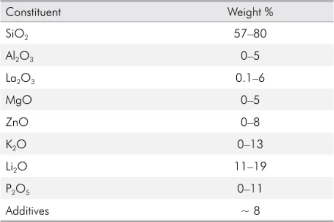
or ingots with composition according to Table 2.44
Depending on the type of piece produced, whether
it was a “blue” block for CAD-CAM system or the
ingot for hot-pressed technique, the crystallization technique of this glass-ceramic changed. However, the
crystallization process was similar in all situations.
Table 1. Mechanical properties for the traditional glass-ceramics in the market.40,41,42
Material
Flexural strength (MPa)
Fracture toughness (MPa.m1/2)
Hardness (GPa)
Leucite glass-ceramic 164 1.03 6.5 Lithium disilicate glass-ceramic 365 2.80 5.3
Table 2. Composition of the glass-ceramic lithium disilicate IPS Empress® 2 raw powder.44
Constituent Weight %
SiO2 57–80
Al2O3 0–5
La2O3 0.1–6
MgO 0–5
ZnO 0–8
K2O 0–13
Li2O 11–19
P2O5 0–11
Briely, the crystallization of the lithium disilicate is controlled by a heating cycle, in which lithium
metasilicate (Li2SiO3) reacts with the glassy phase
(SiO2) to originate lithium disilicate (Li2Si2O5).45 Lithium metasilicate is nucleated from the base glass (Li3PO4, amorphous) at the initial temperatures of
the cycle. Later on, lithium disilicate glass-ceramics
underwent some changes and gave rise to IPS e.max
Lithium Disilicate (Ivoclar Vivadent Ltda., Barueri, Brazil), which has better mechanical properties,
mostly due to the decrease in the size of the
platelet-shaped crystals (length varying from 2.0 to 3.0 µm)
and the increase in interlocking among crystals.46,47
Despite the great acceptance and broad use of lithium
disilicate glass-ceramics, the evolution of dental materials
has attempted to suppress the remaining disadvantages of this ceramic system by means of the development of glass-ceramics reinforced with polycrystalline ceramics. These new glass-ceramics were designed to contain lithium silicate as the main crystalline phase in a vitreous matrix reinforced with zirconium dioxide
crystals (~10%).48 When this material goes through the
crystallization process, the nucleated lithium silicate crystals achieve a mean size (0.5 to 1 μm) that is up to
6 times smaller than that observed for lithium disilicate
crystals present in lithium disilicate glass-ceramics.49
The formation of a smaller and iner crystalline phase
occurs due to the presence of zirconia particles in
the material, which acts as an additive inluencing
the crystallization by hindering crystal growth.50
A microstructure containing smaller crystals guarantees to this material mechanical properties similar to those observed for lithium disilicate ceramics.47 Additionally,
as observed for traditional glass-ceramics, these new
zirconium-reinforced lithium silicate materials maintain
good optical properties, are easily milled in CAD-CAM machines and attain good surface inishing, as they
still have a high amount of glass matrix.51
The two existing commercial examples of lithium
silicate glass-ceramics are: a) Suprinity (Vita Zahnfabrik, Bad Sachingen, Germany), a material marketed in
a partially crystallized state and that requires an additional thermal cycle in a furnace; and b) CELTRA
Duo (Dentisply-Sirona, Bensheim, Germany), a material that is already in its inal crystallization stage. Both
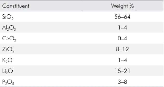
materials have similar composition as shown in Table 3.52
These novel zirconia-reinforced lithium silicate glass-ceramics have good mechanical properties
associated with an excellent esthetic quality, thus being
a valid alterative to lithium disilicate materials for prosthetic rehabilitations with high aesthetic demand. The main advantage of these materials is their timesaving
ability for the production of dental restorations, since
they are faster to be milled in CAD-CAM machines than lithium disilicate glass-ceramics53 and are already
offered in their fully crystallized state (CELTRA Duo, Dentisply-Sirona, Bensheim, Germany) no furnace need) or need a very short crystallization cycle (Suprinity, Bad Sachingen, Germany). A particular advantage of
the lithium silicate ceramic over the lithium disilicate version is the superior polishability due to the smaller crystal sizes in the microstructure.
Polymer infiltrated ceramic networks (PICNs)
In the last decades, the use of CAD-CAM systems in dentistry has increased exponentially, especially
because of the general trends towards high productivity
and aesthetics.54,55 Although CAD-CAM systems were
developed initially for the production of ceramic
restorations, pre-polymerized resin composites blocks
have also been developed to be used with these systems. One of the first resin composites developed as a
CAD-CAM block was ParadigmTM (3MTM, St Paul, USA),
which was considered a fast-milling and wear-friendly
alternative to the use of ceramics. However, problems
commonly related to resin composite systems still
need to be overcome, such as the reduced mechanical
properties and poor wear resistance.54
Recently, a new material has been developed by Vita (VITA Zahnfabrik, Bad Säckingen, Germany) which is marketed as a polymer iniltrated in a porous ceramic,
Table 3. Composition of lithium silicate-based glass-ceramics.52
Constituent Weight %
SiO2 56–64
Al2O3 1–4
CeO2 0–4
ZrO2 8–12
K2O 1–4
Li2O 15–21
generating an interpenetrating network (polymer
iniltrated ceramic network, PICN). This new material was developed based on the glass iniltrated ceramic technology (In-Ceram System, Vita, Bad Sachingen, Germany), which was originally released by Vita in the 90’s.54 The iniltration of a resin into a porous
ceramic preform is signiicantly different from the infiltration of a glass, since the final shrinkage of the polymer after iniltration is almost 5%, i.e., much
greater than the shrinkage experienced upon cooling
of the iniltration glass, which is in the order of 1%.56
PICNs have the advantage of presenting an elastic
modulus that is approximately 50% lower compared
to feldspathic ceramics and hence closer to that of
dentin, they are easier to mill and adjust, and also
can be more easily repaired by composite resins.56
In comparison to dental porcelains, this new material has
been proven to have lower elastic modulus and higher damage tolerance.57 In 2013, the product Enamic (Vita,
Bad Sachingen, Germany) was introduced for dental
restorations. This PICN is based on initial sintering of a
porcelain powder to approximately 70% of its full density, followed by iniltration with a monomer mixture.54,58,59
The material is considered a resin-ceramic composite
material, composed of two interconnected networks: a
dominant ceramic and a polymer. Recent publications showed that the polymeric part of this material is
composed of urethane dimethacrylate (UDMA) and
triethylene glycol dimethacrylate (TEGDMA)
cross-linked polymers.60 Compositional analyses of the
dominant ceramic network revealed a major ceramic
phase, composed (by weight) of SiO2 (58–63%), Al2O3
(20–23%), Na2O (9–11%), K2O (4–6%), B2O3 (0.5–2%), CaO
(<1%) and TiO2(<1%).61 Although being marketed as a
polymer iniltrated ceramic, scientiic analysis has shown
that the inorganic matrix is rather an amorphous glass. A recent publication62 reported that Enamic showed elastic modulus values similar to that reported by the
manufacturer (around 30 GPa), however, the fracture
toughness values measured in this investigation
(0.86 MPa.m1/2) were lower than that reported by the manufacturer (1.5 MPa.m1/2). The fracture toughness
value obtained for PICN was similar to that of the
feldspathic ceramic evaluated. Therefore, the authors
rejected the hypothesis that the presence of a polymer network would create toughening mechanisms in the
microstructure of the material. In addition, this study
showed that PICN had increased susceptibility to SCG compared to a feldsphatic ceramic. This raised the question if the polymer is susceptible to water permeation and degradation.
PICNs have positive properties related to both the
ceramic and composites, with an interesting balance between elasticity and strength, being indicated for single crowns, inlays, onlays and veneers. The polymeric part has a strength below 30 MPa and the ceramic network has a strength around 160 MPa, whereas the inal PICN has strength of 135 MPa. As expected for a composite material, the properties are intermediate between those of ceramics and particle-illed resins.60,61,63
The elastic modulus of these materials is in the
range of 30 GPa, which is half of that reported for
conventional veneering ceramics but closer to what
is usually reported for dentin (15–20 GPa).58,63 Typical
ceramic materials have a higher elastic modulus
values than PICN64. The Vickers hardness of human
enamel (3.43 ± 0.16 GPa) and PICN (3.31 ± 0.11 GPa) are similar,54,65,66,67 and both are higher than the hardness reported for resin composites (0.73 GPa to 1.60 GPa),68,69 and lower than the hardness of zirconia (13.94 GPa)70 and lithium disilicate glass ceramics (10.0 GPa to 11.31 GPa).71 The lexural strength of Enamic (130 MPa)56
is lower than that of a reference lithium disilicate
glass-ceramic material, IPS e.max (342 MPa).72 PICN has a higher tolerance to diamond bur grinding damage
than other CAD/CAM and pressed materials.54,60,73
A study evaluated the damage tolerance of different dental materials and showed that the damage tolerance of PICN was higher when compared to other ceramics
for CAD-CAM, like veneering ceramics.55,73
With respect to optical properties, the shrinkage
of the curing resin results in interfacial stresses occurring between the ceramic framework and the polymer resulting in debonding and leading to a higher opacity because of the gaps developed at
the interface. The selection of resin, the application of high pressure during the curing phase, and the
silanization process enhanced bonding and helped overcoming the aesthetic problems by increasing the
translucency of the material.64 However, Enamic has
been shown to be less translucent than IPS e.max or
works have also shown that the surface of PICN is not as glossy as those obtained for IPS e.max or Lava
Ultimate. Nevertheless, the stain resistance of PICN was superior than that measured for Lava Ultimate
and inferior than that reported for IPS e.max.56,75
Clinical simulations show promising lifetime
results for PICN. A chewing simulation of ive years demonstrated that none of the Enamic crowns failed,
while six IPS e.max CAD had minor cracking and
twelve Vita Mark II restorations revealed signiicant
crack failures.64 In a cyclic fatigue experiment of
500,000 cycles, Enamic performed as well as a lithium
disilicate glass-ceramic.58,76 Based on the reduced
elastic modulus of Enamic, this material is especially
indicated for prosthetic treatments on stiff implants.
Due to the inferior optical properties, PICNs are more
suitable in the molar than in the anterior region.
Novel processing technologies
CAD-CAM refers to a computer system that is used to both design and manufacture a dental restoration.
CAD technology uses a software to deine the shape and dimensions of the restoration, while CAM
technology takes the designed model to a computer numeric control (CNC) machine to manufacture the
restoration, usually from a block made of a dental
material (subtractive manufacturing).
Currently, the production of metal-free restorations
using polycrystalline ceramic infrastructures (e.g.,
Y-TZP) depends on the use of the CAD-CAM systems.77
The introduction of CAD-CAM milling systems for the production of restorations with these polycrystalline ceramics allowed their use in prosthetic restorations
with greater reliability, since the only manufacturing technique available in the past was slip-casting, which
resulted in a greater number of defects and cracks in
the microstructure of the inal restorations.78 CAD-CAM systems have been used in Dentistry
for almost 30 years,79 and during this period different
machines have been launched, as these systems are
constantly evolving and producing restorations with much better adaptation.80,81 Moreover, the evolution of CAD-CAM system have allowed their use to produce restorations with other materials such as veneering
ceramics, resin composites and metal alloys.77,82
Among dental CAD-CAM systems, there are two
types of techniques for producing restorations. The
irst one is the machining of the prosthetic restoration from a block of the sintered material, while the second
consists of machining a block in a partially sintered
state with subsequent inal sintering in a speciic
furnace. Both techniques are used in dentistry and each of them have their advantages and disadvantages. Machining a block of sintered material provides the restoration with a greater precision of its contours
and shape in addition to saving clinical time, since
the restoration does not require an additional heat
treatment. However, when machining a material with high strength like polycrystalline ceramics, both the
wear of the machining unit tools and the machining
time are very high. Also, machining brittle materials
such as dental ceramics can lead to the formation of
microcracks and surface defects.83 On the other hand,
when the restoration is produced from a partially sintered
block, there is the advantage of promoting healing of
machining microcracks during the subsequent sintering process.84 This processing technique is expected to have
a shorter machining time for a less dense material, but one must keep in mind that the inal sintering will promote dimensional changes due to shrinkage, which may lead to prosthetic restoration misit.85
Although the CAD-CAM systems described above
are already well established in the dental market, they
present a major drawback related to the great waste of material upon machining. The waste corresponds
to approximately 90% of the prefabricated block
for a typical restoration86 and leftovers from these
dental restorations are not reusable. Therefore, new
technologies have been developed to overcome this problem. Some of them produce the restoration by means of adding layers instead of grinding pre-fabricated blocks (additive manufacturing).
Addition CAD-CAM systems, also called “solid free-form fabrication”, are still a focus of research and
development for polycrystalline ceramic materials and there are three techniques that have stood out recently.86,87,88,89,90 These techniques are: 1) Selective
Laser Sintering or Melting, 2) Direct 3D Printing and
3) Stereolithography.
The Selective Laser Sintering or Melting is an
but is still in development for polycrystalline ceramics
(BEGO Medifacturing® System, BEGO Medical GmbH, Bremen, Germany). In this technique, the
laser beam sinters thin layers of a ceramic from a
container illed with powder to create a single coping or framework, in which each layer represents a cross
section of the CAD model.86 Direct 3D Printing is
similar to a traditional inkjet printer, performing the direct printing of a ceramic suspension, allowing the generation of dense green bodies with high resolution,
and producing complex shapes.90,91
Stereolithography is frequently used nowadays,
and has already evolved enough to allow production of
more complex ceramic pieces, whereas the previously
mentioned techniques are in the early development stage for dental applications. Stereolithography is
similar to 3D printing, however it makes use of a
suspension containing ceramic particles mixed with a
resin components (acrylates or epoxy monomers).87,88
This resin part is polymerized during printing to shape the solid object and is subsequently removed during the ceramic sintering process. The great advantage associated with all additive techniques is that they provide minimal or no material waste. One still existing disadvantage of all additive methods
to date is the rough surface quality and the poor it
or marginal precision.
Considering the mentioned additive methods, Direct 3D Printing is the technique that stands out,
as the equipment is relatively more accessible and allows for the production of a dense green body ready
for sintering. In 2009,91 using a modiied inkjet printer, a zirconia crown was manufactured with suficient
mechanical properties to be used in the oral cavity. The impression of the posterior dental crown was
performed using a cartridge illed with a 27 vol%
solid content of zirconia-based ceramic suspension. Variations of the Direct 3D Printing technique have
also been studied, the so-called “Robocasting”.89
Both techniques are very similar, differing only in
the way the deposition of the ceramic suspension is
made. Robocasting uses extruded ilaments instead
of ejected droplets to produce the object.
Conclusion
Dental ceramics and processing technologies have
evolved signiicantly in the past ten years, with most of
the evolution being related to new microstructures and
CAD-CAM methods. Also, a trend towards the use of
monolithic restorations has changed the way clinicians
produce all-ceramic dental prostheses, since the more
aesthetic multilayered restorations unfortunately are more prone to chipping or delamination. Composite materials processed via CAD-CAM have become
an interesting option, as they have intermediate
properties between ceramics and polymers and are more easily milled and polished.
1. Marchack BW, Sato S, Marchack CB, White SN. Complete and partial contour zirconia designs for crowns and fixed dental prostheses: a clinical report. J Prosthet Dent. 2011;106(3):145-52. https://doi.org/10.1016/S0022-3913(11)60112-1
2. Schley JS, Heussen N, Reich S, Fischer J,
Haselhuhn K, Wolfart S. Survival probability of zirconia‐ based fixed dental prostheses up to 5 yr: a systematic review of the literature. Eur J Oral Sci. 2010;118(5):443-50. https://doi.org/10.1111/j.1600-0722.2010.00767.x 3. Zhang H, Li Z, Kim B-N, Morita K, Yoshida H,
Hiraga K et al. Effect of alumina dopant on transparency of tetragonal zirconia. J Nanomater. 2012;2012:ID269064. https://doi.org/10.1155/2012/269064
4. Zhang Y. Making yttria-stabilized tetragonal zirconia translucent. Dent Mater. 2014;30(10):1195-203. https://doi.org/10.1016/j.dental.2014.08.375
5. Cheng J, Agrawal D, Zhang Y, Roy R. Microwave sintering of transparent alumina. Mater Lett. 2002;56(4):587-92. https://doi.org/10.1016/S0167-577X(02)00557-8 6. Klimke J, Trunec M, Krell A. Transparent
tetragonal yttria‐stabilized zirconia ceramics: influence of scattering caused by birefringence. J Am Ceram Soc. 2011;94(6):1850-8.
https://doi.org/10.1111/j.1551-2916.2010.04322.x 7. Kim HK, Kim SH. Optical properties of pre-colored dental
monolithic zirconia ceramics. J Dent. 2016;55:75-81. https://doi.org/10.1016/j.jdent.2016.10.001
8. Sedda M, Vichi A, Carrabba M, Capperucci A, Louca C, Ferrari M. Influence of coloring procedure on flexural resistance of zirconia blocks. J Prosthet Dent. 2015;114(1):98-102. https://doi.org/10.1016/j.prosdent.2015.02.001 9. Kim HK, Kim SH. Effect of the number of coloring
liquid applications on the optical properties of monolithic zirconia. Dent Mater. 2014;30(9):e229-37. https://doi.org/10.1016/j.dental.2014.04.008
10. Rinke S, Fischer C. Range of indications for translucent zirconia modifications: clinical and technical aspects. Quintessence Int. 2013;44(8):557-66. https://doi.org/10.3290/j.qi.a29937 11. Kim HK, Kim SH, Lee JB, Han JS, Yeo IS, Ha SR. Effect of
the amount of thickness reduction on color and translucency of dental monolithic zirconia ceramics. J Adv Prosthodont. 2016;8(1):37-42. https://doi.org/10.4047/jap.2016.8.1.37 12. Antonson SA, Anusavice KJ. Contrast ratio of veneering and
core ceramics as a function of thickness. Int J Prosthodont. 2001;14(4):316-20.
13. Stawarczyk B, Frevert K, Ender A, Roos M, Sener B, Wimmer T. Comparison of four monolithic zirconia materials with conventional ones: contrast ratio, grain size, four-point flexural strength and two-body wear. J Mech Behav Biomed Mater. 2016;59:128-38. https://doi.org/10.1016/j.jmbbm.2015.11.040
14. Lawson NC, Janyavula S, Syklawer S, McLaren EA, Burgess JO. Wear of enamel opposing zirconia and lithium disilicate after adjustment, polishing and glazing. J Dent. 2014;42(12):1586-91. https://doi.org/10.1016/j.jdent.2014.09.008
15. Park JH, Park S, Lee K, Yun KD, Lim HP. Antagonist wear of three CAD/CAM anatomic contour zirconia ceramics. J Prosthet Dent. 2014;111(1):20-9. https://doi.org/10.1016/j.prosdent.2013.06.002 16. Jung YS, Lee JW, Choi YJ, Ahn JS, Shin SW, Huh JB.
A study on the in-vitro wear of the natural tooth structure by opposing zirconia or dental porcelain. J Adv Prosthodont. 2010;2(3):111-15. https://doi.org/10.4047/jap.2010.2.3.111 17. Lohbauer U, Reich S. Antagonist wear of monolithic zirconia crowns after 2 years. Clin Oral Investig. 2017;21(4):1165-72. https://doi.org/10.1007/s00784-016-1872-6
18. Hamza TA, Sherif RM. In vitro evaluation of marginal discrepancy of monolithic zirconia restorations fabricated with different CAD-CAM systems. J Prosthet Dent. 2017;117(6):762-6. http://dx.doi.org/10.1016/j.prosdent.2016.09.011
19. Flinn BD, Raigrodski AJ, Mancl LA, Toivola R, Kuykendall T. Influence of aging on flexural strength of translucent zirconia for monolithic restorations. J Prosthet Dent. 2017;117(2):302-9. https://doi.org/10.1016/j.prosdent.2016.06.010
20. Pereira G, Silvestri T, Camargo R, Rippe MP, Amaral M, Kleverlaan CJ et al. Mechanical behavior of a Y-TZP ceramic for monolithic restorations: effect of grinding and low-temperature aging. Mater Sci Eng C. 2016;63:70-7. https://doi.org/10.1016/j.msec.2016.02.049
21. Bömicke W, Rammelsberg P, Stober T, Schmitter M. Short‐term prospective clinical evaluation of monolithic and partially
veneered zirconia single crowns. J Esthet Restor Dent. 2017;29(1):22-30. https://doi.org/10.1111/jerd.12270 22. Sulaiman TA, Abdulmajeed AA, Donovan TE,
Cooper LF, Walter R. Fracture rate of monolithic zirconia restorations up to 5 years: a dental laboratory survey. Prosthet Dent. 2016,116(3):436-9. https://doi.org/10.1016/j.prosdent.2016.01.033 23. Pjetursson BE, Sailer I, Makarov NA, Zwahlen M,
Thoma DS. All-ceramic or metal-ceramic tooth-supported fixed dental prostheses (FDPs)? A systematic review of the survival and complication rates. Partm: multiple-unit FDPs. Dent Mater. 2015;31(6):624-39. https://doi.org/10.1016/j.dental.2015.02.013 24. Lucas LC, Lemons JE. Biodegradation of restorative
metallic systems. Adv Dent Res. 1992;6(1):32-7. https://doi.org/10.1177/08959374920060011301 25. Piconi C, Maccauro G. Zirconia as a ceramic
biomaterial. Biomaterials. 1999;20(1):1-25. https://doi.org/10.1016/S0142-9612(98)00010-6
26. Sailer I, Fehér A, Filser F, Gauckler LJ, Lüthy H, Hämmerle CH. Five-year clinical results of zirconia frameworks for posterior fixed partial dentures. Int J Prosthodont. 2007;20(4):383-8. 27. Guess PC, Bonfante EA, Silva NR, Coelho PG,
Thompson VP. Effect of core design and veneering technique on damage and reliability of Y-TZP-supported crowns. Dent Mater. 2013;29(3):307-16.
https://doi.org/10.1016/j.dental.2012.11.012
28. White SN, Miklus VG, McLaren EA, Lang LA, Caputo AA. Flexural strength of a layered zirconia and porcelain dental all-ceramic system. J Prosthet Dent. 2005;94(2):125-31. https://doi.org/10.1016/j.prosdent.2005.05.007 29. Al-Amleh B, Neil Waddell J, Lyons K,
Swain MV. Influence of veneering porcelain thickness and cooling rate on residual stresses in zirconia molar crowns. Dent Mater. 2014;30(3):271-80. https://doi.org/10.1016/j.dental.2013.11.011
30. Stawarczyk B, Ozcan M, Roos M, Trottmann A, Sailer I, H6ammerle CH. Load-bearing capacity and failure types of anterior zirconia crowns veneered with overpressing and layering techniques. Dent Mater. 2011;27(10):1045-53. https://doi.org/10.1016/j.dental.2011.07.006
31. Schmitter M, Mueller D, Rues S. Chipping behaviour of all-ceramic crowns with zirconia framework and
CAD/CAM manufactured veneer. J Dent. 2012;40(2):154-62. https://doi.org/10.1016/j.jdent.2011.12.007
32. Schmitter M, Schweiger M, Mueller D, Rues S. Effect on in vitro fracture resistance of the technique used to attach lithium disilicate ceramic veneer to zirconia frameworks. Dent Mater. 2014;30(2):122-30. https://doi.org/10.1016/j.dental.2013.10.008 33. Choi YS, Kim SH, Lee JB, Han JS, Yeo IS. In vitro evaluation of
34. Anusavice KJ, Kakar K, Ferree N. Which mechanical and physical testing methods are relevant for predicting the clinical performance of ceramic-based dental prostheses? Clin Oral Implants Res. 2007;18 Suppl 3:218-31. https://doi.org/10.1111/j.1600-0501.2007.01460.x 35. Höland W, Beall GH. Glass ceramic technology. 2nd ed.
Hoboken: John Wiley & Sons; 2012.
36. Höland W, Rheinberger V, Apel E, Hoen C, Höland M, Dommann A et al. Clinical applications of glass-ceramics in dentistry. J Mater Sci Mater Med. 2006;17(11):1037-42. https://doi.org/10.1007/s10856-006-0441-y
37. Ritzberger C, Apel E, Höland W, Peschke A, Rheinberger VM. Properties and clinical application of three types of dental glass-ceramics and ceramics for CAD-CAM technologies. Mater. 2010;3(6):3700-13. https://doi.org/10.3390/ ma3063700
38. MacCulloch WT. Advances in dental ceramics. Br Dent J. 1968;124(8):361-5.
39. Höland W, Schweiger M, Frank M, Rheinberger V. A comparison of the microstructure and properties of the IPS Empress 2 and the IPS Empress glass-ceramics. J Biomed Mater Res. 2000;53(4):297-303. https:// doi.org/10.1002/1097-4636(2000)53:4<297::AID-JBM3>3.0.CO;2-G
40. Lin WS, Ercoli C, Feng C, Morton D. The effect of core material, veneering porcelain, and fabrication technique on the biaxial flexural strength and weibull analysis of selected dental ceramics. J Prosthodont. 2012;21(5):353-62. https://doi.org/10.1111/j.1532-849X.2012.00845.x
41. Quinn JB, Sundar V, Lloyd IK. Influence of
microstructure and chemistry on the fracture toughness of dental ceramics. Dent Mater. 2003;19(7):603-11. https://doi.org/10.1016/S0109-5641(03)00002-2 42. Guazzato M, Albakry M, Ringer SP, Swain MV. Strength,
fracture toughness and microstructure of a selection of all-ceramic materials. Part I. Pressable and alumina glass-infiltrated ceramics. Dent Mater. 2004;20(5):441-8. https://doi.org/10.1016/j.dental.2003.05.003
43. Kern M, Sasse M, Wolfart S. Ten-year outcome of three-unit fixed dental prostheses made from monolithic lithium disilicate ceramic. J Am Dent Assoc. 2012;143(3):234-40. https://doi.org/10.14219/jada.archive.2012.0147
44. Schweiger M, Höland W, Frank M, Drescher H, Rheinberger V. IPS Empress 2: a new pressable high-strength glass-ceramic for esthetic all-ceramic restorations. Quintessence Dent Technol. 1999;22:143-51.
45. Ritzberger C, Schweiger M, Höland W. Principles of crystal phase formation in Ivoclar Vivadent glass-ceramics for dental restorations. J Non Cryst Solids. 2016;432 Part A:137-42. https://doi.org/10.1016/j.jnoncrysol.2015.04.034 46. Kang SH, Chang J, Son HH. Flexural strength
and microstructure of two lithium disilicate glass ceramics for CAD/CAM restoration in the dental clinic. Restor Dent Endod. 2013;38(3):134-40. https://doi.org/10.5395/rde.2013.38.3.134
47. Wendler M, Belli R, Petschelt A, Mevec D, Harrer W, Lube T et al. Chairside CAD/CAM materials. Part 2: flexural strength testing. Dent Mater. 2017;33(1):99-109. https://doi.org/10.1016/j.dental.2016.10.008
48. Rinke S, Pabel A-K, diger M, Ziebolz D. Chairside Fabrication of an all-ceramic partial crown using a zirconia-reinforced lithium silicate ceramic. Case Rep Dent. 2016;2016:ID1354186. http://dx.doi.org/10.1155/2016/1354186 49. Belli R, Wendler M, de Ligny D, Cicconi MR,
Petschelt A, Peterlik H et al. Chairside CAD/CAM materials. Part 1: measurement of elastic constants and microstructural characterization. Dent Mater. 2017;33(1):84-98.
https://doi.org/10.1016/j.dental.2016.10.009
50. Apel E, van’t Hoen C, Rheinberger V, Höland W. Influence of ZrO2 on the crystallization and properties of lithium disilicate glass-ceramics derived from a multi-component system. J Eur Ceram Soc. 2007;27(2-3):1571-77. https://doi.org/10.1016/j.jeurceramsoc.2006.04.103 51. Krüger S, Deubener J, Ritzberger C, Höland W. Nucleation
kinetics of lithium metasilicate in ZrO2‐bearing lithium disilicate glasses for dental application. Int J Appl Glass Sci 2013;4(1):9-19. https://doi.org/10.1111/ijag.12011 52. Gracis S, Thompson VP, Ferencz JL, Silva NR, Bonfante EA.
A new classification system for all-ceramic and ceramic-like restorative materials. Int J Prosthodont. 2015;28(3):227-35. https://doi.org/10.11607/ijp.4244
53. Rinke S, Pabel AK, Rodiger M, Ziebolz D. Chairside Fabrication of an all-ceramic partial crown using a zirconia-reinforced lithium silicate ceramic. Case Rep Dent. 2016;2016:ID1354186. http://dx.doi.org/10.1155/2016/1354186
54. He LH, Swain M. A novel polymer infiltrated ceramic dental material. Dent Mater. 2011;27(6):527-34. https://doi.org/10.1016/j.dental.2011.02.002
55. Coldea A, Fischer J, Swain MV, Thiel N. Damage tolerance of indirect restorative materials (including PICN) after simulated bur adjustments. Dent Mater. 2015;31(6):684-94. https://doi.org/10.1016/j.dental.2015.03.007
56. Chirumamilla G, Goldstein CE, Lawson NC. A 2‐year retrospective clinical study of enamic crowns performed in a private practice setting. J Esthet Restor Dent. 2016;28(4):231-7. https://doi.org/10.1111/jerd.12206 57. Coldea A, Swain MV, Thiel N. In-vitro strength degradation
of dental ceramics and novel PICN material by sharp indentation. J Mech Behav Biomed Mater. 2013;26:34-42. https://doi.org/10.1016/j.jmbbm.2013.05.004
58. Denry I, Kelly JR. Emerging ceramic-based materials for dentistry. J Dent Res. 2014;93(12):1235-42. https://doi.org/10.1177/0022034514553627
59. Ruse ND, Sadoun MJ. Resin-composite blocks for dental CAD/CAM applications. J Dent Res. 2014;93(12):1232-4. https://doi.org/10.1177/0022034514553976
61. Shetty R, Shenoy K, Dandekeri S, Suhaim KS, Ragher M, Francis J. Resin-matrix ceramics: an overview. Int J Rec Sci Res. 2015;6(11):7414-17.
62. Ramos NC, Campos TMB, Paz IS, Machado JPB, Bottino MA, Cesar PF et al. Microstructure characterization and SCG of newly engineered dental ceramics. Dent Mater. 2016;32(7):870-8. https://doi.org/10.1016/j.dental.2016.03.018 63. Della Bona A, Corazza PH, Zhang Y.
Characterization of a polymer-infiltrated ceramic-network material. Dent Mater. 2014;30(5):564-9. https://doi.org/10.1016/j.dental.2014.02.019 64. Swain MV, Coldea A, Bilkhair A, Guess PC.
Interpenetrating network ceramic-resin composite dental restorative materials. Dent Mater. 2016;32(1):34-42. https://doi.org/10.1016/j.dental.2015.09.009 65. Padmanabhan SK, Balakrishnan A, Chu MC, Kim
TN, Cho SJ. Micro-indentation fracture behavior of human enamel. Dent Mater. 2010;26(1):100-04. https://doi.org/10.1016/j.dental.2009.07.015 66. Albero A, Pascual A, Camps I, Grau-Benitez M.
Comparative characterization of a novel cad-cam polymer-infiltrated-ceramic-network. J Clin Exp Dent. 2015;7(4):e495-500. https://doi.org/10.4317/jced.52521 67. Min J, Arola DD, Yu D, Yu P, Zhang Q,
Yu H et al. Comparison of human enamel and polymer-infiltrated-ceramic-network material “ENAMIC” through micro-and nano-mechanical testing. Ceram Int. 2016;42(9):10631-7. https://doi.org/10.1016/j.ceramint.2016.03.160 68. El-Safty S, Akhtar R, Silikas N, Watts DC.
Nanomechanical properties of dental
resin-composites. Dent Mater. 2012;28(12):1292-300. https://doi.org/10.1016/j.dental.2012.09.007 69. Xu HH, Smith DT, Schumacher GE, Eichmiller FC,
Antonucci JM. Indentation modulus and hardness of whisker-reinforced heat-cured dental resin composites. Dent Mater. 2000;16(4):248-54. https://doi.org/10.1016/S0109-5641(00)00014-2 70. Zhang Y, Allahkarami M, Hanan JC. Measuring residual
stress in ceramic zirconia-porcelain dental crowns by nanoindentation. J Mech Behav Biomed Mater. 2012;6:120-7. https://doi.org/10.1016/j.jmbbm.2011.11.006
71. Smith CM, Jiang D, Gong J, Yin L. Determination of the mechanical behavior of lithium disilicate glass ceramics by nanoindentation & scanning probe microscopy. Mater Chem Phys. 2014;148(3):1036-44. https://doi.org/10.1016/j.matchemphys.2014.09.015 72. Leung BT, Tsoi JK, Matinlinna JP, Pow EH. Comparison
of mechanical properties of three machinable ceramics with an experimental fluorophlogopite glass ceramic. J Prosthet Dent. 2015;114(3):440-6. https://doi.org/10.1016/j.prosdent.2015.02.024 73. Tsitrou EA, Northeast SE, Noort R. Brittleness index
of machinable dental materials and its relation to the marginal chipping factor. J Dent. 2007;35(12):897-902. https://doi.org/10.1016/j.jdent.2007.07.002
74. Awad D, Stawarczyk B, Liebermann A, Ilie N. Translucency of esthetic dental restorative CAD/CAM materials and composite resins with respect to thickness and surface roughness. J Prosthet Dent. 2015;113(6):534-40. https://doi.org/10.1016/j.prosdent.2014.12.003 75. Lawson NC, Burgess JO. Gloss and stain resistance
of ceramic‐polymer CAD/CAM restorative blocks. J Esthet Restor Dent. 2016;28 Suppl 1:S40-5. https://doi.org/10.1111/jerd.12166
76. Kelly JR, Rungruanganunt P, Hunter B, Vailati F. Development of a clinically validated bulk failure test for ceramic crowns. J Prosthet Dent. 2010;104(4):228-38. https://doi.org/10.1016/S0022-3913(10)60129-1 77. Raigrodski AJ. Contemporary materials and technologies
for all-ceramic fixed partial dentures: a review of the literature. J Prosthet Dent. 2004;92(6):557-62. https://doi.org/10.1016/j.prosdent.2004.09.015
78. Anusavice KJ. Reducing the failure potential of ceramic-based restorations. Part 2: ceramic inlays, crowns, veneers, and bridges. Gen Dent. 1997;45(1):30-5.
79. Duret F, Blouin JL, Duret B. CAD-CAM in dentistry. J Am Dent Assoc. 1988;117(6):715-20. https://doi.org/10.14219/jada.archive.1988.0096
80. Liu PR. A panorama of dental CAD/CAM restorative systems. Compend Contin Educ Dent. 2005;26(7):507-10.
81. Miyazaki T, Hotta Y, Kunii J, Kuriyama S, Tamaki Y. A review of dental CAD/CAM: current status and future perspectives from 20 years of experience. Dent Mater J. 2009;28(1):44-56. https://doi.org/10.4012/dmj.28.44
82. Stansbury JW, Idacavage MJ. 3D printing with polymers: challenges among expanding options and opportunities. Dent Mater. 2016;32(1):54-64. https://doi.org/10.1016/j.dental.2015.09.018
83. Huang H. Machining characteristics and surface integrity of yttria stabilized tetragonal zirconia in high speed deep grinding. Mater Sci Eng A. 2003;345(1–2):155-63. https://doi.org/10.1016/S0921-5093(02)00466-5 84. Wang H, Aboushelib MN, Feilzer AJ. Strength
influencing variables on CAD/CAM zirconia frameworks. Dent Mater. 2008;24(5):633-8. https://doi.org/10.1016/j.dental.2007.06.030 85. Kohorst P, Junghanns J, Dittmer MP, Borchers L, Stiesch M. Different CAD/CAM-processing routes for zirconia restorations: influence on fitting accuracy. Clin Oral Investig. 2011;15(4):527-36. https://doi.org/10.1007/s00784-010-0415-9
86. Strub JR, Rekow ED, Witkowski S. Computer-aided design and fabrication of dental restorations: current systems and future possibilities. J Am Dent Assoc. 2006;137(9):1289-96. https://doi.org/10.14219/jada.archive.2006.0389
88. Doreau F, Chaput C, Chartier T. Stereolithography for manufacturing ceramic parts. Adv Eng Mater. 2000;2(8):493-6. https://doi.org/10.1002/1527-2648(200008)2:8<493::AID-ADEM493>3.0.CO;2-C 89. Silva NR, Witek L, Coelho PG, Thompson VP,
Rekow ED, Smay J. Additive CAD/CAM process for dental prostheses. J Prosthodont. 2011;20(2):93-6. https://doi.org/10.1111/j.1532-849X.2010.00623.x
90. Özkol E, Zhang W, Ebert J, Telle R. Potentials of the “Direct inkjet printing” method for manufacturing 3Y-TZP based dental restorations. J Eur Ceram Soc. 2012;32(10):2193-201. https://doi.org/10.1016/j.jeurceramsoc.2012.03.006 91. Ebert J, Ozkol E, Zeichner A, Uibel K, Weiss O,