Biomolecules 2020, 10, 717; doi:10.3390/biom10050717 www.mdpi.com/journal/biomolecules Helena Laronha 1,2, Inês Carpinteiro 1, Jaime Portugal 3, Ana Azul 1, Mário Polido 1,
Krasimira T. Petrova 2, Madalena Salema-Oom 1,2 and Jorge Caldeira 1,2,*
1 Centro de Investigação Interdisciplinar Egas Moniz, Instituto Universitário Egas Moniz,
2829-511 Caparica, Portugal; h.laronha@campus.fct.unl.pt (H.L.); icarpinteiro@egasmoniz.edu.pt (I.C.); jaime.portugal@fmd.ulisboa.pt (J.P:) ; aazul@egasmoniz.edu.pt (A.A.); mpolido@egasmoniz.edu.pt (M.P.); moom@egasmoniz.edu.pt (M.S.-O.)
2 UCIBIO and LAQV, Requimte, Faculdade de Ciências e Tecnologia, Universidade Nova de Lisboa,
2829-516 Caparica, Portugal; k.petrova@fct.unl.pt
3 Faculdade de Medicina Dentária Universidade de Lisboa, 1649-003 Lisboa, Portugal;
jaime.portugal@fmd.ulisboa.pt
* Correspondence: jcaldeira@egasmoniz.edu.pt; Tel.: +351-919553592
Received: 9 April 2020; Accepted: 30 April 2020; Published: 5 May 2020
Abstract: Matrix metalloproteinases are enzymes that degrade the extracellular matrix. They have different substrates but similar structural organization. Matrix metalloproteinases are involved in many physiological and pathological processes and there is a need to develop inhibitors for these enzymes in order to modulate the degradation of the extracellular matrix (ECM). There exist two classes of inhibitors: endogenous and synthetics. The development of synthetic inhibitors remains a great challenge due to the low selectivity and specificity, side effects in clinical trials, and instability. An extensive review of currently reported synthetic inhibitors and description of their properties is presented.
Keywords: matrix metalloproteinases; TIMP; synthetic inhibitors
1. Introduction
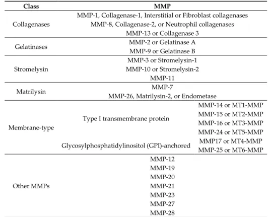
Matrix metalloproteinases (MMPs) are a protein family within the metzincin superfamily, comprising zinc-dependent endopeptidases with similar structural characteristics but with different substrate preferences. MMPs are produced and secreted from cells as inactive proenzymes depending, herein, on a structural alteration for activation [1–6]. In human tissues, there are 23 different types of MMPs expressed and they can be subdivided according to their substrate specificity, sequential similarity, and domain organization [1,2,4,7–17] (Table 1).
MMP-9 or Gelatinase B Stromelysin MMP-3 or Stromelysin-1 MMP-10 or Stromelysin-2 MMP-11 Matrilysin MMP-7 MMP-26, Matrilysin-2, or Endometase Membrane-type
Type I transmembrane protein
MMP-14 or MT1-MMP MMP-15 or MT2-MMP MMP-16 or MT3-MMP MMP-24 or MT5-MMP Glycosylphosphatidylinositol (GPI)-anchored MMP17 or MT4-MMP MMP-25 or MT6-MMP Other MMPs MMP-12 MMP-19 MMP-20 MMP-21 MMP-23 MMP-27 MMP-28
The most common structural features shared by MMPs are [1,2,4,5,7,8,10–14,16,18] (Figure 1) a pro-domain, a catalytic domain, a hemopexin-like domain, and a transmembrane domain for membrane type MMPs (MT-MMPs) although some MMPS do not have all the structural features represented in the figure. The pro-domain keeps MMP inactive by a cysteine switch, which interacts with the catalytic zinc making it impossible to connect the substrate. The catalytic domain has two zinc ions, three calcium ions, and three histidine residues, which are highly conserved [1–9,11–20]. In the terminal zone of the catalytic domain there is a region that forms the outer wall of the S1’ pocket
[1,14,17]. This pocket is the most variable region in MMPs and it is a determining factor for substrate specificity [1,2,6,7,11,17,18]. However, there are six pockets (P1, P2, P3, P1’, P2’, and P3’) and the
fragments of the substrates or inhibitors are named depending on the interaction with these pockets (R1, R2, R3, R1’ or Ra, R2’, and R3’). The linker is proline-rich, of variable length, allowing inter-domain
flexibility and enzyme stability [4,8,12,13]. The hemopexin-like domain is necessary for collagen triple helix degradation and is important for substrate specificity [3,4,7,9,19].
colocalization of MMP-2 with troponin I in cardiac myofilaments [23]. The MMP-2 activity has also been detected in nuclear extracts from human heart and rat liver [23]. The MMPs are involved in many biologic processes, such as tissue repair and remodulation, cellular differentiation, embryogenesis, angiogenesis, cell mobility, morphogenesis, wound healing, inflammatory response, apoptosis, ovulation, and endometrial proliferation [1,2,4,6,8,10,11,13,16–18,20,27]. The deregulation of MMPs activity leads to the progression of various pathologies depending on which enzyme is involved [1,6,10,13–17,20,27]: cancer and metastasis, inflammatory processes, arthritis, ulcers, periodontal diseases, brain degenerative diseases, liver cirrhosis, fibrotic lung diseases, otosclerosis, atherosclerosis, multiple sclerosis, dilated cardiomyopathy, aortic aneurysm, or varicose veins.
Although therapeutic strategies for specific inhibition of MMPs have been long researched, they are difficult to develop because these enzymes are involved in a myriad of pathways [2,5]. However, this inhibition can be done at the biomolecular expression and active enzyme terms [2,5,18]. The MMPs inhibitors can be divided into endogenous inhibitors, which can be specific or non-specific, and synthetic inhibitors [1,2,4,7,10,12–14,16,20,28,29] (Table 2).
Table 2. MMPs inhibitors classification.
Specific Inhibitor Tissue Inhibitor of Metalloproteinases (TIMP)
Endogenous inhibitor
Non-specifics inhibitors
α2-macroglobulin
Tissue factor pathway inhibitor (TFPI) Membrane-bound β-amyloid precursor protein
C-terminal proteinases enhancer protein
Reversion-inducing cystein-rich protein with Kasal domain motifs (RECK)
GPI-anchored glycoprotein
Synthetic inhibitor
Hydroxamate-based inhibitors Non-hydroxamate-based inhibitors Catalytic domain (non-zinc binding) inhibitors
Allosteric and exosite inhibitors Antibody-based inhibitors
2. Specific Endogenous Inhibitor-Tissue Inhibitors of Metalloproteinases (TIMPs)
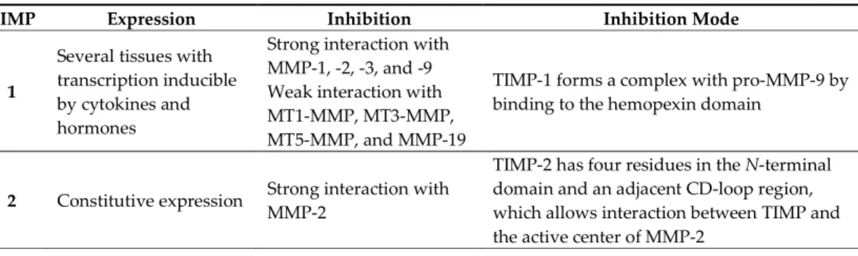
Tissue inhibitors of metalloproteinases (TIMPs) are endogenous proteins responsible for the regulation of MMPs activity, but also of families such as the disintegrin metalloproteinases (ADAM and with thrombospondin motifs ADAMTS) and therefore for maintaining the physiological balance between ECM degradation and MMPs activity [1,2,8,9,18,30]. There are four TIMPs (TIMP-1, -2, -3, and -4) (Table 3), with 22–29 KDa and 41%–52% sequential similarity [2,4,12,13,16,20,31].
Table 3. Tissue inhibitors of metalloproteinases (TIMPs) classification. TIMP Expression Inhibition Inhibition Mode
1
Several tissues with transcription inducible by cytokines and hormones
Strong interaction with MMP-1, -2, -3, and -9 Weak interaction with MT1-MMP, MT3-MMP, MT5-MMP, and MMP-19
TIMP-1 forms a complex with pro-MMP-9 by binding to the hemopexin domain
2 Constitutive expression Strong interaction with MMP-2
TIMP-2 has four residues in the N-terminal domain and an adjacent CD-loop region, which allows interaction between TIMP and the active center of MMP-2
4
expressed in injured tissue
MMP-2 and -14 -
TIMPs consist of a N- and C-terminal domain with 125 and 65 amino acids, respectively, each containing six conserved cysteine residues, which form three conserved disulphide bonds [2,4,7– 9,12,31,32] (Figure 2a). The N-terminal domain is an independent unit, which can be inhibited by MMPs, in a 1:1 ratio [2,4,8–10,12,13,16,20]. This domain has two groups of four residues: Cys-Thr-Cys-Val and Glu-Ser-Val-Cys (Figure 2b), which are connected by disulphide bounds which are important for TIMP activity [7,12]. This is the main domain responsible for MMP inhibition through its binding to the catalytic site in a substrate-like manner [31]. The several domains allow the TIMP and pro-gelatinases interactions [4].
Figure 2. (a) TIMP-1-catalytic domain of the MMP-3 complex. (b) TIMP-1-catalytic domain of the
MMP-3 complex, where two conserved groups, Cys-Thr-Cys-Val and Glu-Ser-Val-Cys, are represented in yellow.
3. Non-Specific Endogenous Inhibitors
Non-specific endogenous inhibitors have been reported to inhibit MMPs (Table 4), however, the inhibition mechanism details have only been partially discovered [7,12].
Table 4. Non-specific endogenous inhibitors [4,7,12,13,33,34].
Non-Specific Inhibitor Inhibition
α2-macroglobulin MMP-2 and -9
Tissue factor pathway inhibitor MMP-1 and -2
Membrane-bound β-amyloid precursor protein MMP-2
C-terminal proteinase enhancer protein MMP-2
Reversion-inducing-cysteine-rich protein with Kasal motifs (RECK) MMP-2, -9, and -14
proteinase enhancer protein and tissue factor pathway inhibitor have sequences with certain similarities to the N-terminal domain of TIMPs [31].
4. Synthetic Inhibitors
MMPs are molecular targets for the development of therapeutic and diagnostic agents [14]. The development of synthetic MMP inhibitors was initially based on the peptide sequence, recognized by proteases, with different chemical functionalities, capable of interacting potently with zinc ion [11– 13,19]. The requirements for an effective inhibitor are [2,11,13,17,19,35]:
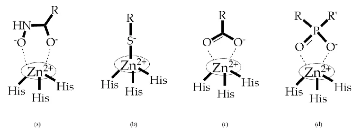
- A functional group able to chelate the zinc ion (II)-zinc binding group (ZBG). The first generation inhibitors used hydroxamate (CONHO−) but the second generation use carboxylate (COO−),
thiolates (S−), phosphonyls (PO2−), for example (Figure 3);
- At least one functional group that promotes hydrogen bonding with the protein backbone; - One or more side chains undergoing Van der Waals interactions with enzyme subsites.
Figure 3. Examples of zinc binding groups (ZBGs). (a) Hydroxamate-based inhibitor; (b)
thiolate-based inhibitor; (c) carboxylate-thiolate-based inhibitor; (d) phosphorous-thiolate-based inhibitor. The R group is the scaffold of inhibitor.
ZBGs have negative charges that prevent their penetration in the cell, restricting their activity to the extracellular space which reduces their cell toxicity [2]. Changes in the ZBG structure or in the point of attachment of the ZBG to the backbone of the MMP inhibitor can change its potency and selectivity [2]. When comparing what selectivity or potency of different ZBGs leaving the structure constant, Castelhano et al. arrived at the following list [36]: hydroxamic acid >> formylhydroxylamine > sulfhydryl > aminocarboxylates > carboxylate.
The selectivity of inhibitor is a primordial goal of MMPs’ inhibitors (MMPis) design to increase efficacy and prevent side effects [2]. This selectivity is based on two molecular characteristics [11]: a chelating capacity for catalytic zinc and the presence of hydrophobic bridges of the active center for the S1’ pocket. Numerous strategies have been suggested for creating selective MMP inhibitors [27]
(Figure 4): endogenous-like inhibitors, exosite targeting inhibitors, a combination of exosite binding and metal chelating inhibitors, and function-blocking antibodies.
Figure 4. Types of MMPs’ inhibitors: (a) endogenous-like inhibitors that chelate the catalytic zinc (II)
ion; (b) exosite targeting inhibitors that alter the conformation of the enzyme; (c) a combination of exosite binding and metal chelating inhibitors; (d) antibodies inhibitors.
The synthetic inhibitors have had a great challenge in their development since first it is necessary to identify the enzymes that are involved in disease progression. Moreover, this goal has an additional difficulty as there are more than 50 human metalloproteinases (23 MMPs, 13 ADAM, and 19 ADAMTS) [2].
4.1. Hydroxamate-Based Inhibitors
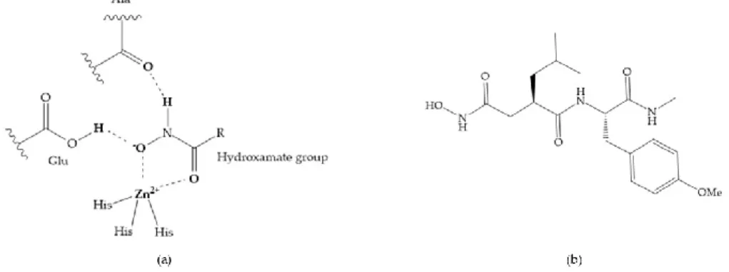
The first generation of MMPis (1995–1999 [16]) were designed based on the knowledge of the triple helix collagen amino acid sequence (cleavage site) and the information derived from specific substrates [6,15,19,31,35,37]. These compounds contain a hydroxamic acid group as ZBG [5,6,15,18,27,28,31,37]. Hydroxamic acids (HA) were first described in 1986 [38,39]. They are easy to synthesize, are monoanionic compounds, bidentate chelating agents, and due the excellent zinc-chelating capability they are the more popular ZBG for MMPs [2,6,17,18,27–29]. The hydroxamic group established interactions with zinc ions, through two oxygens and two hydrogen bonds (NH and OH groups of HA with Ala and Glu, respectively), forming a distorted triangular bipyramid [2,5,18,19,28,29] (Figure 5a). Reich et al., in 1988 [40], used a hydroxamic acid compound, SC-44463 (Figure 5b), to block collagenase and prevent metastasis in a mouse model, which initiated the era of MMP inhibition therapeutics [40].
Figure 5. (a) Interaction between hydroxamate group and catalytic zinc (II) ion. The oxygen of the
quaternary carbonyl group at R1 suggested that [13] (Figure 6):
(i) The stoichiometric orientation of the substituent at R1 position is crucial for the activity; (ii) The phenylpropyl group was established as the best substituent at position R1;
(iii) Hydrophobic substituents at R2’ position and N-metiamides at R3’ position were considered as
the most appropriate.
Figure 6. General structure of hydroxamic acid. OH group: catalytic zinc binding group; Ra: α
substituent; R1: P1’ substituent group and this group is determinant to selectivity and activity; R2: P2’
substituent and this substituent can be cyclized with Ra and R3; R3: P3’ substituent.
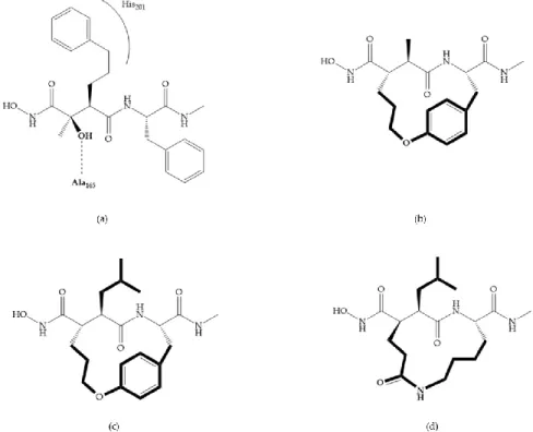
Modification in Ra position (α substituent): a beneficial effect is conferred by lipophilic substituents capable of hydrogen bonding [19]. Analogues of Marimastat have been reported, where the α position is disubstituted by a hydroxyl group and a methyl group [19] (Figure 7a). The X-ray structures of the MMP-3-inhibitor complex showed that there were hydrogen bonds between the hydroxyl group and Ala165 [19]. By binding the positions Ra and R2 in a single chain, forming a cyclic
inhibitor (Figure 7b), there was a substantial increase in water solubility [19,29,41,42]. This strategy led to the discovery of two inhibitors with similar potency to non-cyclized analogues, SE205 and SC903 [41] (Figure 7c,d).
Figure 7. (a) Analogue of Marimastat with the α position disubstituted; (b) analogue of Marimastat
Figure 8. Inhibitors with conformational restrictions between Ra and R1 positions. (a) Inhibitor with
three-membered ring; (b) inhibitor with six-membered ring.
Exploring the depth of S1’ pocket, bulky groups in Rα position confer selective inhibition for
MMP-2, -8, and -9 [29]. An example is the presence of a biphenyl group, which showed higher inhibitory activity against MMP-9 [29].
Modification in R1 position: the incorporation of long groups in the R1 position can promote the
selectivity of MMPis, since pocket S1’ can undergo conformational changes to accommodate certain
substituents [19].
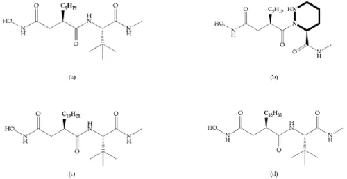
Broadhurst et al. showed that an alkyl chain (C9) at the R1 position reduces the inhibition of
MMP-1, but maintains the inhibitory activity against MMP-2, -3, and -9 [19] (Figure 9a). For matlystatin derivates in the C9 chain, R-94138 (Figure 9b) promotes the inhibition against MMP-9 [44].
The succinyl hydroxamates analogues in the C9 chain promote selectivity for MMP-2, however, a C10
chain (Figure 9c) results in MMP-1 inhibition, while a further increase of the chain to C16 (Figure 9d)
leads to a loss of activity against MMP-1 [19].
Figure 9. Inhibitors with modification of R1 position. (a) Inhibitor with alkyl chain. This inhibitor has
activity against MMP-2, -3, and -9, but the inhibition of MMP-1 is low; (b) R-94138, Matlystatin derivate. The inhibition of MMP-9 is 10 times higher than analogues with C8 or C10 chains; (c) succinyl
hydroxamate analogue with C10 chain, which inhibits MMP-1; (d) succinyl hydroxamate analogue
Figure 10. (a) Succinyl hydroxamate acid with a sulphonamide bond. This compound presents low
inhibitory activity because of the pyramidal nature of the sulphonamide group. (b) Succinyl hydroxamate acid with carbonyl bond.
Modifications in R2 position: modifications in the R2 position led to a modest effect in inhibitory
activity, in vitro, and affects the pharmacokinetic properties [29]. Marimastat and Ro31-9790 (Figure 11) have a good oral activity because the bulky tert-butyl group assists the adjacent amide bond during absorption from an aqueous environment to the lipid environment of the cell membrane [45]. The beneficial combination of the tert-butyl group with the α-hydroxyl group increases the water solubility [19,29]. Babine and Bender suggest that the tert-butyl as R2 group leads to less Van der
Waals interactions, comparing with other groups [46].
Figure 11. (a) Marimastat. The Ra position is substituted with a hydroxyl group (OH). (b) Ro31-9790.
The Ra position has no substituents.
Ikeda et al. described compounds with phenyl R2 substituents (Figure 12) (KB-R7785), which are
active orally, due to the beneficial effect of the R2 phenyl group on absorption, where the amide
shielding and lipophilicity may assist in transepithelial resorption [47]. This inhibitor shows activity against MMP-1 in rats and its effectiveness in arthritis has been demonstrated [47].
Figure 12. KB-R7785 inhibitor.
Modifications in R3 position: the S3’ pocket is an open area and several groups can be introduced
at the R3 position [19]. The introduction of the benzhydryl group leads to compounds with selectivity
to the MMPs-3 and -7 [19].
4.1.1. Succinyl Hydroxamic Acid-Based Inhibitors
Succinyl hydroxamate derivates can be subdivided to peptide derivatives or non-peptide compounds [48]. The N-acetylcysteine has been reported to affect the tumoral invasion process and metastasis by MMP-2 and -9 inhibition [49]. The L-cysteine-2-phenylethylamide is an effective inhibitor, in which the phenyl group fills the S1’ pocket of MMP-8 [50]. Foley et al. prepared several
dipeptides derivatives containing cysteine (RCO-Cys-AA-NH2) and concluded [51]:
- The variation of the acyl group and the second amino acid (AA) leads to the activity against different MMPs.
- The R group interacts with the S1’ pocket.
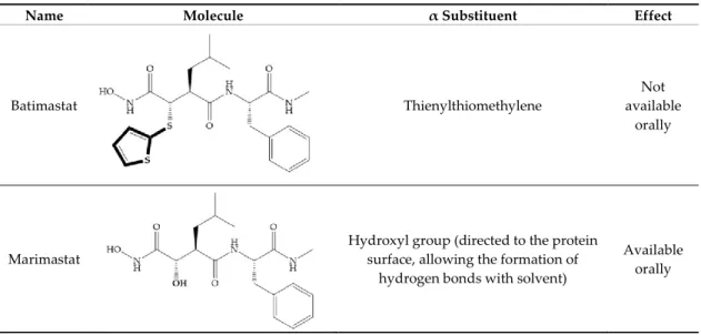
Batimastat (Table 5) was the first MMPi to enter in clinical trials for cancer as it inhibits MMP-1, -2, -7, and -9, but, due to its poor oral bioavailability, it was superseded by Marimastat [28,29,31] (Table 5), which has an alpha-hydroxyl group increasing the aqueous solubility [29]. Marimastat inhibits the activity of MMP-1, -2, -3, -7, -9, -12, and -13 [31]. However, Marimastat failed in clinical trials due to the absence of a therapeutic effect and the patients treated developed musculoskeletal toxicity (MST) [6,31]. Batimastat, marimastat, and ilomastat are examples of succinyl hydroxamates, which have very analogous structure to that of collagen and inhibit MMPs by bidentate chelation of the Zn2+ [2,6,29].
Table 5. Batimastat and Marimastat.
Name Molecule α Substituent Effect
Batimastat Thienylthiomethylene
Not available
orally
Marimastat
Hydroxyl group (directed to the protein surface, allowing the formation of
hydrogen bonds with solvent)
Available orally
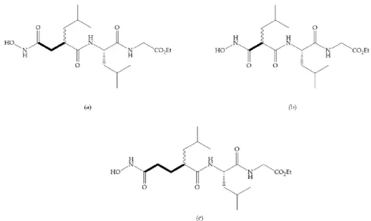
Figure 13. (a) Derivates of succinyl hydroxamic acid; (b) malonyl acid; (c) glutaryl acid.
Marcq et al. [53] developed succinyl hydroxamates derivates selective for MMP-2, by modifications on Ilomastat structure (Figure 14a) to increase the overall hydrophobicity and, consequently, the selectivity [53]. This study resulted in a compound (Figure 14b) with an isobutylidene group of E geometry, which showed a 100-fold greater selectivity for MMP-2 over MMP-3, that is a 70-fold increase compared to Ilomastat [53].
Figure 14. (a) Ilomastat; (b) Ilomastat derivate with isobutylidene group.
Table 6 shows the IC50 and Ki values of some succinyl hydroxamic acid-based inhibitors [6,15–
Marimastat (BB-2516) IC50: MMP-1 = 5 nM; MMP-2 = 6 nM; MMP-3 = 200 nM; MMP-7 = 20 nM; 8 = 2 nM; 9 = 3 nM; 12 < 5 nM; 13 = 0.74 nM; MMP-14 = 1.8 nM Analogue of Marimastat IC50: MMP-1 = 3 nM; MMP-2 = 1.1 nM; MMP-3 = 14 nM; MMP-7 = 0.8 nM; MMP-8 = 0.5 nM; MMP-9 = 0.9 nM; MMP-10 = 0.45 nM; MMP-14 = 94 nM; MMP-15 = 20 nM; MMP-16 = 35 nM SE205 Ki: MMP-1 = 1.2 nM; MMP-3 = 32.7 nM; MMP-9 = 1.8 nM IC50: MMP-1 = 131 nM; MMP-2 = 9.5 nM; MMP-3 = 8.9 nM; MMP-7 = 3.3 nM SC903 Ki: MMP-1 = 2.8 nM; MMP-3 = 24.1 nM; MMP-9 = 2.6 nM IC50: MMP-1 = 4 nM; MMP-2 = 3 nM; MMP-3 = 30 nM; MMP-7 = 20 nM; MMP-8 = 20 nM; MMP-9 = 9 nM
SC-44463 IC50: MMP-1 = 20 nM; MMP-2 = 6 nM; MMP-3 = 30 nM; MMP-7 = 30 nM BB-16 IC50: MMP-1 = 5 nM; MMP-2 = 10 nM; MMP-3 = 40 nM; MMP-7 = 60 nM; MMP-8 = 7 nM Ro-31-9790 IC50: 1 = 10 nM; 2 = 8 nM; MMP-3 = 700 nM; MMP-14 = 1.9 nM Ro-32-0554 IC50: MMP-1 = 0.5 nM; MMP-3 = 9.1 nM; MMP-9 = 4.3 nM IC50: MMP-1 = 10 nM; MMP-2 = 400 nM; MMP-3 = 4.5 μM Ro-32-3555 Ki: MMP-1 = 3 nM; MMP-2 = 154 nM; MMP-3 = 527 nM; MMP-8 = 4 nM; MMP-9 = 59 nM; MMP-13 = 3 nM
IC50: MMP-1 = 40 nM IC50: MMP-1 = 29 μM IC50: MMP-1 = 10 μM IC50: MMP-1 = 9 nM Analogue of Marimastat IC50: MMP-1 = 1 μM; MMP-2 = 15 nM; MMP-3 = 500 nM; MMP-7 = 10 μM; MMP-8 = 30 nM; MMP-9 = 15 nM Ki: MMP-1 = 2 nM; MMP-3 = 3 nM; MMP-9 < 1 nM Ki: MMP-1 = 1.3 nM; MMP-2 = 1.1 nM; MMP-3 = 187 nM Ki: MMP-1 = 6.5 nM; MMP-2 = 20 nM; MMP-3 = 240 nM IC50: 1 = 6 nM; 2 = 30 nM; MMP-3 = 40 nM
IC50: MMP-1 = 375 nM; MMP-2 < 0.15 nM; MMP-3 = 18 nM; MMP-9 = 1.5 nM IC50: MMP-1 = 20 nM; MMP-2 = 2 nM; MMP-3 = 100 nM; MMP-9 = 2 μM IC50: MMP-2 = 20 nM; MMP-3 = 300 nM; MMP-9 = 1 nM KB-R7785 IC50: MMP-1 = 3 nM; MMP-2 = 7.5 nM; MMP-3 = 1.9 nM; MMP-9 = 3.9 nM IC50: MMP-1 = 5.4 nM; MMP-2 = 8.4 nM; MMP-3 = 2.3 nM; MMP-9 = 5 nM; MMP-14 = 2.3 nM IC50: MMP-1 = 5 nM; MMP-2 = 1 nM; MMP-3 = 15 nM; MMP-9 = 1 nM IC50: MMP-1 = 150 nM IC50: MMP-3 = 300 nM Matlystatin B IC50: MMP-2 = 1.7 μM; MMP-9 = 570 nM
R-94138 IC50: MMP-2 = 38 nM; MMP-3 = 28 nM; 7 = 23 nM; 9 = 1.2 nM; MMP-13 = 38 nM Ki: MMP-1 > 8.3 μM; MMP-2 = 3.4 μM; MMP-3 = 1.3 μM; MMP-7 > 8.3 μM; MMP-14 = 7.7 μM Ki: MMP-1 > 12.5 μM; MMP-2 = 1.1 μM; MMP-3 = 100 nM; MMP-7 = 200 nM; MMP-14 = 1.8 nM Ki: MMP-1 = 200 nM IC50: MMP-1 > 5 μM; MMP-2 = 35 nM; MMP-3 = 3.56 nM; MMP-7 > 5 μM; MMP-9 = 304 nM; MMP-12 = 17 nM; MMP-14 = 772 nM; MMP-15 = 60 nM Ki: MMP-1 = 33.16 μM; MMP-2 = 6.3 μM; MMP-8 = 171 nM; MMP-9 = 4.468 μM
Ki: MMP-1 = 5.248 μM; MMP-2 > 3 μM; 3 > 4.5 μM; 9 < 1 nM; MMP-13 > 5 μM BB-1101 IC50: MMP-1 = 10 nM; MMP-2 = 5 nM; MMP-3 = 30 nM; MMP-7 = 30 nM; MMP-8 = 3 nM; MMP-9 = 3 nM IC50: MMP-1 = 3.1 nM; MMP-2 = 4.2 nM; MMP-3 = 25 nM IC50: MMP-1 = 1.1 nM; MMP-2 = 1.1 nM; MMP-3 = 2.3 nM; MMP-7 = 2.2 nM OPB-3206 IC50: MMP-1 = 700 nM; MMP-2 = 5 μM; MMP-3 = 2 μM; MMP-9 = 500 nM IC50: MMP-8 = 300 nM
Batimastat (BB-94) IC50: MMP-1 = 3 nM; MMP-2 = 4 nM; MMP-3 = 20 nM; MMP-7 = 6 nM; MMP-8 = 10 nM; MMP-9 = 1 nM; MMP-13 = 1 nM; MMP-14 = 2.8 nM Ki: MMP-1 = 10 nM; MMP-2 = 4 nM; MMP-3 = 20 nM; MMP-8 = 10 nM; MMP-9 = 1 nM Ilomastat (GM6001; Galardin®) IC50: MMP-1 = 0.4 nM; MMP-2 = 0.4 nM; MMP-3 = 0.19 nM; MMP-14 = 5.2 nM Ki: MMP-1 = 0.4 nM; MMP-2 = 0.39 nM; MMP-3 = 26 nM; MMP-8 = 0.18 nM; MMP-9 = 0.2 nM Analogue of Ilomastat IC50: MMP-2 = 1.3 nM; MMP-3 = 179 nM IC50: MMP-1 = 3 nM; MMP-3 = 280 nM; MMP-7 = 18 nM Ki: MMP-1 = 3 nM IC50: MMP-1 = 8 μM; MMP-2 = 8 μM; MMP-3 = 3.5 μM IC50: MMP-1 = 3.3 μM; MMP-2 = 32 nM; MMP-3 = 57 nM IC50: MMP-3 = 3.4 μM IC50: MMP-3 = 15 nM
IC50: MMP-1 > 50 μM; MMP-2 > 120 μM; MMP-3 = 80 μM; MMP-8 > 120 μM IC50: MMP-2 = 52 μM; MMP-3 = 200 μM; MMP-8 = 1200 μM IC50: MMP-8 = 121 μM PKF 242-484 Ki: MMP-1 = 3.6 nM; MMP-2 = 0.1 nM; 3 = 0.9 nM; 9 = 1 nM; MMP-13 = 4.5 nM CT1746 Ki: MMP-1 = 122 nM; MMP-2 = 0.04 nM; MMP-3 = 10.9 nM; MMP-7 = 136 nM; MMP-9 = 0.17 nM ONO-4817 IC50: MMP-1 = 1600 nM; MMP-9 = 2.1 nM Ki: MMP-2 = 0.73 nM; MMP-3 = 42 nM; MMP-7 = 2500 nM; MMP-12 = 0.45 nM; MMP-13 = 1.1 nM AS 111793# IC50: MMP-1 = 20 nM MMPI-I IC50: MMP-1 = 1 μM; MMP-3 = 150 μM; MMP-8 = 1 μM; MMP-9 = 30 μM
IC50: 1 = 0.1 nM; 3 = 9 nM; 8 = 0.4 nM; MMP-9 = 0.2 nM IC50: MMP-1 = 30 nM; MMP-2 = 20 nM; MMP-3 = 500 nM; MMP-7 = 200 nM; MMP-8 = 20 nM Ki: MMP-1 = 1450 nM; MMP-3 = 15 nM; MMP-8 = 2 nM; MMP-9 = 3 nM Ki: MMP-1 = 8 nM; MMP-3 = 28 nM; MMP-8 < 2 nM; MMP-9 = 1 nM IC50: MMP-1 = 100 nM; MMP-2 = 0.07 nM; MMP-3 = 3 nM; MMP-7 = 700 nM; MMP-8 = 4 nM; MMP-9 = 1 nM IC50: MMP-1 = 11 nM; MMP-3 = 1.04 μM IC50: MMP-1 = 600 nM; MMP-2 = 3 μM; MMP-3 = 50 nM; MMP-7 = 4 nM Ki: MMP-1 = 7.56 μM; MMP-7 = 622 nM; MMP-13 = 7.3 nM
IC50: MMP-1 = 6 μM; MMP-2 = 200 nM; MMP-3 = 100 nM IC50: MMP-2 = 5 nM Ki: MMP-2 = 2.2 nM IC50: MMP-7 = 1 510 μM; MMP-12 = 149 μM Ki: MMP-1 > 4.949 μM; MMP-2 > 3.333 μM; MMP-9 > 2.128 μM Ki: 2 > 15 μM; 8 > 15 μM; MMP-9 > 15 μM; MMP-12 = 410 nM; MMP-13 > 15 μM; MMP-14 = 3.07 μM IC50: MMP-1 = 5.9 μM; MMP-2 = 750 nM; MMP-3=2.1 nM; MMP-9 = 560 nM; MMP-14 = 930 nM IC50: MMP-1 = 51 μM; MMP-2 = 1.79 μM; MMP-3 = 5.9 nM; MMP-9 = 840 nM; MMP-13 = 73 nM; MMP-14 = 1.9 μM Ki: MMP-1 > 4.946 μM; MMP-2 > 3.333 μM; MMP-3 > 4.501 μM; MMP-7 > 6.368 μM; MMP-8 > 3.058 μM; MMP-9 > 2.128 μM; MMP-10 > 5.346 μM; MMP-12 > 6.023 μM; MMP-13 > 5.025 μM; MMP-14 > 5.290 μM; MMP-15 > 7.088 μM
IC50: 1 = 4.6 μM; 2 = 4 nM; 3 = 42 nM; MMP-7 > 10 μM; MMP-9 = 120 nM Ki: MMP-1 > 4.494 μM; MMP-2 > 3.333 μM; MMP-3 = 82 nM; MMP-7 = 25 nM; MMP- 8 > 3.1 μM; MMP-9 > 2.128 μM; MMP-13 > 5.025 μM; MMP-14 > 5.290 μM; MMP-15 > 7.088 μM; MMP-16 > 5.554 μM Ki: MMP-1 > 5 μM; MMP-2 > 3 μM; MMP-3 = 762 nM; MMP-8 = 2.05 μM; MMP-9 > 3 μM; 10 > 1.650 μM; 13 > 5 μM; MMP-14 = 163 nM; MMP-15 = 1.7 μM Ki: 1 > 2 μM; 2 > 2 μM; 3 > 2 μM; 7 = 834 nM; 8 = 126 nM; MMP-9 > 2 μM; MMP-10 > 2 μM; MMP-12 > 2 μM; MMP-13 = 653 nM; MMP-14 > 2 μM; MMP-15 > 2 μM; MMP-16 > 2 μM Ki: MMP-1 = 30 μM; MMP-2 = 2.05 μM; MMP-3 = 141 nM; MMP-7 = 259 nM; 8 = 257 nM; 9 = 10.34 μM; 13 = 1.417 μM; MMP-14 = 15.872 μM; MMP-15 = 3.997 μM; MMP-16 = 1.599 μM
which has good oral availability but did not succeed in clinical trials [56]. The isopropyl group slows down the metabolization of the adjacent hydroxamic acid group and the 3-pyridyl substituent may aid partitioning into the hydrated negatively charged environment of the cartilage [56]. By analysis of the cocrystal structure of this inhibitor and MMP-12, it was possible to conclude that the binding mode between the hydroxamate moiety and the catalytic zinc ion was the same as the binding mode of hydroxamate-based inhibitors [6]. The interaction of CGS-27023A with MMP-3 was possible due to the p-methoxy phenyl substituent occupation of the S1’ pocket and the pyridylmethyl and isobutyl
groups occupation of the S2’ and S1 pockets, respectively [29,56]. The modification of α to form the
thioester derivate led to an increase of the inhibition of the deep pocket of the MMPs [29,56] (Figure 15b).
Figure 15. (a) CGS-27023A; (b) thioester derivate of CGS-27023A.
The NNGH (Figure 16a) (N-Isobutyl-N-(4-methoxyphenylsulfonyl)glycyl hydroxamic acid) was the starting point to many potent MMPis and is accommodated in the entry of the S1’ pocket, but does
not penetrate it [6]. Barta et al. described a series of arylhydroxamate sulphonamides, active against MMP-2 and -13 (Figure 16b) [57]. In this compound, the sulfonyl group formed a single hydrogen bond with Leu160 and the piperidine-O-phenyl moiety extends into the S1’ pocket by Van der Waals
interactions [57]. Noe et al. described a series of 3,3-dimethyl-5-hydroxy pipecolic hydroxamic acid, which possess potent inhibitory activity for MMP-13 [58]. In the first series of compounds, the 3-position of the piperidine ring was explored by the introduction of a polar functionality and it resulted in a compound with excellent activity on MMP-13 (Figure 16c), improved bioavailability, and lower metabolic clearance [58].
Figure 16. (a) NNGH; (b) arylhydroxamate sulphonamide compound; (c)
3-hydroxy-3-methylpipecolic hydroxamates.
The incorporation of a cyclic quaternary center α led to a strong inhibitory effect against MMP-1, -2, -3, -8, -9, -12, and -13 [29]. The RS-113,456 (Figure 17a) is an inhibitor with better oral
bioavailability and metabolic stability compared to the hydroxamate derivates [35]. These two features were improved by shifting the cyclic group to the α-position of the hydroxamic acid (Figure 17b) [29].
Figure 17. (a) RS-113,456; (b) RS-130,830.
Table 7 shows the IC50 and Ki values of some sulfonamide hydroxamic acid-based inhibitors
CGS-27023A (MMI270) IC50: MMP-1 = 33 nM; MMP-2 = 11 nM; MMP-3 = 13 nM; MMP-9 = 8 nM; MMP-12 = 7.7 nM; MMP-13 = 6 nM Ki: MMP-1 = 3 nM; MMP-2 = 20 nM; MMP-3 = 148 nM; MMP-8 = 1.9 nM Analogue of CGS-27023A IC50: MMP-1 = 104 nM; MMP-2 = 0.7 nM; MMP-3 = 0.7 nM; MMP-9 = 2.5 nM; MMP-13 = 12 nM Ki: MMP-1 = 770 nM; MMP-2 = 620 nM; MMP-3 = 4.1 μM; MMP-9 = 620 nM CGS-25966 Ki: MMP-3 = 92 nM NNGH IC50: MMP-12 = 72 nM Ki: MMP-10 = 0.6 nM IC 50: MMP-1 = 2 nM; MMP-2 = 20 nM; MMP-3 = 30 nM; MMP-7 = 20 nM; MMP-9 = 7 nM IC50: MMP-1 = 6 nM; MMP-2 = 900 nM; MMP-3 = 200 M; MMP-7 = 200 nM; MMP-8 = 200 nM; MMP-9 = 2μM; MMP-13 = 400 nM
Prinomastat (AG3340) Ki: MMP-1 = 8.3 nM; MMP-2 = 0.05 nM; MMP-3 = 0.3 nM; 7 = 54 nM; 9 = 0.26 nM; MMP-13 = 0.03 nM; MMP-14 = 0.33 nM CP-471,474 IC50: MMP-1 = 1170 nM; MMP-2 = 0.7 nM; MMP-3 = 16 nM; MMP-9 = 13 nM IC50: MMP-1 > 10 μM; MMP-2 = 3.3 nM; MMP-13 = 12 nM IC50: MMP-1 = 196 nM; MMP-2 = 0.01 nM; MMP-9 = 1 nM RS-113,456 IC50: MMP-3 = 5.2 nM Ki: MMP-1 = 70 nM; MMP-2 = 0.054 nM; MMP-7 = 240 nM; MMP-8 = 0.13 nM; MMP-9 = 0.065 nM; MMP-12 = 0.15 nM; MMP-13 = 0.17 nM; MMP-14 = 0.089 nM RS-130,830 Ki: MMP-1 = 590 nM; MMP-2 = 0.22 nM; 3 = 9.3 nM; 7 = 1.2 μM; MMP-9 = 0.58 nM; MMP-13 = 0.52 nM RS-104,966 Ki: MMP-1 = 23 nM; MMP-13 = 0.13 nM IC50: MMP-1 = 3 245 nM; MMP-9 = 7 nM; MMP-13 = 4 nM IC 50: MMP-2 = 960 nM; MMP-13 = 1.17 μM; MMP-14 = 3.41 μM
IC50: MMP-1 = 310 nM IC50: MMP-1 = 920 nM; MMP-13 = 0.95 nM IC50: MMP-1 = 841 nM; MMP-9 = 33 nM; MMP-13 = 29 nM IC50: MMP-1 = 763 nM; MMP-9 = 2 nM; MMP-13 = 2 nM MMP-8 inhibitor I IC50: MMP-8 = 4 nM Ro-32-7315 IC50: MMP-1 = 500 nM; MMP-2 = 250 nM; MMP-3 = 210 nM; MMP-7 = 310 nM; MMP-9 = 100 nM; MMP-12 = 11 nM; MMP-13 = 110 nM IC50: MMP-1 = 346 μM; MMP-9 = 24 μM
PGE-4410186 IC50: MMP-1 = 24 nM; MMP-3 = 18.4 nM; MMP-7 = 30 nM; MMP-9 = 2.7 nM MMP-9 inhibitor I IC50: MMP-1 = 1.05 nM; MMP-9 = 5 nM; MMP-13 = 113 nM Ki: MMP-1 = 1.085 μM; MMP-2 = 1 nM; MMP-9 = 10 nM; MMP-13 = 3 nM
MMPI-II (MMP-2/MMP-9 inhibitor II)
IC50: MMP-1 = 970 nM; MMP-2 = 17 nM; MMP-3 > 1000 nM; 7 = 800 nM; 9 = 30 nM; MMP-14 = 17 nM IC50: MMP-1> 50 μM; MMP-2 = 12 nM; MMP-3 = 4.5 μM; MMP-7 > 50 μM; MMP-9 = 200 nM IC50: MMP-1 = 147 nM; MMP-2 = 0.09 nM; MMP-3 = 50 nM; MMP-7 > 1 μM; MMP-8 = 1.6 nM; MMP-9 = 6.7 nM; MMP-14 = 9.8 nM
Ki: MMP-1 = 2 μM; MMP-2 = 10 nM; MMP-3 = 500 nM IC50: MMP-1 = 200 nM; MMP-9 = 0.43 nM IC50: MMP-1 = 2.471 μM; MMP-7 = 961 nM; MMP-8 = 35 nM; MMP-9 = 777 nM; MMP-13 = 96 nM; MMP-14 = 582 nM IC50: MMP-1 = 37.3 μM; MMP-2 = 664 nM; MMP-9 = 5.5 μM; MMP-13 = 2.277 μM; MMP-14 = 24 μM IC50: MMP-1 = 8.78 μM; MMP-2 = 355 nM; MMP-9 = 1.67 μM; MMP-13 = 230 nM; MMP-14 = 4.71 μM Ki: (S enantiomer) MMP-3 = 19 nM (R enantiomer) MMP-3 = 36 nM IC50: MMP-1 = 2.268 μM; MMP-9 = 152 nM; MMP-13 = 18 nM IC50: MMP-1 = 14 μM; MMP-2 = 529 nM; MMP-3 = 1 nM; MMP-9 = 2.42 μM; MMP-14 = 20.1 μM
that accommodates into the S1’ pocket and by the phosphinamide oxygen, which establishes the
hydrogen bonds with NH of Leu164 and Ala165 [29]. However, this group is susceptible to hydrolysis
at low pH, limiting the inhibitory activity [29].
Figure 18. Inhibitor with phosphinamide group.
All hydroxamate-based inhibitors are very potent and they inhibit MMPs at low concentrations [18]. On the other hand, the hydroxamate acids have poor oral bioavailability, inhibit multiple MMPs, and cause side effects [2,17,27,28,35]. Additionally, this functional group may be metabolized via dehydroxylation or may be cleaved by endopeptidases and releasing hydroxylamine, can be hydrolyzed to carboxylic acids or reduced to O-glucuronyl or O-sulfate, leading to decreased effective inhibitor concentration and reducing its potency in vivo [18,27,28,35].
Table 8 shows the IC50 and Ki values of some phosphamides hydroxamic acid-based inhibitor
[6,15–19,29,35,37,54,55].
Table 8. IC50 and Ki values of phosphamides hydroxamic acid-based inhibitors.
IC50 (X = H; Y = (CH2)2C6H5; Z = Me; R = Ph): MMP-1 = 252 nM; MMP-3 = 700 nM IC50 (X = H; Y = (CH2)2C6H5; Z = Ph; R = Ph): MMP-1 = 854 nM; MMP-3 = 1.75 μM IC50 (X = Me; Y = CH2C6H5; Z = Me; R = Ph): MMP-1 = 120 nM; MMP-3 = 67.9 nM IC50 (X = Me; Y = CH2C6H5; Z = Et; R = Ph): MMP-1 = 608 nM; MMP-3 = 700 nM IC50 (X = Me; Y = CH2C6H5; Z = Ph; R = Ph): MMP-1 = 6.79 μM; MMP-3 = 10.3 μM IC50 (X = CH2i-Pr; Y = CH2C6H5; Z = Me; R = Ph): MMP-1 = 20.5 nM; MMP-3 = 24.4 nM
IC50 (X = CH2i-Pr; Y = CH2C6H5; Z = Me; R = Me): MMP-1 = 518
nM; MMP-3 = 1.04 μM IC50 (X = CH2i-Pr; Y = H; Z = CH3): R isómer, MMP-1 = 2.51 μM; MMP-3 = 2.55 μM S isomer: MMP-1 > 100 μM; MMP-3 = 130.5 μM IC50 (X = CH2i-Pr; Y = CH2C6H5; Z = CH3): R isomer, MMP-1 = 20.5 nM; MMP-3 = 24.4 nM S isomer, MMP-1 = 7.12 μM; MMP-3 = 9.17 μM IC50 (X = CH3; Y = CH2C6H5; Z = C2H5): R isomer, MMP-1 = 608 nM; MMP-3 = 700 nM S isomer, MMP-1 = 33.3 μM; MMP-3 = 49.3 μM
IC50 (X = CH2i-Pr; Y = CH2C6H5): MMP-1 = 20.5 nM; MMP-2 =
13.3 nM; MMP-3 = 24.4 nM; MMP-7 = 886 nM; MMP-8 = 5.3 nM; MMP-9 = 20.6 nM; MMP-13 = 7.4 nM
4.2. Non-Hydroxamate-Based Inhibitors
The side effects caused by hydroxamate-based inhibitors, due to the lack of selectivity and in vivo lability, have been fostering the development of new compounds with alternative ZBGs [5,6,11,16,17,27–29]. The second generation of MMPis (1999–2003 [16]) was designed with a wide variety of peptidomimetic and non-peptidomimetic structures with higher selectivity and exploiting the deep S1’ pocket present in some MMPs [6,15,16,18,59–61]. These compounds include carboxylic
acids, sulfonylhydrazides, thiols, aminomethyl benzimidazole, phosphorous-based, nitrogen-based and heterocycles bidentate chelators, and can be monodentade, bidentade, and tridentade chelates [2,6,18,28,29].
The non-hydroxamate-based inhibitors open up a wide spectrum of affinities for the zinc ion from the catalytic site and new opportunities for targeting and inhibiting the active center [18,28]. They have weak Zn2+ chelating ability and the rates of severe side effects, such as the musculoskeletal
syndrome (MSS) decreased dramatically compared with the hydroxamate inhibitors [28]. 4.2.1. Thiolates-Based Inhibitors
The ability of the monodentate binding of thiols to zinc ion in proenzymes has served as inspiration for the design of several MMPis [5,29]. The potency of thiol inhibitors is intermediate between that of hydroxamate- and carboxylate- based inhibitors [29]. The first example of inhibitor thiol-based for MMP-1 is a bipeptidic analogue, where the incorporation of a thiol group as α substituent leads to improvement of activity (Figure 19a) [19]. Derivates with “linker” substituent between P1-P1’ positions show a total loss of activity (Figure 19b) [19,62]. On the contrary
incorporation of a methyl carboxylate group leads to a significant increase in activity (Figure 19c) [19]. The increased activity of these compounds may be a consequence of beneficial interactions between S1, the carbonyl ester, and the thiol group, participating in the bidentate coordination of the
zinc [19].
Figure 19. (a) Thiol-based inhibitor with the thiol group as α-substituent. The stoichiometric is S when
the thiol group is present. In its absence, the compound with R stoichiometric is more active than the S analogue; (b) thiol-based inhibitor with “linker” substituent; (c) thiol-based inhibitor with methyl carboxylate group.
Montana et al. have identified a series of inhibitors with mercaptoacyl, obtaining moderate inhibitors (Figure 20a) against a wide variety of enzymes with a deep pocket shown to be orally active in mouse models with arthritis [63]. The thiol and acyl carbonyl groups could cooperate in binding
Figure 20. (a) Inhibitor with mercaptoacyl, where the thiol and acyl carbonyl groups could cooperate
in binding to the zinc ion. (b) Variant of Montana compounds. n = 0, the compound does not have activity against MMPs-2, 3, and -12. n = 1, the compound has low activity against MMP-3.
The β-mercaptoacilamide represented in Figure 21 is active against the MMP-9 in vitro and exhibits oral activity in rats [19]. The 4-alcoxy substituent of cyclohexane group improved the activity against all MMPs [19]. Replacement of the 4-ethoxy substituent with 4-propyloxy leads to a significant reduction in MMP-1 activity and improves selectivity for MMP-3 [19]. The equivalent cyclopentyl compounds are inactive [19]. The mercaptoamide is unstable in solution hence, to overcome this issue, Campbell and Levin have prepared a series of mercaptoalcohols and mercaptoketones inhibitors [64]. The mercaptoalcohols have exhibited modest activity against MMP-1, -3, and -9, while the equivalent mercaptoketones could be optimized to active broad-spectrum inhibitors [64].
Figure 21. β-mercaptoacilamide inhibitor.
In 2005, Hurst et al. [65] reported a series of mercaptosulphides inhibitors that targeted MMP-1 [65]. The structure–activity relationship indicates that the five-membered ring increases the stability of the inhibitor compared to the linear structure, which can be quickly oxidized and lose its potency [65].
IC50 (R = H): MMP-1 = 360 nM IC50 (R = Me): MMP-1 = 220 nM IC 50: MMP-1 = 2.5 nM IC50: MMP-3 = 260 nM; MMP-8 = 50 nM; MMP-9 = 90 nM D2163 IC50: MMP-1 = 25 nM; MMP-2 = 41 nM; MMP-3 = 157 nM; MMP-8 = 10 nM; MMP-9 = 25 nM; MMP-13 = 4 nM Rebimastat (BMS-275231) IC50: MMP-1 = 25 nM; MMP-2 = 41 nM; MMP-3 = 157 nM; MMP-9 = 25 nM; MMP-13 = 4 nM Ki (n = 0): MMP-2 = 1.2 nM; MMP-3 = 39 nM; MMP-12 = 18 nM Ki (n = 1): MMP-3 = 210 nM
IC50: MMP-3 = 45 nM; MMP-8 = 3 nM; MMP-9 = 5 nM Ki: MMP-1 = 49 nM; MMP-2 = 1.1 nM; MMP-3 = 470 nM; MMP-7 = 40 nM; MMP-9 = 0.57 nM; MMP-14 = 24 nM Ki: MMP-8 = 1.2 μM IC50 (X = CH): MMP-1 = 30 nM IC50 (X = N): MMP-1 > 100 μM IC50: MMP-3 = 2.5 μM IC50: MMP-3 = 600 nM IC50: MMP-1 = 890 nM; MMP-3 = 4.6 μM; MMP-9 = 4.5 μM IC50: MMP-1 = 15 nM; MMP-3 = 16 nM; MMP-9 = 0.3 nM IC50: MMP-1 > 10 μM; MMP-3 = 36 nM; MMP-9 = 20 nM
IC50 (X,Y = O): MMP-1 = 10 nM; MMP-2 = 8 nM; MMP-9 = 0.1 nM IC50 (X = OH; Y = H): MMP-1 = 140 nM; MMP-3 = 430 nM; MMP-9 = 12 nM IC50 (X = H; Y = O): MMP-1 = 5 nM; MMP-3 = 9 nM; MMP-9 = 0.14 nM IC50: MMP-1 = 823 nM; MMP-3 = 207 nM; MMP-9 = 26 nM IC 50: MMP-1 = 70 nM; MMP-13 = 0.1 nM IC50: MMP-1 = 1.5 μM; MMP-3 = 500 nM; MMP-8 = 4 nM; MMP-13 = 0.5 nM Ki: MMP-2 = 46 nM; MMP-3 = 10 μM; MMP-9 = 100 nM; MMP-14 = 210 nM Ki: MMP-2 = 1.3 μM; MMP-7 > 2.5 μM; MMP-8 = 2.7 μM; MMP-9 = 6.3 μM; MMP-13 = 1.7 μM PNU-141803
Ki: MMP-2 = 49.5 μM; MMP-3 = 310 nM Ki: MMP-2 = 3 μM; MMP-3 = 18 nM PNU-142372
IC50: MMP-1 = 15 μM CP-271485 IC50: MMP-9 = 5.1 μM; MMP-12 > 100 μM SB-3CT Ki: MMP-1 = 206 μM; MMP-2 = 14 nM; MMP-3 = 15 μM; MMP-7 = 96 μM; MMP-9 = 600 nM Ki: MMP-1 = 11 μM; MMP-2 = 50 nM; MMP-14 = 590 nM Ki: MMP-2 = 16 nM; MMP-3 = 3.6 μM; MMP-7 = 295 μM; MMP-9 = 180 nM; MMP-14 = 900 nM Ki: MMP-1 = 5.4 μM; MMP-2 = 110 nM; MMP-3 = 12.2 μM; MMP-7 = 39 μM; MMP-9 = 130 nM; MMP-14 = 680 nM
is present in several MMPis that contain large lipophilic groups, such as biphenyls, since they fit in the S1’ pocket [5,6]. These ZBGs are particularly appreciated for their high stability in vivo and their
great positive effects on solubility, bioavailability, and selective properties [5,17]. The hydroxamate-based inhibitors are more potent in physiological conditions than carboxylate inhibitors, due to differences in acidity constants [29]. The carboxylate inhibitors bind more tightly to MMPs at low pH, while hydroxamate-based ones have a wider range of pH from 5 to 8 [29]. Fray et al. [66] compared the inhibition profiles of hydroxamates and carboxylic inhibitors (Figure 21a) and observed that the substitution of a carboxylate by a hydroxamate causes a 10-fold increase in potency of the inhibitor towards MMP-3 but decreases the selectivity against MMP-1, -2, -9, and -14 [66]. This effect is attributed to the fact that the strong zinc (II) affinity to the hydroxamic acid group is the main determinant of the binding energy, while in carboxylates this energy relies to a bigger extent on specific interactions with the specific pockets [66].
Hagmann et al. [67] described a series with N-carboxyalkyl group substituents, which presented inhibition for MMP-1, -2, and -3 [67] (Figure 21b). However, the substitution of the phenethyl group, in P1’ position, for a linear alkyl chain removes the inhibitory activity for MMP-1 but it does not affect
the activity for MMP-2 and -3 [67]. A similar effect was achieved by the 4-substitution of the phenyl ring of the phenethyl group with a small linear alkyl group [67] (Figure 22b). A similar range of P3′ esters has been identified with “phthalamidobutyl” (Figure 22b), increasing activity against MMP-3 and further increasing selectivity [67].
Figure 22. (a) Fray et al. inhibitors. R = NH(OH), hydroxamate-based inhibitor. R = OH,
carboxylate-based inhibitor. (b) Hagmann inhibitors. If X = H and Y = Me the compound presents inhibition to MMP-1, -2, and -3. When X = C4H9 and Y = Me, the inhibitor has a similar effect to the previous one.
The inhibitor with X = H and Y = Phthbutyl (phthalamidibutyl) shows activity against MMP-3.
The interaction of the P1′ biphenyl substituent with pocket S1′ is an important factor contributing
to the binding of the inhibitor [19]. The X-ray structure of the acyclic compound with MMP-3 revealed an important interaction between the phenyl terminal of the biphenyl group and the side chain of histidine (His224) [19]. The carboxylic acids derived from “D-valine” have a selective inhibition for
MMP-2 and -3 [19]. The 4-substitution of the biphenyl ring helped to increase potency compared to the unsubstituted analogue and also helped to improve the pharmacokinetic properties [19].
With the aim of development inhibitors with high selectivity for a single MMP, Wyeth published, in 2005, a series of biphenyl compounds with carboxylates sulphonamides (Figure 23a). These compounds were tested for the treatment of osteoarthritis and indeed presented selectivity against MMP-13 [18]. Wyeth research developed a series of carboxylic acids-based inhibitors, which were potent and selective against MMP-13, with the carboxylate function connected to a benzofuran
Figure 23. (a) Inhibitor of Wyeth with carboxylate sulphonamide; (b) inhibitor of Wyeth with the
carboxylate function connected to a bezofuran via biphenyl sulphonamide spacer.
Tanomastat (Bayer) inhibits MMP-2, -3, -9, and -13 but not MMP-1 [6,29]. This inhibitor participated in clinical trials, proving tolerable and no serious MSS, but the efficacy was negative in small-cell lung cancer because the median overall survival of patients treated did not increase [6,29]. Table 10 shows the IC50 and Ki values of some carboxylates-based inhibitors [6,15–
L-758,354 Ki: MMP-2 = 17 nM; MMP-3 = 10 nM Ki: MMP-3 = 42 nM Ki: MMP-3 = 21 nM Tanomastat (BAY 12-9566) Ki: MMP-1 > 5 μM; MMP-2 = 11 nM; MMP-3 = 134 nM; MMP-9 = 301 nM; MMP-13 = 1.47 μM Ki: MMP-1 = 0.9 nM; MMP-3 = 15 nM; MMP-9 = 3 nM AG 3067 Ki: MMP-1 > 1000 nM; MMP-2 = 16 nM; MMP-3 = 2 nM; MMP-7 = 614 nM
IC50: MMP-2 = 34.2 μM; MMP-3 = 23 nM; MMP-9 = 30.4 μM; MMP-13 = 2.3 μM; MMP-14 = 66.9 μM AG 3365 Ki: MMP-2 = 0.04 nM; MMP-3 = 1.5 nM; MMP-7 = 305 nM; MMP-13 = 0.05 nM AG 3433 Ki: MMP-2 = 0.9 nM; MMP-3 = 19 nM; MMP-7 = 4545 μM; MMP-13 = 3.3 nM An-1 IC50: MMP-2 = 9.3 nM; MMP-9 = 201 nM IC50: MMP-1 > 98 μM; MMP-2 = 4.52 μM; MMP-3 > 98 μM; MMP-7 > 98 μM; MMP-12 = 520 nM; MMP-13 = 12 μM; MMP-14 = 43.5 μM IC50: MMP-1 > 98 μM; MMP-12 = 62 nM; MMP-13 = 970 nM
IC50: MMP-1 = 3.2 μM; MMP-2 = 5 nM; MMP-3 = 12 nM; MMP-9 = 8.3 μM IC50: MMP-1 > 98 μM; MMP-12 = 1.150 μM; MMP-13 = 26.1 μM PD-0359601 IC50: MMP-2 = 6.6 μM; MMP-3 = 3.2 nM; MMP-8 = 160 nM; MMP-12 = 1.7 nM IC50: MMP-9 = 91 nM Ki: MMP-1 = 20 nM; MMP-3 = 91 nM; MMP-9 = 91 nM IC50 (X = NH): MMP-1 = 90 nM IC50 (X = CH2): MMP-1 = 380 nM IC50: MMP-1 > 400 μM; MMP-2 = 132 nM; MMP-3 = 81 nM; MMP-7 = 1.1 μM; MMP-8 = 42 nM; MMP-9 > 7 μM; MMP-13 = 1.8 nM; MMP-14 = 5 μM IC50: MMP-13 = 6.72 nM
PF-00356231 IC50: MMP-2 > 100 μM; MMP-3 = 390 nM; MMP-8 = 1.7 μM; MMP-9 = 980 nM; MMP-12 = 14 nM; MMP-13 = 270 nM MMP 408 IC50: MMP-1 > 6 μM; MMP-3 = 351 nM; MMP-7 > 6 μM; 9 = 1.3 μM; 12 = 2 nM; 13 = 120 nM; MMP-14 = 1.1 μM IC50: MMP-3 = 50 nM Ki: MMP-1 > 10 μM; MMP-2 > 1.06 μM; MMP-3 = 3.88 μM; MMP-7 = 2.01 μM; MMP-8 = 410 nM; MMP-9 > 10 μM; MMP-12 = 1 nM; MMP-13 = 684 nM; MMP-14 = 3.01 μM Ki: MMP-1 = 127 nM; MMP-3 = 5.819 μM; MMP- = 671 nM; MMP-9 = 2.232 μM; MMP-12 = 2.5 nM; MMP-13 = 501 nM; MMP-14 = 968 nM
Ki (X = H; Y = Me): MMP-1 = 760 nM; MMP-2 = 200 nM; MMP-3 = 470 nM Ki (X = C4H9; Y = Me): MMP-1 = 5.9 μM; MMP-2 = 3.5 nM; MMP-3 = 18 nM Ki (X = H; Y = Phthbutyl): MMP-1 = 720 nM; MMP-2 = 86 nM; MM-3 = 8 nM Ki (Y = [H2]-Phthbutyl): MMP-1 > 10 μM; MMP-2 = 6 nM; MMP-3 = 0.36 nM Ki (Y = Me): MMP-1 > 10 μM; MMP-2 = 310 nM; MMP-3 = 68 nM Ki: MMP-1 > 25 μM; MMP-2 > 25 μM; MMP-3 > 25 μM; MMP-7 > 25 μM; MMP-8 > 25 μM; MMP-9 > 25 μM; MMP-12 > 25 μM; MMP-13 = 4.4 nM; MMP-14 > 25 μM; MMP-15 > 25 μM; MMP-16 > 25 μM; MMP-24 > 25 μM; MMP-25 > 25 μM; MMP-26 > 25 μM IC50: MMP-1 > 1000 nM; MMP-2 = 19 nM; MMP-3 > 1000 nM; MMP-7 > 1000 nM; MMP-9 = 32 nM Ki: MMP-8 = 205 nM; MMP-12 = 3.3 nM; MMP-13 = 18 nM; MMP-14 = 1.054 μM
Ki: MMP-2 = 57 nM; MMP-3 = 2.164 μM; MMP-8 = 5.3 nM; MMP-13 = 338 nM Ki: MMP-1 = 2.5 μM; MMP-2 = 8.1 μM; MMP-3 = 13.5 μM; MMP-8 = 17 nM; MMP-9 = 6.6 μM Ki: MMP-1 = 67 μM; MMP-2 = 192 nM; MMP-3 = 40 nM; MMP-7 = 626 nM; MMP-8 = 271 nM; MMP-9 = 1.265 μM; MMP-11 = 18.4 μM; MMP-12 = 0.19 nM; MMP-13 = 49 nM; MMP-14 = 140 nM
of the peptide-analogous inhibitors can be phosphonates/phosphonic acids, phosphoramidates, phosphonamidates, and phosphinates/phosphinic peptides [27]. The phosphinic acid (PO(OH)-CH2)
mimics the transition state obtained in substrate degradation, where each oxygen atom can coordinate both the catalytic zinc and the catalytic Glu [6,19,27]. The phosphinic acids are monodentate chelates [27]. In contrast to hydroxamate compounds, the phosphinic compounds interact with both the primed and unprimed side of the catalytic site [17,27,35] due to the placement of the ZBG in the middle of the scaffold and not at its N- or C-terminal, as in the cases of hydroxamate and carboxylate inhibitors [17]. Another advantage of phosphinic acids is the improved metabolic stability compared with hydroxamate acids [27].
The effectiveness of phosphoric acid inhibitors has been studied and it has been found that the three pockets unprimed are connected to obtain the maximum performance [19]. The S1-S2 pockets
can be exploited using aromatic groups [19,68], that is why Reiter et al. prepared compounds with 4-benzyl (Figure 24) as a substituent to fill S2 pocket. They found that in the absence of this substituent
or its replacement by small aliphatic or cyclohexyl methyl groups led to a loss of activity [19,68].
Figure 24. Compound prepared by Reiter et al., with a 4-benzyl substituent. The 4-benzyl group fills
the S2 pocket and if the benzyl group was omitted or replaced by a small aliphatic or cyclohexyl
methyl group, the activity is lost. The isobuthyl group fills the S1’ pocket in a manner similar to other
substrate-like inhibitors.
Matziari et al. [69] synthesized a series of phosphinic pseudopeptides bearing long P1’ side
chains, compounds that contain groups at the ortho-position of the phenyl ring and are selective for MMP-11 by the interaction of these groups with residues located at the entrance of the S1’ cavity [69].
These results suggest that the development of compounds able to probe the entrance of the S1’ cavity
might represent an alternative strategy to gain selectivity [69].
Other phosphorus-based ZBGs are the carbamoyl phosphates, in which the two oxygens form a five membered ring with the zinc ion [18]. The negative charge of these inhibitors prevents their penetration into the cell and restrain them for extracellular space, contributing to low cytotoxicity [18]. Pochetti et al. [70] described a compound with high affinity to MMP-8 (Ki = 0.6 nM) but inhibits also MMP-2 (Ki = 5 nM) and MMP-3 (Ki = 40 nM) (Figure 25). The R enantiomer is more potent (1000 time more) than the S enantiomer (Ki = 0.7 μM) [70].
Ki: MMP-2 = 30 μM; MMP-8 = 38.9 μM; MMP-9 = 35.6 μM;
MMP-11 = 230nM; MMP-13 = 15.7 μM; MMP-14 = 160 μM
Ki: MMP-2 = 4.65 μM; MMP-8 = 18.4 μM; MMP-9 = 3.91
μM; MMP-11 = 0.11 μM; MMP-13 = 4.7 μM; MMP-14 = 30.1 μM
Cyclohexylamine salt of (S/R)-1-(3′-methylbiphenyl-4-sulfonylamino)-methylpropyl phosphonic acid
IC50 (S isomer): MMP-3 > 100 μM IC50 (R isomer): MMP-1 = 150 nM; MMP-2 = 1.5 nM; MMP-3 = 52 nM; MMP-7 = 460 nM; MMP-8 = 1.4 nM; MMP-9 = 8 nM; MMP-13 = 2.6 nM; MMP-14 = 79 nM Ki (S isomer): MMP-2 = 1.2 μM; MMP-8 = 0.7 μM Ki (R isomer): MMP-2 = 5 nM; MMP-3 = 0.04 μM; MMP-8 = 0.6 nM IC50: MMP-1 = 160 nM; MMP-2 = 20 nM; MMP-3 = 150 nM; MMP-7 = 1.4 μM; MMP-8 = 1.1 nM; MMP-9 = 59 nM; MMP-13 = 13 nM; MMP-14 = 32 nM IC50: MMP-1 > 100 μM; MMP-2 = 0.02 μM; MMP-3 = 90 μM; MMP-8 = 20 μM; MMP-9 > 100 μM IC50: MMP-1 > 100 μM; MMP-2 = 4 μM; MMP-3 > 100 μM; 8 > 100 μM; 9 = 20 μM; 12 > 100 μM; MMP-13 > 100 μM
IC50 (X = CH2; Y = Ph(CH2)2; Z = iC4H9; R = Ph): MMP-1 > 10 μM; MMP-2 = 20 nM; MMP-3 = 1.4 nM Ki (X = NH; Y = iC4H9; Z = 2-naphthyl; R = Me): MMP-3 = 7 nM Ki: MMP-1 = 3 μM; MMP-3 = 6 nM IC50: MMP-1 = 270 nM IC50 (R = PhO(CH2)3): MMP-1 > 30 μM; MMP-13 = 30 nM IC50 (R-PhCH2): MMP-1 = 690 nM; MMP-3 = 1.2 μM; MMP-13 = 14 nM IC50: MMP-1 > 5 μM; MMP-2 = 1.5 nM; MMP-7 > 2.5 μM; MMP-9 = 13 nM; MMP-13 = 1.6 nM IC50: MMP-1 = 20 nM IC50: MMP-1 > 100 μM; MMP-2 > 100 μM; MMP-9 > 100 μM IC50: MMP-1 = 180 nM
the Food and Drug Administration (FDA) and its metabolic availability and bioavailability are well described [2,18]. This ZBG type binds to Zn2+ using the nitrogen atom and the carbonyl oxygen
adjacent to nitrogen, which favors the formation of an enol because it is established by two hydrogen bonds [18].
The pyrimidine-2,4,6-trione inhibitors were published in 2001 by Hoffman-LaRoche. These compounds show relative specificity to gelatinases and potential usefulness as anticancer drugs [2]. The derivatization of position 5 of this compound promotes access to S1’ and S2’ pockets [18]. In
development of osteoarthritis drugs, the pyrimidine-2,4,6-trione inhibitors have been optimized to inhibit MMP-13 [2].
4.2.5. Heterocyclic Bidentate-Based Inhibitors
Heterocyclic bidentate ZBGs have better biostability and higher catalytic zinc ion binding capacity than hydroxamic acids, due to ligand rigidity [2]. Compared heterocycles bidentate and acetohydroxamic acid, the first are more potent to inhibit MMP-1, -2, and -3 and show low toxicity in cell viability assays [2].
Pyrones are biocompatible and they present good aqueous solubility [16]. The arylic portion was added to fit the MMP-3 hydrophobic S1’ pocket, resulting in the compounds more potent than the
corresponding hydroxamate-based inhibitors [16].
Table 12 shows the IC50 and Ki values of some heterocyclic bidentate-based inhibitors [6,15–
Ro-206-0222 IC50: MMP-1 = 4.310 μM; MMP-9 = 2 nM Ro-28-2653 IC50: MMP-1 = 16 μM; MMP-2 = 12 nM; MMP-3 = 1.8 μM; MMP-8 = 15 nM; MMP-9 = 16 nM; MMP-14 = 10 nM IC50: MMP-1 = 2.4 μM; MMP-2 = 397 nM; MMP-3 = 17 μM; MMP-8 = 394 nM; MMP-9 = 540 nM; MMP-12 = 619 nM; MMP-13 = 0.36 nM; MMP-14 = 540 nM IC50: MMP-13 = 0.87 nM; MMP-14 = 23 nM IC50: MMP-13 = 1 nM; MMP-14 = 220 nM IC50: MMP-8 = 107 nM; MMP-9 = 20 nM
Ki: MMP-1 > 5 μM; MMP-2 = 1.8 nM; MMP-9 = 1.9 nM; MMP-13 = 0.33 nM IC50: MMP-2 = 0.14 μM; MMP-8 = 0.14 μM; MMP-12 = 0.22 μM; MMP-13 = 0.36 nM Ki: MMP-2 = 2.17 μM; MMP-3 > 4.50 μM; MMP-7 > 6.37 μM; MMP-12 = 1.02 μM GW-3333 IC50: MMP-1 = 19 μM; MMP-3 = 20 nM; MMP-9 = 16 nM S-3304 IC50: MMP-2 = 2 nM; MMP-9 = 10 nM AZD-126 IC50: MMP-9 = 4.5 nM; MMP-12 = 6.1 nM IC50: MMP-1 > 50 μM; MMP-2 = 610 nM; MMP-3 = 10 nM IC50: MMP-1 > 50 μM; MMP-2 > 50 μM; MMP-3 = 19 nM
IC50: MMP-1 > 50 μM; MMP-2 = 4.4 μM; MMP-3 = 77 nM; MMP-7 > 50 μM; MMP-8 = 245 nM; MMP-9 = 32.3 μM; MMP-12 = 85 nM; MMP-13 = 6.6 μM IC50: MMP-1 > 50 μM; MMP-2 = 9.3 μM; MMP-3 = 0.24 μM; 7 > 50 μM; 8 = 64 nM; MMP-9 > 50 μM; MMP-12 = 22 nM; MMP-13 = 20.6 μM IC50: 1 > 50 μM; 2 = 16.5 μM; 3 = 41.7 μM; MMP-7 > 50 μM; MMP-8 = 3.8 μM; MMP-9 > 50 μM; MMP-12 = 1.2 μM; MMP-13 = 16.5 μM IC50: MMP-1 > 50 μM; MMP-2 = 7.6 μM; MMP-3> 50 μM; MMP-7 > 50 μM; MMP-8 = 5.0 μM; MMP-9 > 50 μM; MMP-12 = 6.7 μM; MMP-13 = 6.7 μM IC50: MMP-1 > 50 μM; MMP-2 = 0.92 μM; MMP-3 = 0.56 μM; 7 > 50 μM; 8 = 86 nM; MMP-9 = 27.1 μM; MMP-12 = 18 nM; MMP-13 = 4.1 μM IC50: MMP-1 > 400 μM; MMP-2 = 135 nM; MMP-3 = 81 nM; MMP-7 = 1.1 μM; MMP-8 = 42 nM; MMP-9 > 7 μM; MMP-13 = 1.8 nM; MMP-14 = 5 μM IC50: MMP-1 = 14 μM; MMP-2 = 529 nM; MMP-3 = 1 nM; MMP-9 = 2.42 μM; MMP-14 = 20.1 μM KI: MMP-1 > 500 μM; MMP-9 = 6 μM IC50: MMP-1 > 1 μM; MMP-2 = 5 nM; MMP-3 = 56 nM; MMP-9 = 2.4 nM; MMP-12 = 2.5 nM
IC50: MMP-1 = 30 nM; MMP-2 = 9.8 nM; MMP-3 = 1.7 μM; MMP-7 = 475 nM; MMP-9 = 3 nM; MMP-14 = 17 μM IC50: MMP-1 > 100 μM; MMP-2 > 100 μM; MMP-3 > 100 μM; MMP-7 > 100 μM; MMP-8 > 100 μM; MMP-9 > 100 μM; MMP-12 > 100 μM; MMP-13 > 100 μM; MMP-14 > 100 μM IC50: MMP-1 > 10 μM; MMP-7 > 10 μM; MMP-9 > 10 μM; MMP-13 = 12 nM; MMP-14 > 10 μM IC50: MMP-1 > 30 μM; MMP-2 > 30 μM; MMP-3 > 30 μM; MMP-7 > 30 μM; MMP-8 > 100 μM; MMP-9 > 100 μM; MMP-12 > 100 μM; MMP-13 = 0.67 nM; MMP-14 > 30 μM; MMP-17 > 30 μM IC50: MMP-1 > 10 μM; MMP-2 > 10 μM; MMP-3 > 2.5 μM; MMP-7 > 10 μM; MMP-8 = 7.4 nM; MMP-9 > 10 μM; MMP-12 > 10 μM; MMP-14 > 10 μM IC10 μM; MMP-8 = 720 nM; MMP-9 = 10 μM; MMP-10 = 160 nM; 50: MMP-1 > 10 μM; MMP-2 = 5.3 μM; MMP-3 = 4 μM; MMP-7 > MMP-13 = 0.0039 nM; MMP-14 > 10 μM
IC50: MMP-1 > 10 μM; MMP-2 > 2.5 μM; MMP-7 > 10 μM; MMP-8 = 57 nM; MMP-9 > 10 μM; MMP-14 > 10 μM IC50: 1 > 21.5 μM; 7 > 21.5 μM; MMP-13 = 430 nM IC50: MMP-1 > 10 μM; MMP-7 = 3.025 nM; MMP-13 = 0.5 nM IC50: MMP-1 > 22 μM; MMP-2 = 18 μM; MMP-3 > 22 μM; MMP-7 > 22μM; MMP-8 > 22 μM; MMP-9 = 8.9 μM; MMP-10 = 16 μM; MMP-12 > 22 μM; MMP-13 = 1 nM; MMP-14 = 8.3 μM IC50: MMP-1 > 18.6 μM; MMP-7 > 18.6 μM; MMP-13 = 620 nM; MMP-14 > 62 μM IC50: MMP-13 = 6.6 μM Ki: MMP-2 > 3.333 μM; MMP-3 > 4.501 μM; MMP-7 > 636 nM; MMP-12 > 6.023 μM; MMP-13 = 4.314 μM IC50: MMP-12 > 22 μM; MMP-13 > 100 μM
because they reach higher plasma levels for prolonged periods, consequently require less frequent administration, cause less gastrointestinal side effects, and have promising anti-proliferative and anti-metastatic activity [2,16,29]. The CMT binds to pro- or active MMPs, disrupt the native conformation of the protein, and leave the enzymes inactive [29]. In the search for new anticancer agents, the first series of CMT was obtained by removal of the dimethylamino group from the carbon-4 position, resulting in a compound without antimicrobial activity but with anticollagenolytic activity, in vitro and in vivo [16]. Preclinical studies demonstrated that CMT can inhibit gelatinases, stromelysins, collagenases, and MT-MMPs, by downregulating the expression of gelatinases, reducing the production of enzymes and inhibiting the activation of gelatinases and pro-collagenases [16,29].
Doxycycline (Figure 26a) is a semi-synthetic tetracycline that inhibits MMP-2, -9, -7, and -8 and is the only compound approved as an MMP inhibitor for the treatment of periodontitis [2,6]. The COL-3 (Figure 26b) showed specificity for MMP-2, -9, and -14, by decrease trypsinogen-2 and inducible nitric oxide (iNO) production, which are regulators of MMP activity [16]. Although COL-3 is currently being evaluated in clinical phase II trials, it showed poor solubility and stability [16].
Figure 26. (a) Doxycycline; (b) COL-3.
Table 13 shows the IC50 values of some tetracyclines-based inhibitors [6,15–19,29,35,37,54,55,71].
Table 13. IC50 values of tetracyclines-based inhibitors.
Doxycycline
IC50: MMP-1 > 400 μM; MMP-7 = 28 μM; MMP-3 = 30 μM;
MMP-13 = 2 μM
CMT-1
Minocycline
IC50: MMP-9 = 272 μM
4.2.7. Mechanism-Based Inhibitors
The mechanism-based inhibitors coordinate with catalytic zinc ion, in a monodentate mode, allowing the nucleophilic attack by a conserved glutamic residue on the active site and forming a covalent bond [2,17]. This attack causes a conformational change in the catalytic site environment [17] preventing dissociation of the inhibitor and decreasing the rate of catalytic turnover and the amount of inhibitor needed to saturate the enzyme [2].
In 2000, Mobashery et al. [72] were the first to report this novel type of MMPi that blocks gelatinases with a unique mechanistic mode [72]. The thiirane inhibitor showed a mechanism-based, slow-binding inhibition for MMP-2 and MMP-9 [72]. Bernardo et al. [73] also reported a slow-binding thiirane-containing inhibitor, (Figure 27), selective for MMP-2 and -9, where the sulfur group coordinates with the catalytic zinc ion, activates the thiirane group to interact with the active site glutamate, by nucleophilic attack causing a loss of activity [73]. These inhibitors are the first example of a suicide-inhibitor of MMPs [73].
Figure 27. Inhibitor developed by Bernardo el al. The sulfur group coordinates with the catalytic zinc
ion and the activation of the thiirane group happened with interactions between the active site glutamate, by nucleophilic attack.
“suicide inhibitor” in which a functional group is activated, leading to covalent modification of the active site [2]. SB-3CT also shows slow-binding kinetics with MMP-2, -3, and -9, contributing to slow dissociation of the MMP-inhibitor complex, but it is a reversible inhibitor which differentiates it from the truly irreversible suicide inhibitors [2]. O SB-3CT has potential benefits in brain damage caused by cerebral ischemia and has anti-cancer effects in T-cells lymphoma and prostate cancer models [2]. 4.3. Catalytic Domain (Non-Zinc Binding) Inhibitors
The catalytic domain of MMPs contains other regions that can be exploited [17]. The first 3D-structure of the complex MMP-1 (catalytic domain)-synthetic inhibitor was reported in 1994 by Glaxo researchers [35]. Thereafter, other complexes have been studied and it was found that the S1’ pockets
have different depths among MMPS and this difference has been utilized in developing selective MMPis [28,35].
Stockman and Finel optimized two distinct series of MMP-3 inhibitors: PNU-141803 (amide, Figure 28a) and 142372 (urea, Figure 28b) [19]. The connection between MMP-3 and PNU-142372 shows that the aromatic ring from the inhibitor extends to the S3 pocket (hydrophobic) and
the thiadiazole sulfur group interacts with the catalytic zinc [19]. Moreover, the two nitrogen atoms form hydrogen bonds with Ala164 and Glu202 residues [19]. The alkylation of nitrogen atom or its
replacement for carbon leads to the removal activity [19]. The replacement of a tyrosine for a serine within the S3 pocket (present in MMP-1) leads to the removal of inhibitory activity and explains the
absence of activity against collagenases [19].
Figure 28. (a) PNU-141803; (b) PNU-142372.
Sanofi-Aventis developed a compound (Figure 29) for MMP-13 (IC50 = 6.6 μM), with very high
selectivity [6]. This compound binds deeply to the S1’ pocket and to a side pocket that has not been
identified for other MMPs [6]. The pyridyl moiety is towards to the entrance of the S1’ pocket, without
interacting with the catalytic Zn(II) ion and the oxygen atoms neither from the amide (peptidic) bonds of the main chain (between Thr245 and Thr247) nor from hydroxyl group from the Thr247 side chain in
the S1’ pocket [6].
manifest anti-angiogenic, anti-proliferative, and anti-tumor effects [28].
Although the natural MMPis are more biocompatible and less toxic, they have disadvantages such as the effective dosages are in micromolar scale, which is thousands of times higher than synthetic inhibitors and are difficult to patent, making the pharmacological companies and investors reluctant to sponsor large-scale clinical trials [28].
4.4. Allosteric and Exosite Inhibitors
The catalytic zinc ion is common in all MMPs, therefore, if interactions of the substrates with this ion are minimized this would improve the inhibitor selectivity [2]. The hemopexin-like domain can move relatively to the catalytic domain and allosterically manipulate enzymatic activity by conformation deformation [28]. The allosteric drugs have a non-competitive inhibition mode [16,28], they bind and lock the MMP active site, forcing it to take less favorable conformation for substrate binding [2,16], avoiding off-target inhibition [28] and preventing the occurrence of side effects [28]. Exosite inhibitors are another alternative for selective MMPis since these inhibitors bind to alternative sites of MMPs [16,28].
Remacle et al. reported NSC405020, a small molecule that binds selectively to the hemopexin-like domain of MMP-14 [28]. This molecule inhibits the MMP-14 homodimerization and the interaction between the hemopexin-like domain and catalytic domain, preventing the type I collagen degradation [28].
Dufour et al. developed a peptide targeting the MMP-9 hemopexin-like domain, which blocks MMP-9 dimer formation and cell migration [28]. Scannevin et al. identified a highly selective compound (JNJ0966), which binds to the MMP-9 pro-peptide domain, inhibiting its activation, but not affecting the activity of MMP-1, -2, and -14 [28].
Xu et al. synthesized a peptide which inhibits the hydrolysis of type I and IV collagen by MMP-2, through binding to its collagen binding domain [28].
4.5. Antibody-Based Inhibitors
Antibodies are selective and have high affinity to MMP [27]. Clinical trials utilizing antibodies have provided evidence that selective MMP inhibitors do not induce MSS [27]. The therapeutic potential of anti-MMP antibodies has yet to be realized [27]. The antibodies may also undergo proteolysis, may be removed from circulation rapidly, and are costly [27].
Antibodies are large Y-sharped proteins which bind to an antigen via the fragment antigen-binding (Fab variable region) [28]. Monoclonal antibodies are highly specific, and they have affinity to MMPs [2,29]. The hemopexin domain can be a potential target for MMPs antibodies [2].
REGA-3G12 and REGA-2D9 are antibodies specific to MMP-9 [2,27,28] but not MMP-2 [28]. The inhibition mode of the REGA-3G12 involves the catalytic domain, the N-terminal region Trp116-Lys214
[28], and not the catalytic zinc ion or the fibronectin region [2]. The AB0041 and AB0046 are monoclonal anti-MMP-9 antibodies, which showed inhibition to tumor growth and metastasis in a model of colorectal carcinoma [27].
Andecaliximab (GS-5745), the humanized version of AD0041 [27], is a highly selective antibody and exerts allosteric control over tumor growth and metastasis in a colorectal carcinoma model (IC50
= 0.148 nM) [28]. This antibody is the only inhibitor that has undergone clinical trials [27,28] and it inhibits the pro-MMP-9 activation and inhibits non-competitively the MMP-9 activity [27].
DX-2400 is an antibody isolated from phage and it targets MMP-14 and the MMP-14-pro-MMP-2 complex, decreasing MMP activity [MMP-14-pro-MMP-28]. DX-MMP-14-pro-MMP-2400 inhibits the metastasis in a breast cancer xenograft mouse model [27]. However, the LAM-2/15 is the only selective inhibitor that inhibits MMP-14
![Table 4. Non-specific endogenous inhibitors [4,7,12,13,33,34].](https://thumb-eu.123doks.com/thumbv2/123dok_br/15212593.1019546/4.918.192.725.457.756/table-non-specific-endogenous-inhibitors.webp)