INTRODUCTION
T
he better understanding of the tissue healing process has led to new directions. One of them is to interpret it through the immunology, which investigates the intrinsic tissue and biochemical reactions in the repair of tissue aggression. However, there are still few studies that use immunohistochemical markers for this purpose, thus opening a rewarding path for the development of surgical research. Of the recognized markers with this potential, three stand out: metalloproteinases and matrix metalloproteinases 9 (MMP-9), transforming growth factor beta (TGF-β) and myofibroblast and smooth muscle alpha-actin(ACTA2)1-4.Metalloproteinases (MMPs) form a family of Zn2+-dependent endopeptidases, which promote the
degradation of extracellular matrix, and may also be called matrixins. All members of this family are secreted as pro-enzymes released by neutrophils, monocytes, macrophages and fibroblasts. They can also be secreted by tumor cells in response to various stimuli5. The first
report on metalloproteinases was published in 1962 by Gross and Lapiére6, who found an active enzyme in skin
culture that degraded type-I collagen.
The transforming growth factor beta (TGF-β) and its role in healing is more recent. Desmouliére
et al.1, in 1993, demonstrated that TGF-β plays an
important role in myofibroblastic differentiation during fibrocontractive and cicatricial phenomena, by regulating ACTA2 expression in these cells. In turn, TGF-β1 is a multifunctional regulator of cell growth and differentiation during development and repair, influencing the synthesis of extracellular matrix components, such as collagens, fibronectin, laminin and glycosaminoglycans7. In addition, it is important
to modulate the synthesis of membrane receptors, the integrins, thus increasing cell-cell and cell-matrix interaction. TGF-β1 is probably the most important growth factor in the induction of ACTA21,8-10.
Myofibroblasts were for many years characterized by fibroblastic cells located only in the granulation tissues that exhibited an important
Comparative efficacy of immunohistochemical markers in
surgical healing
Avaliação da eficácia de três marcadores imunoistoquímicos envolvidos nos
diferentes tempos da cicatrização da ferida cirúrgica
OctáviO AntOniO AzevedOdA cOstA FilhO, AcBc-PR1; JuRAndiR MARcOndes RiBAs FilhO, tcBc-PR1; BRunO luiz ARiede1; teRezA cA -vAlcAnti1; JOãO GuilheRMe seiFeRt scAPini1; cAMilA vitOlA PAsettO1.
1 - Evangelical Faculty of Paraná, Evangelical University Hospital of Curitiba, Institute of Medical Research, Post-Graduation Program in Principles of Surgery, Curitiba, PR, Brazil.
A B S T R A C T
Objective: to evaluate the efficacy of three immunohistochemical markers involved in the wound healing process. Methods: experimen-tal study of 40 Wistar rats of the markers meexperimen-talloproteinases and matrix meexperimen-talloproteinase 9 (MMP-9), beta transforming growth factor (TGF-β) and myofibroblasts and smooth muscle actin alpha (α-MLA) markers, studied from fragments of surgical scar of abdominal incision involving skin, aponeurosis and peritoneum. The animals were divided into four subgroups of ten according to the day of death, scheduled in three, seven, 14 and 21 days. Results: MMP-9 expression showed a progressive increase of its concentration, more evident from 7th to 14th days, varying the tissue immunoexpression between 2.65% and 11.50% . TGF- β showed expression at high level on the 3rd day, fell in the 7th, rising again in the 14th, with a small decrease in the 21st day, varying the tissue immunoexpression between 0.03% and 2.92%. The α-AML presented levels with little variation and a slight increase, varying the tissue immunoexpression between 0.88% and 3.23%. Conclusion: MMP-9 presented as the best marker, followed by TGF-β. However, α-AML was not a good indicator of the evolution of tissue repair.
cytoplasmic microfilament apparatus. Currently, these cells are also found in specialized normal tissues, where they have constitutive and mechanical function, also being present in several pathological conditions2,11.
They play a key role in wound retraction and in the synthesis and secretion of extracellular matrix proteins2,9. The main microfilaments that constitute
the cytoplasm of the myofibroblast are actin and/or desmin and proteins associated with smooth muscle cells, being mainly characterized by the presence of the smooth muscle α-actin isoform, typically located in the smooth muscle cells of the blood vesselwalls.
The aim of the study was to evaluate the efficacy of these three immunohistochemical markers involved in the wound healing process.
METHODS
We carried out this study at the Medical Research Institute of the Postgraduate Program in Principles of Surgery of the Evangelical Faculty of Paraná, Curitiba, PR, Brazil, It was approved by the Ethics in Research Committee of the Evangelical Beneficent Society of Curitiba.
We used forty, three-month-old, adult,male Wistar rats (Rattus norvergicus albinus, Rodentia mammalia), weighing 250-300 g. We subdivided them into four subgroups of ten, with death scheduled at three, seven, 14 and 21 days. The animals remained in light/dark cycles of 12 h, at steady temperature. We held tham in cages, and fed them with chow suitable for the species and water ad libitum. For the operative procedure, they were fasted preoperatively for 12 h. Anesthesia was done with 10% ketamine and 2% xylazine hydrochloride. After anesthesia and fixation at the extremities, we performed ventral abdominal wall tricotomy and local antisepsis with 10% polypodid iodine. We initiated the surgical procedure by the abdominal cutaneous incision for exposure of the alba line in 7cm. We then incised the aponeurosis to reach the peritoneum at 6cm. Weclosed the aponeurosis and the peritoneum with continuous polypropylene suture and the skin with mononylon, both 5-0.
We returnedthe animals to the same preoperative housing conditions and recorded the
procedure date and the scheduled death in each subgroup of ten. We used oral paracetamol 200mg/ ml for postoperative analgesia, in the dose of 40 drops for each 50ml of water, for 48h. All subjects had daily wound evaluations. Following the schedule, weeuthanized them by intraperitoneal injection of anesthetics at twice the usual anestheticdose. Once the death was confirmed, we analyzed the abdominal wall to check for complications such as hematoma, infection, suture dehiscence and adhesions12,13.
Afterwards, we made the skin and subcutaneous incision in the cranial-caudal direction, with resection of the entire ventral wall in 8x6 cm, encompassing the entire incision. The macroscopic evaluation considered the occurrence of complications as present or absent.
Evaluation by immunohistochemical markers
Preparation for immunohistochemical analysis
We used the standardized Tissue Micro Array (TMA) at the midpoint of the scar, and a paraffin-shaped donor block. We removed the samples for analysis with a punch, resulting in 4mm diameter tissue cylinders with full abdominal wall thickness.
The cylinders were introduced into a receptor block and each sample was marked in the form of X and Y axes. We thus obtained a receptor block with ten independent samples that underwent sequential histological preparation, numbered on taped slides for multiple reactions. Each receptor block contained a subgroup with ten samples corresponding to the programmed death in three, seven, 14 and 21 days.
Preparation for immunohistochemical staining
The paraffin blocks were cut into a 3-μm microtome and distended on histological slides previously prepared with Organosilane (Sigma-Aldrich A3648) to promote greater adhesion of the cuts to the slides, avoiding material loss during the immunohistochemical procedure. The slides were stored in an oven with a temperature between 55 and 58° C for 24 h, and transferred to vertical glass vats to start the process.
times of three minutes), 95º alcohol and 85º alcohol. Endogenous peroxidase blockade (blocking of free radicals from the fixative agent) was carried out with hydrogen peroxide solution and 5% methanol (to dilute the peroxide in methanol), and washed with distilled water.
Heat antigen retrieval allowed the release of the antigenic epitopes from the tissue. The slides were immersed in the ImmunoRetriver (Dako®) water bath at 99° C for 40 min, allowed to cool to room temperature. Washing was performed in distilled water by identifying each slide with hydrophobic pen (Dako®). The cut area was delimited prior to dripping the antibody aliquots and left in TBS tris pH 7.3 buffer to prevent it from drying.
Aliquots of the monoclonal primary antibodies MMP-9, TGF-β and ACTA2 were then dripped and taken to the humid chamber for 18h at 4°C (overnight). The slides were washed in TBS buffer pH 7.3 and left for 15min. We washed them again in buffer, and when they were dry, we dripped Advance link (Dako®) and waited for 30min. After another washing in buffer and we dripped Advance enzyme (Dako®), secondary ligand (marker) and waited for 30min.
The slides were washed in buffer. After being considered dry, we added the DAB chromogen (1:1) until we visualized the brown color and washed quickly in distilled water. Next, we counterstained them with Harris haematoxylin for 5 min, for background imaging; we then washed them in tap water and awaited 5 min; we dehydrated them with absolute ethyl alcohol (3x1 min),diaphanizedthem with xylene (3 x 5min) and assembled them.
Reading of the slides
The immunoblotted slides were read through the Olympus® BX50 optical microscope (Tokyo, Japan) coupled to a Dinoeye video camera and computer with Image Pro Plus™ (Maryland, USA) image analysis software. Four images were captured in Large Magnification Field (LMF=400x), with a total area of 115,226.1μm2 and a resolution of 1024x768 pixels.
The positive control of the reaction was digitized and a LMF image was chosen as a mask, containing the positivity appropriate for the chosen biomarker. The
mask was then superimposed on the digital images of the cases. Based on the optimal immunopositivity of the mask, the software found the immunopositive areas in the study samples, transforming this data into an immunopositive area per square micrometer (μm2). The area in μm2 generated by this method was
then divided by the constant 115,226.1μm2, which
is the total area of the field evaluated, generating a percentage of immunopositive area by LMF. We calculated the mean percentage with four LMFs for each case.
Statistical analysis
We described quantitative variables using the mean and standard deviation. For paired comparison of the moments, we used theStudent’s t-test for independent samples, and the analysis of variance model with variation source to estimate the variance within the evaluation moments. We used the Bonferroni procedure to maintain the level of global significance. In the case of rejection of the normality hypothesis, we investigated a transformation in the data that allowed attending said condition. We used the chi-square test to compare the moments of evaluation within each subgroup regarding the classification of hematoma, dehiscence and adhesion. For the comparison of the groups at each moment of evaluation in relation to adhesion classification, we used the Fisher’s exact test. We applied the non-parametric Mann-Whitney test to analyze the variance of the collagen type and its concentration. We analyzed the data using the IBM SPSS Statistics v.20 software, and p-values lower than 0.05 indicated statistical significance.
RESULTS
Macroscopic evaluation
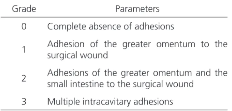
There was neither animal deathfrom the procedure nor infection or suture dehiscence. One animal of the three-day subgroup presented with a hematoma, with no statistical significance (p=1). Regarding adhesions, we used the parameters of Gonçalves et al.13, which classifies them according to
We observed only grade-1adhesions (Table 2).
Immunohistochemical analysis
Matrix Metalloproteinase 9 (MMP-9)
In the evolutionary follow-up, there was a progressive increase of MMP-9 expression, more evident from the seventh to the 14th day, with a lower
progression up to the 21st, with statistical significance
at the 3rd, 7th and 21st days (Table 3).
We observed MMP-9 expression in all cases of the samples, itstissue immunoexpression varying between 2.65% and 11.50% (Figure 1).
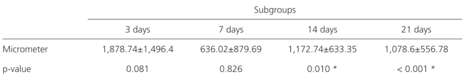
Transforming growth factor beta (TGF- β)
TGF-β expression showed a high level on day 3, fell on the 7th, returning on the 14th, with a small
drop on the 21st day, with statistical significance at the
14th and 21st days (Table 4).
We observed expression of TGF-β in all cases, its tissue immunoexpression varying between 0.03% and 2.92% (Figure 2).
Smooth muscle alpha-actin (ACTA2)
ACTA2 expression presented slightly variable levels in the series, with a slight increase between the 3rd and 7th days, adiscrete decrease from the 7th to
14th days, and slight increase until the 21st day, with
statistical significance at day 14 (Table 5).
We observed expression of ACTA2 in all cases (Figure 3), its tissue immunoexpression varying between 0.88% and 3.23%.
DISCUSSION
Immunomarker research in healing has a very large and polymorphous universe. Many years will be necessary to define the most varied processes of tissue repair in face of the most diverse aggressions. These may or may not be associated with the infectious processes, which naturally alter the normal reaction. The amplitude of the research in surgical healing can be estimated by the numerous immunological forms of reaction only by interpreting the three markers studied here.
The MMP family includes about 25 proteins that can be divided into collagenases (MMP-1, 8 and
Table 1. Classification of abdominal adhesions13.
Grade Parameters
0 Complete absence of adhesions
1 Adhesion of the greater omentum to the surgical wound
2 Adhesions of the greater omentum and the small intestine to the surgical wound
3 Multiple intracavitary adhesions
Figure 1. Photomicrograph of MMP-9 immunostainnig (200x): case with a 2.65% tissue immunoexpression area – subgroup 3 days.
Table 2. Adhesion types according to time.
Grip 3 days 7 days 14 days 21 days
Grade 0 9 9 9 10
Grade 1 1 1 1 0
Grade 2
Grade 3
13), gelatinases 2 and 9), stromelysins (MMP-3, 7 and 10), matrilysins(MMP-7 and 26), membrane-type MMPs (MMP-14, 15, 16, 17 and 24) and other MMPs, which are classified by substrate specificity and, mainly, according to their structure4. The participation
of MMPs in several biological events is due to their potential influence on cellular behavior through some actions, such as cleavage of proteins that make cell-cell adhesion, release of bioactive molecules on the cell surface or by cleavage of molecules present on the cell surface, which transmit signals in the extracellular environment. Several biological processes occur with the participation of metalloproteinases, such as determination of the architecture of the extracellular matrix14, embryonic development, blastocyst
implantation, organ morphogenesis, nervous system development, ovulation, cervical dilatation, postpartum uterine regression16, development and remodeling
of oral tissue, healing, angiogenesis and apoptosis. In this study, similar to Nagase et al.16 data, MMP-9
expression presented significance at the 3rd, 7th and 21st days, proving to be a good indicator, since in only one subgroup, day 14, did not present statistical significance.
The TGF-β superfamily includes several growth factors. Actins are only present in eukaryotic cells, being involved in various cellular functions
including muscle contraction, motility, cell adhesion and division, as well as maintenance of cellular morphology. In vertebrates, it constitutes a family of six proteins, expressed in specific patterns in the development of each tissue17. There are four isoforms
restricted to tissue types, such as α-actin-skeletal, cardiac α-actin, smooth muscle α-actin and smooth muscle γ-actin, respectively in skeletal muscles, cardiac muscle, vascular and enteric smooth muscles. Smooth muscle α-actin is a microfilament (5 to 8 nm in diameter) that composes the cytoskeleton of all mammalian smooth muscle cells, myoepithelial cells, and is particularly abundant in smooth muscle cells of vessel walls17, having important contractile
function in these cells and being considered a marker of the myofibroblastic phenotype18,19.
It is also present in conjunctive cells, such as fibroblasts1,20and myofibroblasts21. Ultrastructurally,
they have well-developed rough endoplasmic reticulum and Golgi complex, pinocytosis vesicles, gap junctions, and organized arrangement of actin myofilament bundles focallylocated along the cell axis (stress fibers) or binding to extracellular fibronectin domains through specialized membrane proteins (integrins), characterizing membrane specializations called fibronexes10,21. Functionally,
this mechano-transducer system is able to transmit the forces generated in the extracellular
Table 3. Mean, standard deviation and statistical analysis in square micrometers by LMF (MMP-9).
Subgroups
3 days 7 days 14 days 21 days
Micrometer 4,739.95±1,676.9 6,633.82±1,867.31 10,560.1±1,653.4 11,533.89±1,161.7
p-value < 0.001 * < 0.001 * 0.099 0.001 *
* Student t Test for independent samples; p<0.05.
Figure 2. Photomicrographs of histological samples immunostained with TGFβ (200x) in a medium tissue immunoexpression area: A) case with 0.08% at day 14; B) case with 1.85% at day 3; C) case with 2.92% at day 3.
matrix22, being important in the process of tissue
repair. Although controversial, the most widely accepted hypothesis in the literature on the origin of myofibroblasts is from quiescent fibroblasts, which once activated, synthesize an abundance of rough endoplasmic reticulum cisternae for matrix production, and transduction of genes that encode transcription of smooth muscle actin myofilaments, important in cellular contractility. Alliot-Licht et al.23 demonstrated the presence of ACTA2-positive
cells in mineralized culture of human dental pulps. Ultrastructurally,these cells had numerous well-developed fibronexes and fibers, as well as edentulous nuclei and intercellular gap junctions, characterizing them as myofibroblasts. They suggested that some growth factors present in the cell culture medium, combined or not with extracellular matrix proteins,
would be responsible for the differentiation of pulp cells in myofibroblasts. The action of cytokines and growth factors, represented by the family of TGF (growth transforming factors), particularly TGF-β1, is essential to differentiate fibroblasts in myofibroblasts. In this study, in contrast to Tomasek
et al.2 findings, ACTA2 expression displayed statistical
significance only on the 14th day, revealing to be aninferior signal method compared with the ones previously studied, MMP-9 and TGF-β.
Thus, among the three markers studied, MMP-9 presented statistical significance in the 3rd,
7th and 21stday subgroups, displaying the best results,
followed by TGF-β, with statistical significance at 14 and 21 days. ACTA2, on its turn,was not a good indicator of the evolution of tissue repair, presenting statistical significance only at the 14th day.
Table 4. Mean, standard deviation and statistical analysis in square micrometers by LMF (TGF-β).
Subgroups
3 days 7 days 14 days 21 days
Micrometer 1,878.74±1,496.4 636.02±879.69 1,172.74±633.35 1,078.6±556.78
p-value 0.081 0.826 0.010 * < 0.001 *
* Student t Test for independent samples; p<0.05.
Table 5. Mean, standard deviation and statistical analysis in square micrometers by LMF (ACTA2).
Subgroup
3 days 7 days 14 days 21 days
Micrometer 1,836.81±724.47 2,011.21±516.91 1,879.22±721.32 2,022.17±1,007.34
p-value 0.665 0.140 0.011 * 0.091
* Student t Test for independent samples; p<0.05.
Objetivo: avaliar a eficácia de três marcadores imunoistoquímicos envolvidos no processo de cicatrização de ferida cirúrgica. Métodos:
estudo experimental em 40 ratos da raça Wistar, dos marcadores metaloproteinases e metaloproteinase da matriz 9 (MMP-9), fator de transformação do crescimento beta (TGF-β) e miofibroblasto e alfa actina de músculo liso (α-AML), estudados a partir de fragmentos de cicatriz cirúrgica de incisão abdominal envolvendo pele, aponeurose e peritônio. Os animais foram distribuídos em quatro subgrupos de dez de acordo com o dia da morte, programada em três, sete, 14 e 21 dias. Resultados: na expressão da MMP-9 ocorreu aumento progressivo de sua concentração, mais evidente do 7º ao 14º dias variando a imuno-expressão tecidual entre 2,65% e 11,50%.TGF- β mostrou expressão em nível alto no 3º dia, caiu no 7º, voltando a subir no 14º, com pequena queda no 21º dia variando a imuno-ex-pressão tecidual entre 0,03% e 2,92%. A α-AML apresentou níveis com pouca variação e discreto aumento variando a imuno-expressão tecidual entre 0,88% e 3,23%. Conclusão: a MMP-9 se apresentou como melhor marcador, seguido pela TGF-β. Já o α-AML não se mostrou um bom sinalizador da evolução da reparação tissular.
Descritores: Cicatrização. Cirurgia Geral. Imuno-Histoquímica. Fatores de Crescimento de Fibroblastos.
REFERENCES
1. Desmoulière A, Geinoz A, Gabbiani F, Gabbiani G. Transforming growth factor-beta 1 induces al-pha-smooth muscle actin expression in granulation tissue myofibroblasts and in quiescent and growing cultured fibroblasts. J Cell Biol. 1993;122(1):103-11. 2. Tomasek JJ, Gabbiani G, Hinz B, Chaponnier C, Brown
RA. Myofibroblasts and mechano-regulation of con-nective tissue remodelling. Nat Rev Mol Cell Biol. 2002;3(5):349-63.
3. Eyden B. The myofibroblast: an assessment of contro-versial issues and a definition useful in diagnosis and research. Ultrastruct Pathol. 2001;25(1):39-50. 4. Visse R, Nagase H. Matrix metalloproteinases and
tis-sue inhibitors of metalloproteinases: structure, func-tion, and biochemistry. Circ Res. 2003;92(8):827-39. 5. Woessner JF Jr. Matrix metalloproteinases and their
inhibitors in connective tissue remodeling. FASEB J. 1991;5(8):2145-54.
6. Gross J, Lapiere CM. Collagenolytic activity in amphib-ian tissues: a tissue culture assay. Proc Natl Acad Sci U S A. 1962;48:1014-22.
7. Pais-Costa SR, Farah JF, Artigiani-Neto R, Martins SJ, Goldenberg A. Evaluation of P53, E-cadherin, Cox-2, and EGFR protein immunoexpression on prognostic of resected gallbladder carcinoma. Arq Bras Cir Dig. 2014;27(2):126-32.
8. Grégoire M, Lieubeau B. The role of fibroblasts in tumor behavior. Cancer Metastasis Rev. 1995;14(4):339-50. 9. Serigiolle LC, Barbieri RL, Gomes HM, Rodrigues DA,
Studart SV, Leme PL. Critical analysis of experimental model for study of adhesions after incisional hernias in-duced in rats’ and repair of abdominal wall with differ-ent biomaterials. Arq Bras Cir Dig. 2015;28(3):178-82. 10. Magalhães CR, Malafaia O, Torres OJ, Moreira LB,
Tefil SC, Pinherio MR, et al. Liver regeneration with l-glutamine supplemented diet: experimental study in rats. Rev Col Bras Cir. 2014;41(2):117-21.
11. Eyden B. Fibroblast phenotype plasticity: relevance for understanding heterogeneity in “fibroblastic” tumors. Ultrastruct Pathol. 2004;28(5-6):307-19. 12. Kagueyama FM, Nicoli FM, Bonatto MW, Orso IR.
Importance of biopsies and histological evaluation in patients with chronic diarrhea and normal colonos-copies. Arq Bras Cir Dig. 2014;27(3):184-7.
13. Gonçalves RM, Esquerdo CRM, Petroianu A, Barbo-sa AJA. Influência de aderências peritoneais e fio
ci-rúrgico na tensão de ruptura da parede abdominal em ratos. Rev Col Bras Cir. 2000;27(3):147-52. 14. Yong VW, Zabad RK, Agrawal S, Goncalves da Silva
A, Metz LM. Elevation of matrix metalloproteinases (MMPs) in multiple sclerosis and impact of immuno-modulators. J Neurol Sci. 2007;259(1-2):79-84. 15. de Oliveira RP, Portari Filho PE, Iglesias AC, de
Olivei-ra CA, Pannain VL. CompaOlivei-rative study of the differ-ent degrees of risk of gastrointestinal stromal tumor. Rev Col Bras Cir. 2015;42(1):32-6.
16. Nagase H, Woessner JF Jr. Matrix metalloproteina-ses. J Biol Chem. 1999;274(31):21491-4.
17. McHugh KM, Crawford K, Lessard JL. A comprehen-sive analysis of the developmental and tissue-specif-ic expression of the isoactin multigene family in the rat. Dev Biol. 1991;148(2):442-58.
18. Vaughan, MB, Howard, EW, Tomasek, JJ. Transform-ing growth factor-β1 promotes the morphological and functional differentiation on the myofibroblast. Exp Cell Res. 2000;257(1):180-9.
19. Hinz B, Phan SH, Thannickal VJ, Galli A, Bochaton-Pial-lat ML, GabbianI G. The myofibroblast: one function, multiple origins. Am J Pathol. 2007;170(6):1807-16. 20. Sloan AJ, Smith AJ. Stimulation of the dentine-pulp
complex of rat incisor teeth by transforming growth factor-beta isoforms 1-3 in vitro. Arch Oral Biol. 1999;44(2):149-56.
21. Schürch W, Seemayer TA, Gabbiani G. The myofi-broblast: a quarter century after its discovery. Am J Surg Pathol. 1998;22(2):141-7.
22. Dugina V, Fontao L, Chaponnier C, Vasiliev J, Gabbiani G. Focal adhesion features during myofibroblastic dif-ferentiation are controlled by intracellular and extracel-lular factors. J Cell Sci. 2001;114(Pt 18):3285-96. 23. Alliot-Licht B, HurtreL D, Gregoire M.
Characteriza-tion of alpha-smooth muscle actin positive cells in mineralized human dental pulp cultures. Arch Oral Biol. 2001;46(3):221-8.
Received in: 21/12/2016
Accepted for publication: 28/04/2017 Conflict of interest: none.
Source of funding: none.
Mailing address:
Jurandir Marcondes Ribas Filho E-mail: [email protected]