Description and molecular phylogeny of one new and one
known needle nematode of the genus Paralongidorus
(Nematoda: Longidoridae) from grapevine in Portugal using
integrative approach
Carlos Gutiérrez-Gutiérrez&Manuel Mota& Pablo Castillo&Margarida Teixeira Santos& Juan E. Palomares-Rius
Accepted: 11 October 2017 / Published online: 18 October 2017 # Koninklijke Nederlandse Planteziektenkundige Vereniging 2017
Abstract A new and a known longidorid nematode, Paralongidorus lusitanicus n. sp. and Paralongidorus plesioepimikis, are described and illustrated from popula-tions extracted from soil associated with grapevine (Vitis vinifera L.) from Escaroupim and Pó (central-Western Portugal), respectively. The new needle nematode P. lusitanicus n. sp. is characterised by a very large body size (8072–12,022 μm), an expanded and rounded lip region, ca 30μm wide, with a clear constriction followed by a depression posterior to the amphidial aperture, amphidial fovea very large (11.0–19.0 μm), stirrup-shaped, with conspicuous slit-like aperture as shown in
scanning electron microscopy studies, a very long and flexible odontostyle (180.0–223.0 μm), guiding ring lo-cated at 28.0–41.5 μm from anterior end, vulva anterior to the mid-body (34–41%), a dorsally convex-conoid tail with rounded terminus (29–42 μm long), bearing two or three pairs of caudal pores and males common (ratio 1:1.6 females) with spicules ca 80μm long. Morphological and morphometric traits for P. plesioepimikis fit well with the original description, and is reported for the first time in Portugal. Integrative diagnosis of both species was com-pleted with molecular data obtained using D2-D3 expan-sion segments of 28S rDNA, ITS1-rDNA and partial 18S–rDNA. The phylogenetic relationships of these spe-cies with other Paralongidorus spp. using these three molecular markers indicated that P. lusitanicus n. sp. clustered together with other Paralongidorus spp. forming a sister clade with P. plesioepimikis, both of them sharing a large body, long odontostyle, an anteriorly lo-cated vulva and an expanded and rounded lip region with a clear constriction followed by a depression posterior to the amphidial aperture.
Keywords Bayesian inference . Longidorids . Paralongidorus . Phylogeny . rDNA . Taxonomy
Introduction
Longidoridae is one of the largest families of soil nem-atodes in term of species diversity given that they
https://doi.org/10.1007/s10658-017-1364-9
C. Gutiérrez-Gutiérrez (*)
:
M. MotaNemaLab/ICAAM, Instituto de Ciências Agrárias e Ambientais Mediterrânicas & Departamento de Biologia, Universidade de Évora, Núcleo da Mitra, Ap. 94, 7002-554 Évora, Portugal e-mail: carlosg@uevora.pt
M. Mota
Departamento Ciências da Vida, Universidade Lusófona de Humanidades e Tecnologias, EPCV, C. Grande 376, 1749-024 Lisbon, Portugal
P. Castillo
:
J. E. Palomares-RiusInstitute for Sustainable Agriculture (IAS), Spanish National Research Council (CSIC), Campus de Excelencia Internacional Agroalimentario, ceiA3, Avenida Menéndez Pidal s/n, 14004 Córdoba, Spain
M. T. Santos
Instituto Nacional de Investigação Agrária e Veterinária (INIAV), Quinta do Marquês, 2780-159 Oeiras, Portugal
comprise a total of seven genera and more than 750 species (Decraemer & Robbins2007). The taxonomic and systematic position of the genus Paralongidorus Siddiqi, Hooper & Klan, 1963 within the family Longidoridae (Thorne, 1935) Meyl, 1961 appears to be accepted by the scientific community, however the species composition are continuously being subject of debate (Hunt1993, Siddiqi et al.1993, Coomans1996, Escuer & Arias 1997, Decraemer & Robbins 2007). This genus, member of the commonly known needle nematodes, is quite diverse with about 90 valid species of migratory ectoparasites that parasitise a wide range of agronomic crops, ornamentals, and forest trees (Taylor & Brown 1997, Decraemer & Robbins 2007, Palomares-Rius et al.2013, Kornobis et al.2015, Barsi & De Luca2017). This group of phytopathogenic spe-cies are of global interest because they cause directly damage on the roots of the host plant attributable to their ectoparasitic feeding and one species is able to transmit damaging nepoviruses (Taylor & Brown 1997, Decraemer & Robbins2007). Paralongidorus maximus (Bütschli 1874) Siddiqi 1964 has important phytopath-ological implications because it is a vector of Raspberry ringspot virus (RpRSV) (Jones et al. 1994, Taylor & Brown1997). This species is considered as a major pest in Europe and other many parts of the world (Taylor & Brown1997), and for this reason is included in the A2 list of pest recommended regulation of the European and Mediterranean Plant Protection Organization (EPPO,
www.eppo.int/QUARANTINE/).
An accurate and detailed description of needle nem-atodes is essential to establish unequivocal diagnosis in order to discern virus vectors and/or quarantine patho-gen species, select appropriate management strategies for preventing the spread of them and establish efficient control measures. Morphometric and morphological identification within this genus at species level is mainly based on characteristics of adult females (Escuer & Arias1997).However, the high intraspecific variability of some diagnostic features and the great diversity in phenotypic plasticity make species identification based on gross morphology and internal anatomical features a technically difficult task even for experts. Recently, the sequencing of RNA-based markers is an increasingly powerful approach for the molecular diagnostics and for understanding their inter- and intra-genetic variability (Palomares-Rius et al.2013, Kornobis et al.2015, Barsi & De Luca 2017). Several ribosomal RNA (rRNA) genes are used for molecular characterizations of these
nematodes: partial 28S rRNA gene (He et al. 2005, Palomares-Rius et al.2008,2013, Pedram et al.2012, Kornobis et al. 2015, Barsi & De Luca 2017), 18S rRNA gene (Palomares-Rius et al.2008,2013, Pedram et al. 2012, Kornobis et al. 2015) and internal tran-scribed spacer (ITS) region of the rRNA genes (Palomares-Rius et al.2008,2013, Pedram et al.2012, Kornobis et al.2015, Barsi & De Luca2017). Kornobis et al. (2015) designed a PCR–RFLP of the D2D3 region of 28S rRNA gene with five restriction enzymes to establish diagnostic profiles from P. rex Andrássy, 1986. Recently, Barsi & De Luca (2017) designed a PCR–RFLP of ITS region based on six restriction en-zymes for genotyping of species-specific variations for P. francolambertii Barsi & De Luca, 2017. D2-D3 ex-pansion segments of 28S rRNA and ITS rRNA have proved to be more effective in species identification when compared to partial 18S rRNA, as both these markers display more molecular variability than partial 18S rRNA (He et al.2005, Palomares-Rius et al.2008, 2013, Pedram et al.2012, Kornobis et al.2015, Barsi & De Luca 2017). Additionally, the mitochondrial DNA (mtDNA) markers have been used for species identifi-cation, phylogeny and phylogeography in a large num-ber of species inside Longidoridae (Lazarova et al. 2006,2016, Gutiérrez-Gutiérrez et al.2011, 2013a,b, Kumari 2014, Subbotin et al. 2014, 2015), however there is a lack of the mitochondrial molecular data on Paralongidorus spp. Only a small fraction of Paralongidorus species (nine identified species) has so far sequences in the GenBank database. These se-quences should be obtained by using voucher specimens from the described species in order to perform molecular species comparison and to differentiate molecularly cryptic species (Palomares-Rius et al.2014). Based on our current knowledge, eleven valid Paralongidorus species have been reported in several cultivated and wild plants in Europe and in the Mediterranean Basin (Bravo & Lemos1997, Escuer & Arias1997, Taylor & Brown 1997, Lišková & Brown2003, Barsi & De Luca2017): i) P. maximus is the most common Paralongidorus spp. in Europe and it is frequently found parasitizing a large number of forest trees, grapevine, chestnut tree (Castanea spp.), walnut trees (Juglans regia L.) and a wide range of vegetable crops in Europe(Heyns, 1975; Lamberti et al., 1983; Roca et al. 1988; Macara 1994; Lišková, 1995; Bravo & Lemos 1997; Taylor & Brown1997; Lišková & Brown,1998,1999, 2003; Gutiérrez-Gutiérrez et al. 2016), ii) P. iberis
Escuer & Arias, 1997 and P. monegrensis Escuer & Arias, 1997 in the rhizosphere of plants in the Rhamno-Cocciferelum ecosystem in Spain (Escuer & Arias1997), iii) P. litoralis Palomares-Rius, Subbotin, Landa, Vovlas & Castillo 2008, in the rhizosphere of mastic tree (Pistacia lentiscus L.) in Spain (Palomares-Rius et al.2008), iv) P. paramaximus Heyns, 1965 on soil around plants in citrus orchards (Citrus aurantium L.) in Spain (Palomares-Rius et al.2008), v) P. epimikis Dalmasso, 1969 in grapevine in Algeria (Dalmasso 1969), vi) P. plesioepimikis Palomares-Rius, Cantalapiedra-Navarrete, Gutiérrez-Gutiérrez, Liébanas & Castillo,2013in the rhizosphere of stone pine (Pinus pinea L.) in Spain (Palomares-Rius et al. 2013), vii) P. georgiensis (Tulaganov,1937) Luc & Doucet 1984, in a vegetable garden soil in Egypt (Tulaganov1937), viii) P. remeyi Altherr 1963 in an unknown host in France (Altherr,1963), ix) P. rex is commonly found infesting grapevine and oak forest in Hungary (Andrássy1986, Barsi et al. 2007), forest tree species and grasses in Poland and Ukraine (Kornobis et al. 2015), and x) P. francolambertii in the rhizosphere of silver lime trees (Tilia tomentosa Moench) and juniper trees (Juniperus communis L.) in a forest in Serbia (Barsi & De Luca 2017). In Europe the low species diversity of Paralongidorus and Longidorus and their current geographical distribution can be attributed to their center of origin, which is supposed to be located in South African-Indian division when it was only one supercontinent (Pangea), before the separation of major plate tectonics (Coomans,1985).
In order to establish the species diversity of the genus Paralongidorus in Portugal and its distribution, we sampled several vineyards in the Lisbon and Tejo wine regions from Spring to Autumn of 2016 in central-Western Portugal, and as a result, a population of an unknown Paralongidorus species was found in the rhi-zosphere of grapevine (Vitis vinifera L.) from an old vineyard at Escaroupim (Salvaterra de Magos, Ribatejo province) and another population resembling P. plesioepimikis was found in a new vineyard at Pó (Bombarral, Estremadura province). One of the uniden-tified populations (Escaroupim) was visually difficult to identify using the polytomous identification key pro-posed by Escuer & Arias (1997) because shared a large number of diagnostic characters with a group of Paralongidorus species characterized by a large body and very long odontostyle, and an expanded lip region, set off from the rest of the body by a clear constriction
followed by a usually slight depression posterior to the amphidial aperture (Palomares-Rius et al.2013). These traits prompted us to undertake a detailed morphological and molecular sequence-based comparative study with previously described Paralongidorus species. These observations indicated that one of the detected species appeared to be morphologically and morphometrically unrelated to nominal Paralongidorus species and did not fit with their descriptions. Detailed studies using light and scanning electron microscopy (SEM) and mo-lecular analyses indicated that this population should be assigned to a new species and is described herein as Paralongidorus lusitanicus n. sp.
The objectives of the present study were: (i) to char-acterise two Portuguese populations of Paralongidorus morphologically and genetically using the D2-D3 ex-pansion segments of 28S rRNA, ITS1 rRNA and the partial 18S rRNA gene sequences; and (ii) to explore the phylogenetic relationships of these needle nematode populations within Paralongidorus spp.
Material and methods
Nematode population sampling, extraction and morphological identification
Specimens of the unidentified Paralongidorus species detected in this study were isolated from infested sandy soil samples collected from the rhizosphere of grapevine (Vitis spp.) from an old vineyard at Escaroupim (Salvaterra de Magos, Ribaltejo province, Portugal) and a new vineyard at Pó (Bombarral, Estremadura province, Portugal). Nematodes were extracted from soil by a modification of Cobb’s decanting and sieving method (Flegg1967).
Specimens for light microscopy (LM) study were killed and fixed in an aqueous solution of 4% formal-dehyde +1% glycerol, dehydrated using alcohol-saturated chamber and processed to pure glycerine using Seinhorst’s method (Seinhorst1966). Specimens were examined using an Olympus BX50 light microscope with differential interference contrast at magnifications up to 1,000× magnification. Photographs of nematodes were taken by an Olympus DP70 camera and Cell software (Olympus Software Imaging for Life Sci-ences). All measurements were expressed in microme-ters (μm). For line drawings of the new species, light micrographs were imported to CorelDraw ver. X6 and
redrawn. All other abbreviations used are as defined in Jairajpuri and Ahmad (1992).
For SEM studies, fixed specimens were dehydrated in a graded ethanol series, critical point dried, mounted on SEM stubs, sputter-coated with a thin layer of gold (Abolafia et al. 2002), and observed with a Hitachi S-3700 N microscope coupled with a Bruker XFlash5010 SDD detector.
Nematode molecular identification
For molecular analyses and in order to avoid mistakes in the case of mixed populations in the same sample, two live nematodes from each population were temporarily mounted in a drop of 1 M NaCl containing glass beads to ensure specimens conformed to the unidentified pop-ulations of Paralongidorus. Morphometric measure-ments and photomicrographs recorded during this initial study were not used as part of the morphological study and morphometric analyses. Following morphological confirmation, the slides were dismantled and DNA ex-tracted. Nematode DNA was extracted from single fe-male individuals and PCR assays were conducted as described by Castillo et al. (2003). One nematode spec-imen of each sample was transferred to an Eppendorf tube containing 16μl ddH2O, 2μl 10× PCR buffer and
2 μl proteinase K (600 μg/ml) (Promega, Benelux, The Netherlands) and crushed during 2 min with a micro-homogeniser, Vibro Mixer (Zürich, Switzerland). The tubes were frozen at −80 °C for (15 min) and incubated at 65 °C (1 h), then at 95 °C (10 min). One μl of extracted DNA was transferred to an Eppendorf tube containing: 2.5 μl 10X NH4 reaction buffer, 0.75 μl MgCl2 (50 mM), 0.25 μl dNTPs mixture (10 mM each), 0.75μl of each primer (10 mM), 0.2 μl BIOTAQ DNA Polymerase (BIOLINE, UK) and ddH2O to a final volume of 25μl. The D2-D3 expansion
segments of 28S rRNA was amplified using the D2A
(5′-ACAAGTACCGTGAGGGAAAGTTG-3′) and
D3B (5′-TCGGAAGGAACCAGCTACTA-3′) primers (De Ley et al. 1999). The ITS1 region was amplified using forward primer 18S (5’TTGATTACGTCCCT GCCCTTT-3′) (Vrain et al.1992) and reverse primer rDNA1 (5′-ACGAGCCGAGTGATCCACCG-3′) (Cherry et al.1997). Finally, the portion of 18S rRNA was amplified using primers 988F (5′-CTC AAA GAT TAA GCC ATG C-3′), 1912R (5′-TTT ACGGTC AGA ACT AGG G-30), 1813F (5′-CTG CGT GAG
AGGTGA AAT-3′) and 2646R (50-GCT ACC TTG TTA CGA CTT TT-3′) (Holterman et al.2006).
PCR cycle conditions were: one cycle of 94 °C for 2 min, followed by 35 cycles of 94 °C for 30 s, anneal-ing temperature of 55 °C for 45 s, 72 °C for 3 min, and finally 72 °C for 10 min. PCR products were purified after amplification using ExoSAP-IT (Affmetrix, USB products), quantified using a Nanodrop spectrophotom-eter (Nanodrop Technologies, Wilmington, DE, USA) and used for direct sequencing in both directions using the primers referred to above. The resulting products were purified and run on a DNA multicapillary sequenc-er (Model 3130XL genetic analyssequenc-er; Applied Biosystems, Foster City, CA, USA), using the BigDye Terminator Sequencing Kit v.3.1 (Applied Biosystems, Foster City, CA, USA), at the Stab Vida sequencing facilities (Caparica, Portugal). The newly obtained se-quences were submitted to the GenBank database under accession numbers indicated on the phylogenetic trees (KY750560-KY750569).
Phylogenetic analyses
D2-D3 expansion segments of 28S rRNA, ITS1 region, and partial 18S rRNA sequences of different Longidorus spp. and Paralongidorus spp. from GenBank were used for phylogenetic reconstruction. Outgroup taxa for each dataset were chosen following previous published studies (He et al.2005, Holterman et al.2006, Gutiérrez-Gutiérrez et al.2013b, Palomares-Rius et al.2013, Archidona-Yuste et al.2016a). Multiple sequence alignments of the different genes were made using the Q-INS-i algorithm of MAFFT V.7.205 (Katoh & Standley2013), which accounts for secondary RNA structure. Sequence alignments were visualised using BioEdit (Hall1999) and edited by Gblocks ver. 0.91b (Castresana 2000) in Castresana Laboratory server
(http://molevol.cmima.csic.es/castresana/Gblocks_
server.html) using options for a less stringent selection (minimum number of sequences for a conserved or a flanking position: 50% of the number of sequences +1; maximum number of contiguous non-conserved posi-tions: 8; minimum length of a block: 5; allowed gap positions: with half). Percentage similarity between se-quences was calculated using a sequence identity matrix using BioEdit. For that, the score for each pair of se-quences was compared directly and all gap or place-holding characters were treated as a gap. When the same position for both sequences had a gap it was not treated
as a difference. Phylogenetic analyses of the se-quence datasets were based on Bayesian inference ( B I ) u s i n g M r B a y e s 3 . 1 . 2 ( R o n q u i s t & Huelsenbeck 2003). The best-fit model of DNA evolution was obtained using JModelTest V.2.1.7 (Darriba et al. 2012) with the Akaike Information Criterion (AIC). The best-fit model, the base fre-quency, the proportion of invariable sites, and the gamma distribution shape parameters and substitu-tion rates in the AIC were then given to MrBayes for the phylogenetic analyses. Unlinked general time-reversible model with invariable sites and a gamma-shaped distribution (GTR + I + G) for the D2-D3 expansion segments of 28S rRNA, and for the ITS1 region, and the partial 18S rRNA under a transitional model with invariable sites and a gamma-shaped distribution (TIM2 + I + G). These BI analyses were run separately per dataset using four chains for 2 × 10⁶ generations for the D2-D3, and 1 × 10⁶ generations for the rest of molecular markers. The Markov Chains were sam-pled at intervals of 100 generations. Two runs were conducted for each analysis. After discarding burn-in samples and evaluating convergence, the remaining samples were retained for further analy-ses. The topologies were used to generate a 50% majority-rule consensus tree. Posterior probabilities (PP) are given on appropriate clades. Trees from all analyses were visualised using TreeView (Page 1996) and FigTree software V.1.42 (http://tree.bio. ed.ac.uk/software/figtree/).
Results
Paralongidorus lusitanicus1n. sp. (Figs.1,2,3and4, Table1)
Female: Body very long and rather robust, slightly tapering towards posterior end, usually assuming an open close C to spiral shape, always more coiled in the posterior half, when heat relaxed. Cuticle usually smooth under low magnifications, 4.6 ± 0.8 (3.5–6.5) μm thick at mid-body, but thicker 11.7 ± 0.7 (8.5–16.5) μm at tail end. A variable number of body pores in the
pharyngeal region; 5–7 lateral, 1–2 ventral and 1–2 dorsal pores. Lip region wide expanded, anteriorly slightly convex, almost flat at the end and widely round-ed in lateral view, clearly set off by a constriction followed by a depression as shown by SEM, 29.9 ± 1.5 (27.0–33.0) μm wide and 13.1 ± 1.1 (13.5– 14.5) μm high. Amphidial fovea very large, stirrup-shaped, with conspicuous aperture ca two-third as wide as lip region as shown by SEM. Twelve cephalic papil-lae appearing as small apertures, an inner line and other line outer, both lines composed by six labial papillae, each outer labial papillae located just anterior to a dis-tinct cephalic lobe 3.1 ± 0.7 (2.0–4.5) μm long (Figs.1, 2, and3, Table1). A single guide ring, 6.0–8.5 μm wide, located 0.9 ± 0.1 (0.8–1.1) times the lip region diam. From anterior end. Lateral chord 16.0 (11.0–26.0) μm wide, ca 24.4% of corresponding body diam. at mid-body. Odontostyle very long and flexible, 2.0 (1.3–3.1) times as long as odontophore which is weakly devel-oped, with rather faint basal swellings (Figs. 1-2, Ta-ble 1). Nerve ring encircling part of pharynx near odontophore base, at 355.6 (236–461) μm from the anterior end. Basal bulb long and cylindrical, 149.5 ± 16.8 (104.0–177.0) μm long or ca 4.1 times the total pharynx length, and 26.3 ± 4.9 (22.0–38.0) μm in diam. Glandularium 129.7 ± 13.3 (111.0–140.0) μm long. Dorsal pharyngeal gland nucleus (DN) located at 18.1 ± 3.2 (13.8–25.9) % and ventrosublateral nuclei (SVN) near middle of bulb, 45.9 ± 7.5 (30.7–54.8) % of distance from anterior end of pharyngeal bulb. Nucleoli of DN one-fourth times larger than nucleolus of SVN. Cardia conoid-rounded, 15.1 (8.0–25.0) μm long (Figs. 1-2, Table1). Reproductive system typical of genus with both genital branches equally developed; anterior branch 383–1559 μm long and posterior branch 382– 1346 μm long.. Ovaries paired and reflexed, usually equally developed and with a single row of oocytes, variable in length (183–251 μm long), vulva in form of a transverse slit, located anteriorly to mid-body (33.0–41.0% of towards the front of the body), vagina perpendicular to body axis, 40–58 μm long, ca 65% of corresponding body diam., surrounded by well-developed muscles. Anterior and posterior oviduct of similar size. Uteri 248–499 μm long, with presence of sperm cells in some females; conspicuous muscle sphincter between uterus and oviduct. Prerectum vari-able in length, 531.3 ± 180.5 (408.0–701.0) μm long or ca 6.1–10.6.5 times anal body diameter, and rectum ca 0.7–1.2 times as long as anal body diameter. Tail short,
1The species epithet refers to Lusitania, the ancient roman province
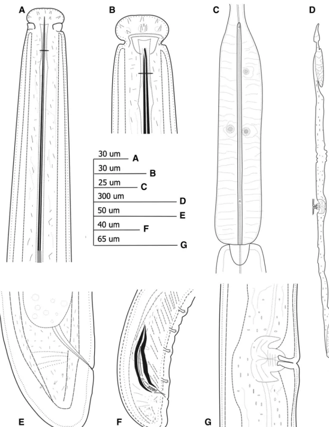
Fig. 1 Line drawings of Paralongidorus lusitanicus n. sp. paratypes from the rhizosphere of grapevine (Vitis from Escaroupin, Portugal (A-F). A: female neck region; B: female lip
region; C: detail of basal pharyngeal bulb; D: Detail of genital branches; E: female tail region; F: male tail region; G: vulva region
convex-conoid with a broadly rounded terminus (Figs. 1,2and3, Table1).
Male Common (ratio 1:1.6 females). Morphologically similar to female except for genital system, but with
posterior region strongly curved ventrally when heat relaxed. Genital tract diorchic with testes opposed, oc-cupying ca 51.6% of body length containing multiple rows of different stages of spermatogonia. Tail conoid-rounded, with broad blunt terminus and thickened outer
Fig. 2 Light micrographs of Paralongidorus lusitanicus n. sp. female paratypes from the rhizosphere of grapevine (Vitis sp.) from Escaroupim, Portugal (A-M). A. Female anterior regions; B-C. Lip regions showing amphidial fovea at different focus; D: Detail of basal bulb; E: Vulva region; F: sperm cells; G: Male tail region; H: Female tail region; I: First-stage juvenile anterior region; J-M:
Tail of J1, J2, J3 and J4, respectively. Abbreviations: a = anus; af = amphidial fovea; cd = cardia; gr = guiding ring; ost = odontostyle; ov = ovary; rost = replacement odontostyle; V = vulva. (Scale bars: A, G, L = 20μm; B, C, F, H, M = 22.5 μm; D-E: 17μm; I = 50 μm; J: 25 μm; K: 30 μm)
cuticular layer. Spicules arcuate, robust, 77–85 μm long; lateral guiding pieces 17–24 μm long, more or less straight or with curved proximal end. One pair of adcloacal supplements and a variable number of midventral supplements (11–13) (Figs.1-2, Table1). Juveniles All four juvenile stages were found and dis-tinguished by relative lengths of body, functional and replacement odontostyle (Table1, Fig.4; Robbins et al. 1995,1996) and genital primordium. Body usually as-suming a J-shape, a C-shape to spiral C-shape in J2 and J3 and similar to adults in J4 when heat relaxed. Lip region of all juvenile stages similar to that of adults. First-stage juveniles (J1) characterised by a bluntly conoid-rounded tail with ventral peg with a c’ ratio 1.6–1.8, odontostyle length 103–110 μm long, and slightly shorter distance from anterior end to stylet
Fig. 3 SEM micrographs of Paralongidorus lusitanicus n. sp. A-D: Female anterior end in lateral, ventro-lateral and frontal view showing cephalic lobe (cl), amphidial aperture (af), inner labial papillae (ip) and outer labial papillae (op); E-F: Female tail, oblique and lateral view showing caudal pores (cp) and anus (a). (Scale bars: A-D = 30μm; B-D = 35μm.) 60 80 100 120 140 160 180 200 220 240 Odontostyle length (m) 0 2000 4000 6000 8000 10000 12000 Body length (m) Ost-J1 rOst-J1 Ost-J2 rOst-J2 Ost-J3 rOst-J3 Ost-J4 rOst-J4 Ost-female
Fig. 4 Relationship of body length to length of functional and replacement odontostyle (Ost and rOst, respectively) length in all developmental stages from second-stage juveniles (J2) to mature females of Paralongidorus lusitanicus n. sp.
guiding ring to the next juvenile stages. Tail shape of J2 conoid-rounded. J3 and J4 tail shape barely dorsally con-vex-conoid, but more elongate than that of female, shorter body length, shorter odontostyle length, and shorter dis-tance from anterior end to guiding ring (Fig.2, Table1). Type habitat and locality
Rhizosphere of grapevine (Vitis sp. at Escaroupim, lo-cality of Salvaterra de Magos, Ribatejo province,
Portugal (GPS coordinates: N 39° 4′ 35.922″ E 8° 43′ 58.471″).
Type material
Holotype female (slide LLP001) and seven female paratypes (LLP002-LLP008) together with six male paratypes (LLP009-LLP014) deposited in the Nema-tode Collection of the Nematology Lab, Institute for Mediterranean Agricultural and Environment Sciences,
Table 1 Morphometrics of Paralongidorus lusitanicus n. sp. from the rhizosphere of grapevine (Vitis vinifera L.) from Escaroupim, Portugal. All measurements inμm and in the format: mean ± s.d. (range)*
Character Holotype Paratype Females Paratype Males J1 J2 J3 J4
n 1 16 9 4 4 2 7 L 10,649 10,627 ± 1024 (8072–12,022) 9357 ± 1022 (7809–10,663) 1833 ± 109 (1729–1977) 2583 ± 123 (2409–2700) 3614, 4196 6362 ± 672 (5575–7442) a 162.0 163.0 ± 22.7 (116.5–192.4) 165.4 ± 23.1 (123.0–198.9) 54.9 ± 4.0 (50.6–60.2) 61.7 ± 4.3 (56.5–66.9) 73.8, 88.6 116.3 ± 19.1 (97.4–143.1) b 18.9 18.0 ± 3.9 (13.8–31.1) 15.5 ± 1.6 (12.8–18.0) 5.9 ± 4.0 (5.6–7.9) 7.2 ± 0.6 (6.7–7.9) 9.7, 9.8 11.0 ± 1.3 (9.6–13.4) c 317.7 302.9 ± 34.5 (240.3–369.4) 252.4 ± 37.3(192.2–327.4) 42.7 ± 2.1(41.1–45.6) 67.4 ± 2.1(65.8–70.5) 86.0, 108.4 176.1 ± 20.4(156.6–216.6) c´ 0.7 0.8 ± 0.1 (0.7–0.9) 0.9 ± 0.1 (0.7–1.2) 1.6 ± 0.1 (1.6–1.8) 1.2 ± 0.02 (1.1–1.2) 1.0, 1.1 0.8 ± 0.1 (0.7–0.9) V or T 39.3 37.6 ± 2.5 (33.0–40.9) 51.6 ± 2.3 (48.4–55.0) – – – – G1 5.5 6.6 ± 2.8 (3.2–13.4) – – – – – G2 4.4 6.0 ± 2.2 (3.2–12.4) – – – – – Odontostyle length 188.5 194.2 ± 12.4 (180.0–223.0) 180.4 ± 26.8(117.0–206.0) 107.7 ± 3.2(103.0–110.0) 117.8 ± 3.4(114.0–121.0) 133.0, 135.0 166.1 ± 13.6(159.0–189.0) Replacement odontostyle length – – – 121.7 ± 1.2 (120.5–123.0) 142.0 ± 5.8(134.0–148.0) 155.0, 163.0 189.9 ± 15.3(181.0–214.0) Odontophore length 97.0 102.9 ± 18.6 (72.0–140.0) 95.1 ± 10.4 (74.0–107.0) 59.6 ± 2.8 (57.0–63.0) 68.4 ± 12.9 (50.0–80.0) 64.0, 84.5 91.0 ± 14.6 (65.0–98.0) Lip region width 29.0 29.9 ± 1.5
(27.0–33.0) 29.5 ± 2.9 (29.0–31.0) 14.9 ± 0.7 (14.0–16.0) 19.5 ± 1.5 (18.0–21.0) 24.0, 24.5 26.8 ± 2.2 (25.0–29.0) Oral aperture-guiding ring 33.0 34.8 ± 3.9
(28.0–41.5) 34.9 ± 2.5 (31.9–43.8) 21.2 ± 0.7 (20.4–21.9) 24.0 ± 1.3 (22.0–25.0) 26.5, 28.0 30.2 ± 1.5 (29.0–33.0) Tail length 33.5 35.3 ± 3.5 (29.0–42.0) 37.4 ± 4.2 (32.0–44.0) 42.9 ± 1.7 (41.1–45.0) 38.3 ± 2.2 (36.0–41.0) 38.7, 42.0 36.2 ± 3.3 (32.0–39.0) Spicules – – 80.3 ± 2.7 (77.0–85.0) – – – –
Lateral accessory piece – – 21.0 ± 2.3 (18.0–24.0) – – – – J 111.0 11.7 ± 2.5 (8.6–16.5) 12.3 ± 2.4 (8.5–16.0) 13.0 ± 1.5 (11.0–14.0) 9.5 ± 0.9 (9.0–11.0) 10.0, 11.0 11.6 ± 1.0 (9.5–14.0) *
ICAAM, University of Évora, Évora, Portugal, and three female (LLP015- LLP017) and one male paratypes (LLP018) in the Institute for Sustainable Ag-riculture, IAS-CSIC, Córdoba, Spain. Two female and one male paratypes deposited at each of the following collections: Royal Belgian Institute of Natural Sciences, Brussels, Belgium, and Istituto per la Protezione delle Piante (IPP) of Consiglio Nazionale delle Ricerche (C.N.R.), Sezione di Bari, Bari, Italy. Specific D2-D3, ITS1-rRNA, and partial 18S rDNA sequences were deposited in GenBank with accession numbers KY750560-KY750562, KY750565-KY750566, and KY750569, respectively.
Diagnosis and relationships
Paralongidorus lusitanicus n. sp. is characterised by a large body size (8.07–12.02 mm), an expanded and rounded lip region, ca 30 μm wide, with a clear con-striction followed by a depression posterior to the amphidial aperture, amphidial fovea large, stirrup-shaped, with conspicuous slit-like aperture, a very long and flexible odontostyle (180.0–223.0 μm), a guiding ring located at 28.0–41.5 μm from anterior end, normal arrangement of the pharyngeal gland nuclei, dorsal pha-ryngeal gland nucleus in anterior part of bulb, a pair of ventrosublateral pharyngeal glands near the middle of bulb, vulva anterior to the mid-body (33–41%), and a dorsally convex-conoid tail with a rounded terminus (29–42 μm long), from 0.7 to 0.9 times longer than width, bearing two or three pairs of caudal pores and male common; and specific D2-D3, ITS1, and partial 18S–rRNA sequences. According to the polytomous key of Escuer & Arias (1997), the new species has the following codes (codes in parentheses are exceptions): A1, B1, C3, D2, E2, F6, G7, H23, I2(3), J1(2), K56, L(3)4, M(2)3, N2, O2.
On the basis of amphidial fovea, lip region, body length, odontostyle length, ratios a, c, and c’, distance from oral aperture to guiding ring, bulb length, tail length and shape, spicules length, number of supple-ments in male tail and tail morphology of the J1, P. lusitanicus n. sp. is close to P. epimikis, P. deborae (Jacobs & Heyns1982) Luc & Doucet, 1984, P. iranicus Pedram, Pourjam, Namjou, Reza Atighi, Cantalapiedra-Navarrete, Liébanas, Palomares-Rius & Castillo 2012, P. francolambertii, P. litoralis, P. maximus, P. rex and P. plesioepimikis. Morphologically and morphometrically, P. lusitanicus n. sp. can be distinguished from these
species by several features as indicated below. From P. epimikis it differs by a longer lip region diam. (27.0–33.0 vs 23.0–24.0), a different shape of amphidial fovea (stirrup vs funnel), a slightly smaller a ratio (116.5–192.4 vs 184–211), a vulva more anteriorly lo-cated (33.0–41.0 vs 44–46%), a different tail shape (dorsally convex-conoid vs conoid-rounded) and abun-dance of males (frequent vs rare) having usually a longer spicules (77.0–85.0 vs 57–58 μm) (Dalmasso 1969). First-fourth juvenile stages of the new species and P. epimikis show morphologically a high similarity; how-ever, the J1 stages can be differenced by a lower c’ratio (1.6–1.8 vs 2.22–2.23) and a shorter tail length (41.0– 45.0 vs 49.0–50.0 μm). On the other hand, P. lusitanicus n. sp. differs from P. deborae in having lower V ratio (33.0–41.0 vs 42–48%), a longer lip region diam. (27.0– 33.0 vs 25.0–27.0 μm), a longer odontostyle (180.0– 223.0 vs 156.0–166.0 μm), a different female tail shape (dorsally convex-conoid vs conoid-rounded) and longer spicules length (77.0–85.0 vs 63–72 μm) (Jacobs & Heyns1982). From P. iranicus it differs in a, c, c´ ratios (116.5–192.4 vs 101.9–137.7, 240.3–369.4 vs 221.3– 314.8, 0.7–0.9 vs 0.5–0.7, respectively), V ratio (33–41 vs 37–44%), lip region diam. (27.0–33.0 vs 25.0– 30.0 μm), type of depression between lip region and body contour (less marked in the new species), odontostyle length (180.0–223.0 vs 155.0–184.0 μm), odontophore length (72.0–139.6 vs 82.0–100.0 μm), and tail length and shape (29.0–42.0 vs 25-37 μm, dor-sally convex-conoid vs broadly rounded) (Pedram et al. 2012). From P. francolambertii it can be differentiated mainly in a, c, and V ratios (116.5–192.4 vs 143.0– 197.0, 116.5–192.4 vs 227.0–344.0, 33.0–41.0 vs 42.0–49.0%, respectively), lip region diam. (27.0–33.0 vs 18.0–20.0 μm), female tail shape and length (dorsally convex-conoid vs convex-conoid with a terminal round-ed point and thickenround-ed outer cuticular layer, 29.0–42.0 vs 22–29), type of depression between lip region and body contour (less marked in the new species), body length (8.0–12.0 vs 5.9–8.3 mm), odontostyle length (180.0–223.0 vs 131.0–149.0 μm) and spicules length (77.0–85.0 vs 49.0–58.0 μm) (Barsi et al.2017). First juvenile stages of the new species and P. francolambertii are highly similar morphologically, in spite they can be differentiated by the tail shape (bluntly conoid-rounded tail with ventral peg vs elongate-conoid without peg). From P. litoralis it can be differentiated mainly by means of a and V ratios (116.5–192.4 vs 113.7–164.4, 33.0–41.0 vs 37.0–44.0%, respectively), lip region
diam. (27.0–33.0 vs 25.0–30.0 μm), female tail shape (dorsally convex-conoid vs bluntly rounded) and spic-ules length (77.0–85.0 vs 63.0–72.0 μm) (Palomares-Rius et al.2008). All juvenile stages of the new species and P. litoralis are highly similar morphologically, in fact the J1 stages of them are almost identical, only having small anatomical differences such as tail shape (bluntly rounded tail with ventral peg vs conoid-rounded with a central peg). From P. maximus it differs in a, c, c´ ratios (116.5–192.4 vs 77.0–133.0, 240.3– 369.4 vs 178.0–320.0, 0.7–0.9 vs 0.44–0.60, respective-ly), lip region diam. (27.0–33.0 vs 34.0–39.0 μm), odontostyle length (180.0–223.0 vs 152.0–187.0 μm), distance from oral aperture to guiding ring (28.0–41.5 vs 37–47 μm), tail shape (dorsally convex-conoid vs blunt-ly rounded), abundance of males (frequent vs rare) and spicules length (77.0–85.0 vs 63.0–72.0 μm) (Heyns 1975). All juvenile stages of the new species and P. maximus are highly similar morphologically, in spite of that the J1 stage can be differentiated by a bigger c´ ratio (1.6–1.8 vs 1.06–1.25). From P. rex it differs main-ly in a, c, c´ ratio (116.5–192.4 vs 106.0–111.0, 240.3– 369.4 vs 221.3–314.8, 0.7–0.9 vs 0.5–0.6 respectively), V ratio (33–41 vs 47%), amphidial fovea shape (stirrup vs funnel), odontostyle length (180.0–223.0 vs 178.0– 180.0 μm), female tail length and shape (29–42.0 vs 40.0–45.0 μm, convex-conoid vs conoid-rounded) and abundance of males (frequent vs not found) (Andrassy 1986). All juvenile stages of the new species and P. rex
showed anatomically and morphologically a high simi-larity with P. lusitanicus n. sp., however the J1 stages can be differentiated by a higher c´ ratio (1.6–1.8 vs 1.1– 1.3) and a longer tail length (41.0–45.0 vs 29.0– 36.0μm). From P. plesioepimikis it differs only in the odontophore length (72.0–140.0 vs 66.0–87.0 μm), abundance of males (frequent vs not found) (Palomares-Rius et al.2013). All juvenile stages of the new species and P. plesioepimikis are almost indistin-guishable, however the J1 stages can be differentiated by a bigger c´ ratio that the new species (1.6–1.8 vs 1.3– 1.6). Furthermore, the new species can be clearly sepa-rated from all other sequenced Paralongidorus spp. and similar morphologically and morphometrically (P. iranicus, P. francolambertii, P. litoralis, P. paramaximus, P. maximus, P. plesioepimikis, P. bikanerensis (Lal & Mathur, 1987) Siddiqi, Baujard & Mounport,1993and P. rex) by using the three molecular markers of D2-D3 expansion segments of 28S rDNA, ITS1 rRNA and partial 18S rRNA (Figs.6,7, and8). Paralongidorus plesioepimikis Palomares-Rius et al. 2013(Fig.5, Table2)
The Portuguese population of this species from the rhizosphere of grapevine (V. vinifera L.) in the locality of Pó (Bombarral, Estremadura province) (Table2) was characterised by the absence of males; females exhibited
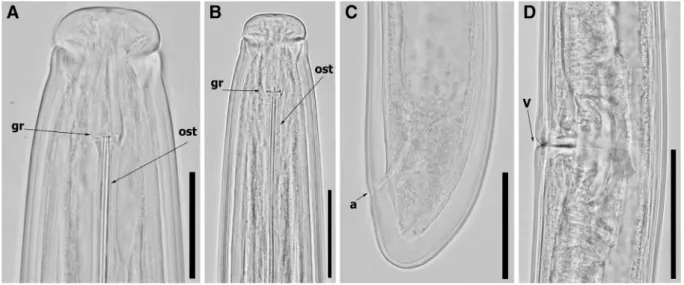
Fig. 5 Light micrographs of Paralongidorus plesioepimikis Palomares-Rius et al.2013females from the rhizosphere of grape-vine (Vitis vinifera L.) from Pó, Bombarral, Portugal (A-D). A: Lip regions showing amphidial fovea at different focus; B: Female
anterior regions; C: Female tail region; D: Vulva region. Abbrevi-ations: a = anus; gr = guiding ring; ost = odontostyle; V = vulva. (Scale bars: A-B = 20μm; C = 40 μm; D: 75 μm)
a robust and very large body length (10.3–11.9 mm), lip region rather expanded, 29.0–33.0 μm wide and 14.0– 14.5μm high, oval-rounded in lateral view and clearly set off by a clear constriction followed by a depression posterior to amphidial aperture, ca 19.0 μm long. Amphidial fovea ca two-third as wide as lip region, stirrup-shaped, with conspicuous slit-like aperture. Odontostyle very long ca 1.6 times as long as odontophore, which is weakly developed, with rather weak basal swellings (Fig.5, Table2). Cuticle of mid-body 5.0–6.0 μm thick, 15.0–15.5 μm thick at tail tip, marked by very fine superficial transverse striae. Basal bulb cylindrical occupying 169.0–183.5 μm long or 22.5 (21.5–23.5) % of total pharynx length. Dorsal pharyngeal gland nucleus in anterior part of bulb, one subventral pair of nuclei near middle of basal bulb. Glandularium 147.2 ± 2.1 (146.0–149.0) μm long. Car-dia clearly visible, elongate and conoid-rounded, 21.1 ± 1.6 (20.0–22.0) μm long (Fig.5, Table2). Gen-ital tracts typical of the genus, with both genGen-ital branches equally developed, anterior branch 410– 548μm long and posterior branch 382–480 μm long, ovaries paired and reflexed, usually equally developed occupying 37.4–56.4% of the total branch length, vulva in form of a transverse slit, usually located anteriorly to mid-body (34.2–37.6%), vagina perpendicular to body axis, 40.5–49.0 μm long, ca 59% of corresponding body
diam., surrounded by visible well-developed muscles. Anterior and posterior oviduct of similar size, separated of uterus by a conspicuous muscle sphincter. Uteri large, 146–176 μm long, where spermatic cells can usually be seen. Prerectum 846 (830–862) μm long and rectum 56.4 (55.0–58.0) μm long. Tail dorsally convex-conoid, broadly rounded terminus, with two or three pores each side of the tail and characterised by 39.0–41.5 μm long and c’ ca 0.7 (Fig. 5, Table 2). Morphological and morphometric traits of this population agree with the original description of this species by Palomares-Rius et al. (2013). In fact, morphological features and mor-phometric measurements of females from the Portu-guese population were within the range of the original description from Palomares-Rius et al. (2013), except by some slight differences in tail length, c´ ratio and lip region diameter, which may be due to geographical intraspecific variability, such as reported in others spe-cies of Paralongidorus (Kornobis et al.2015). This is the first report of this species for Portugal and confirms a wider distribution of this specie in the Iberian Peninsula and Mediterranean Basin since it has only been reported from Spain (Palomares-Rius et al. 2013). The alpha-numeric codes for P. plesioepimikis to be applied to the polytomic identification key for the Paralongidorus species by Escuer & Arias (1997): A1, B1, C3, D2, E1, F6, G7, H2, I23, J1, K56, L34, M3, N-, O-.
Phylogenetic relationships of Paralongidorus lusitanicus n. sp. and P. plesioepimikis within the genera
Longidorus and Paralongidorus
Amplification of D2-D3 expansion segment of 28S rRNA, ITS1 rRNA, and the partial 18S rRNA from Paralongidorus lusitanicus n. sp. and P. plesioepimikis yielded a single fragment of ca 800, 1100, and 1700 bp, respectively. Sequences from P. lusitanicus n. sp. matched well with the Paralongidorus spp. sequences deposited in GenBank being clearly different from all of them. Four new D2-D3 of 28S rRNA gene sequences were obtained in the present study. D2-D3 expansion segments of 28S rRNA sequences of P. lusitanicus n. sp. (KY750560-KY750562) showed a 93–97% similarity values (differed in a range from 21 to 54 nucleotides) w i t h s e v e r a l P a r a l o n g i d o r u s s p p . s u c h a s P. p l e s i o e p i m i k i s ( J Q 6 7 3 4 0 3 ) , P. m a x i m u s (KF412826), P. paramaximus (EU026156), P. litoralis (EU026155), P. rex (KJ427793), P. iranicus (JN032587) and P. francolambertii (LT669805). ITS1
Table 2 Morphometrics of Paralongidorus plesioepimikis Palomares-Rius et al.2013 from the rhizosphere of grapevine (Vitis vinifera) from Pó, Portugal. All measurements inμm and in the format: mean ± s.d. (range)*
Character Females n 2 L 10,353, 11,948 a 141.5, 152.7 b 16.4, 19.8 c 169.8, 287.8 c´ 0.7, 0.7 V or T (34.2, 36.8) Odontostyle length 215.0, 220.0 Odontophore length 132.0, 135.0 Lip region width (29.0, 33.0) Oral aperture-guiding ring (36.0, 40.0) Tail length (38.0, 41.5)
J (15.0, 15.5)
*
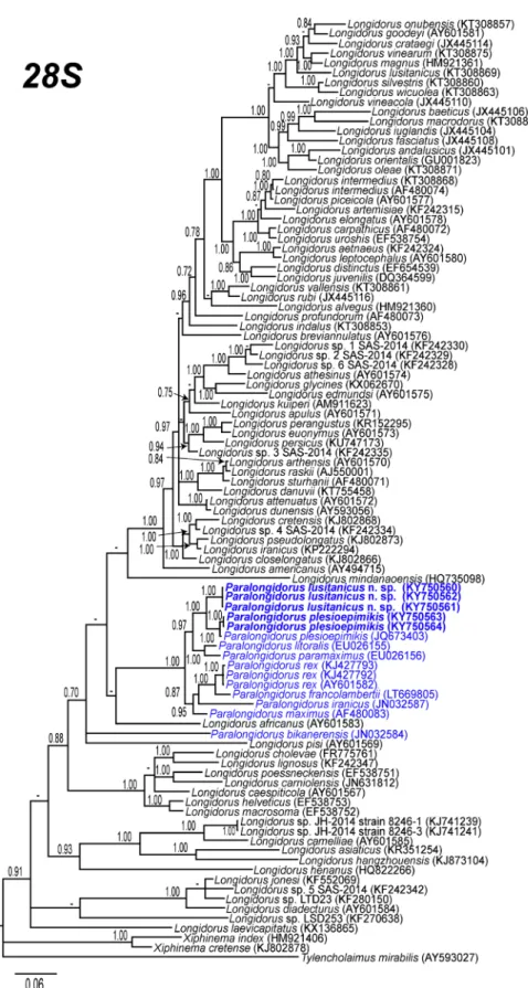
Fig. 6 Phylogenetic relationships of Paralongidorus lusitanicus n. sp. and P. plesioepimikis Palomares-Rius et al.2013within the genus Longidorus and Paralongidorus. Bayesian 50% majority rule consensus trees as inferred from D2-D3 expansion segments of 28S rRNA sequences alignments under the GTR + I + G model. Posterior probabilities more than 70% are given for appropriate clades. Newly obtained sequences in this study are in bold
showed some moderate similarity, 83% with P. p l e s i o e p i m i k i s ( J Q 6 7 3 4 0 7 ) , 8 1 % , w i t h P. paramaximus (JQ673410), and 80%, with P. litoralis (JQ673409), differing in a range from 120 to 136 nucleotides. The partial 18S rRNA sequence for P. lusitanicus n. sp. (KY750569) showed high similarity (99% similar) with several Paralongidorus spp., such as P. plesioepimikis (JQ673405), P. litoralis (EU026158), P. paramaximus (EU026157), P. maximus (AJ875152), differing in a range from 3 to 8 nucleotides. Intraspecific sequence diversity (uncorrected p-distance) of ribosom-al markers among the studied specimens for P. lusitanicus n. sp. was small. In fact, D2-D3 showed no intraspecific sequence diversity and ITS1 region only varied 1% (3 bp, 3 indels). Finally, the Portuguese population of P. plesioepimikis showed some high sim-ilarity for D2-D3 and ITS1 region (99% (5 nucleotides) and 97% (34 nucleotides), respectively) with P. plesioepimikis from Spain (JQ673403, JQ673407).
Phylogenetic trees reconstructed by the BI method for the three rRNA markers (D2-D3 expansion regions
of 28S rRNA gene, ITS1 region and the partial 18S rRNA) are presented in Figs. 6,7 and 8. The D2-D3 domains of 28S rRNA gene tree based on a multiple edited alignment (97 sequences) of 765 total characters revealed a major clade for the majority of the Paralongidorus species, including P. lusitanicus n. sp. (Fig.6). This tree is similar to the most recent phyloge-netic analysis showed by Palomares-Rius et al. (2013). Paralongidorus lusitanicus n. sp. is well-related phylo-genetically to other species described and/or cited in the Iberian Peninsula such as P. plesioepimikis (JQ673403), P. litoralis (EU026155), P. maximus (AF480083) and P. paramaximus (EU026156). However, as in other studies (Palomares-Rius et al. 2008, 2013, Barsi and De Luca2017), P. bikanerensis (JN032584) showed a position outside of the main clade for Paralongidorus.
Similarly, the 50% majority rule consensus BI tree of a multiple alignment including 15 18S rRNA sequences with a1654 bp alignment length (Fig.7); and 17 ITS1 region sequences with a 984 bp alignment length (Fig.8) showed a clear phylogenetic relationship of
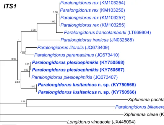
Fig. 7 Phylogenetic relationships of Paralongidorus lusitanicus n. sp. and P. plesioepimikis Palomares-Rius et al.2013within the genus Paralongidorus. Bayesian 50% majority rule consensus trees as inferred from ITS1 rRNA sequences alignments under
the GTR + I + G model. Posterior probabilities more than 70% are given for appropriate clades. Newly obtained sequences in this study are in bold
P. lusitanicus n. sp. with P. plesioepimikis in both datasets and also with P. litoralis and P. paramaximus in the partial 18S rRNA.
The description of P. lusitanicus n. sp. in the rhizo-sphere of grapevine suggests that the diversity of these nematodes in Southern Europe is still not fully clarified, and requires further study. Interestingly, the phylogenetic relationship among Iberian Peninsula species could pro-vide insight into the speciation of some of these species, additionally of the main centre of origin in other parts of the world as suggested by Coomans (1985). However, this idea must be studied with more sequences from other species of Paralongidorus, because extensive sampling for Longidoridae in Southern Spain showed a scarce presence of this genus (Archidona-Yuste et al.2016a,b). This point has also been documented for several species of Longidorus, but not for Xiphinema in an extensive sam-pling and phylogenetic study in Southern Spain (Archidona-Yuste et al.2016a,b).
In summary, the present study extends our knowledge on the species diversity of the genus Paralongidorus by integrating morphological and molecular characteriza-tions and elucidates phylogenetic relacharacteriza-tionships with oth-er Paralongidorus spp. The molecular markoth-ers obtained could be used for precise and unequivocal diagnosis of this species which may help for effective quarantine
inspection and appropriate application of exclusion principles.
Acknowledgements This research was financially supported by FCT - Foundation for Science and Technology postdoctoral fel-lowship SFRH/ BPD/95315/2013 and FEDER Funds through the Operational Programme for Competitiveness Factors - COMPETE and National Funds through FCT under the Strategic Projects PEst-C/AGR/UI0115/2011 and PEst-OE/ AGR/UI0115/2014 (Portugal). This research is partially supported by grant AGR-136 from‘Consejería de Economía, Innvovación y Ciencia’ from Junta de Andalucía, and Union Europea, Fondo Europeo de Desarrollo regional, BUna manera de hacer Europa^. Authors thank A. Candeias from HERCULES Laboratory, Universidade de Évora and J. A. Paulo Mirão from Instituto de Ciências da Terra (ICT) for their help with SEM studies, I. Ferreira and G. Albarrán Madrigal from Instituto de Ciências Agrárias e Ambientais Mediterrânicas (ICAAM)-Universidade of Évora for the excellent technical assistance and C. Cantalapiedra-Navarrete (IAS-CSIC) for critical reading of the manuscript before submission and for the excellent technical assistance.
Compliance with Ethical Standards
Conflict of interest All authors certify that 1) they do not have any actual or potential conflict of interest, 2) the study described is original and has not been published previously, and is not under consideration for publication elsewhere, 3) all prevailing local, national and international regulations and conventions, and normal scientific ethical practices, have been respected. We also certify Fig. 8 Phylogenetic relationships of Paralongidorus lusitanicus
n. sp. and P. plesioepimikis Palomares-Rius et al.2013within the genus Longidorus and Paralongidorus. Bayesian 50% majority rule consensus trees as inferred from 18S rRNA sequences
alignments under the TIM2 + I + G model. Posterior probabilities more than 70% are given for appropriate clades. Newly obtained sequences in this study are in bold letters
that all authors have reviewed the manuscript and approved the final version of manuscript before submission.
Research involving Human Participants and/or Animals No specific permits were required for the described fieldwork studies. Permission for sampling the vineyards was granted by the land-owner. The sites are not protected in any way.
Informed Consent All the authors certify that the work carried out in this research followed the principles of ethical and profes-sional conduct have been followed. The funders had no role in study design, data collection and analysis, decision to publish, or preparation of the manuscript.
References
Abolafia, J., Liébanas, G., & Peña-Santiago, R. (2002). Nematodes of the order Rhabditida from Andalucia Oriental, Spain. The subgenus Pseudacrobeles Steiner, 1938, with description of a new species. Journal of Nematode Morphology and Systematic, 4, 137–154. Altherr, E. (1963). Contribution a la connaissance de la faune des
sables submerges en Lorraine. Nematodes. Ann. Speleologie, 18, 53–98.
Andrássy, I. (1986). Egy új tufonálféreg faj Magyarországról: Paralongidorus rex sp. n. (Nematoda: Longidoridae). Állattani Közlemények, 73, 115–118.
Archidona-Yuste, A., Navas-Cortés, J. A., Cantalapiedra-Navarrete, C., Palomares-Rius, J. E., & Castillo, P. (2016a). Unravelling the biodiversity and molecular phylogeny of needle nematodes of the genus Longidorus (Nematoda: Longidoridae) in olive and a description of six new species. PLoS One, 11, e0147689.https://doi.org/10.1371/journal. pone.0147689.
Archidona-Yuste, A., Navas-Cortés, J. A., Cantalapiedra-Navarrete, C., Palomares-Rius, J. E., & Castillo, P. (2016b). Remarkable diversity and prevalence of dagger nematodes of the genus Xiphinema Cobb, 1913 (Nematoda: Longidoridae) in olives revealed by integrative approaches. PLoS One, 11, e0165412.https://doi.org/10.1371/journal.pone.0165412. Barsi, L., & Luca, F. D. (2017). Morphological and molecular
characterisation of Paralongidorus francolambertii sp. n. (Nematoda: Longidoridae) from Serbia. Nematology, 19, 681–695.https://doi.org/10.1163/15685411-00003080. Barsi, L., Répási, V., Nagy, P., Agostinelli, A., & Coiro, M. I.
(2007). A new record of Paralongidorus rex Andrássy, 1986 from Hungary and comments on head morphology of P. maximus (Bütschli, 1874) Siddiqi, 1964 (Nematoda: Dorylaimida). Nematologia Mediterranea, 35, 61–67. Bravo, M. A., & Lemos, R. M. (1997). Longidorus,
Paralongidorus and Xiphinema in Portugal. In M. S. Santos, A. de, I. O. Abrantes, D. J. F. Brown, & R. M. Lemos (Eds.), An introduction to virus vector nematodes and their associated viruses. Instituto do Ambiente e Vida (pp. 421–441). Coimbra: Universidade de Coimbra.
Castillo, P., Vovlas, N., Subbotin, S., & Troccoli, A. (2003). A new root-knot nematode, Meloidogyne baetica n. sp. (Nematoda: Heteroderidae), parasitizing wild olive in Southern Spain. Phytopathology, 93, 1093–1102.
Castresana, J. (2000). Selection of conserved blocks from multiple alignments for their use in phylogenetic analysis. Molecular Biology and Evolution, 17, 540–552.
Cherry, T., Szalanski, T. C., Todd, T. C., & Powers, T. O. (1997). The internal transcribed spacer region of Belonolaimus (Nemata: Belonolaimidae). Journal of Nematology, 29, 23– 29.
Coomans, A. (1985). A phylogenetic approach to the classification of the Longidoridae (Nematoda: Dorylaimida). Agriculture, Ecosystems and Environment, 12, 335–354.
Coomans, A. (1996). Phylogeny of the longidoridae. Russian Journal of Nematology, 4, 51–60.
Dalmasso, A. (1969). Étude anatomique et taxonomique des genres Xiphinema, Longidorus et Paralongidorus (Nematoda: Dorylaimida). Mémoires du Muséum National d’Histoire Naturelle, Nouvelle Série, A. Zoologie, 61, 34–82.
Darriba, D., Taboada, G. L., Doallo, R., & Posada, D. (2012). jModelTest 2: more models, new heuristics and parallel com-puting. Nature Methods, 9, 772.
De Ley, P., Félix, M. A., Frisse, L. M., Nadler, S. A., Sternberg, P. W., & Thomas, K. W. (1999). Molecular and morphological characterization of two reproductively isolated species with mirror-image anatomy (Nematoda: Cephalobidae). Nematology, 1, 591–612.
Decraemer, W., & Robbins, R. T. (2007). The who, what and where of Longidoridae and Trichodoridae. Journal of Nematology, 39, 295–297.
Escuer, M., & Arias, M. (1997). Paralongidorus iberis sp. n. and P. monegrensis sp. n. from Spain with a p o l y t o m o u s k e y t o t h e s p e c i e s o f t h e g e n u s Paralongidorus Siddiqi, Hooper & Khan, 1963 (Nematoda: Longidoridae). Fundamental and Applied Nematology, 20, 135–148.
Flegg, J. J. M. (1967). Extraction of Xiphinema and Longidorus species from soil by a modification of Cobb’s decanting and sieving technique. Annals of Applied Biology, 60, 429–437.
Gutiérrez-Gutiérrez, C., Bravos, M. A., Santos, M. T., Vieira, P., & Mota, M. (2016). An update on the genera Longidorus, Paralongidorus and Xiphinema (Family Longidoridae) in Portugal. Zootaxa, 4189, 99–114.
Gutiérrez-Gutiérrez, C., Cantalapiedra-Navarrete, C., Montes-Borrego, M., Palomares-Rius, J. E., & Castillo, P. (2013a). Molecular phylogeny of the nematode genus Longidorus (Nematoda: Longidoridae) with description of three new species. Zoological Journal of the Linnean Society, 167, 473–500.
Gutiérrez-Gutiérrez, C., Cantalapiedra-Navarrete, C., Remesal, E., Palomares-Rius, J. E., Navas-Cortés, J. A., & Castillo, P. (2013b). New insight into the identification and molecular phylogeny of dagger nematodes of the genus Xiphinema (Nematoda: Longidoridae) with description of two new spe-cies. Zoological Journal of the Linnean Society, 169, 548– 579.
Gutiérrez-Gutiérrez, C., Palomares-Rius, J. E., Cantalapiedra-Navarrete, C., Landa, B. B., & Castillo, P. (2011). Prevalence, polyphasic identification, and molecular phylog-eny of dagger and needle nematodes infesting vineyards in southern Spain. European Journal of Plant Pathology, 129, 427–453.
Hall, T. A. (1999). BioEdit: a user-friendly biological se-quence alignment editor and analysis program for Windows 95/98NT. Nucleic Acids Symposium Serial, 41, 95–98.
He, Y., Subbotin, S., Rubtsova, T. V., Lamberti, F., Brown, D. J. F., & Moens, M. (2005). A molecular phylogenetic approach to Longidoridae (Nematoda: Dorylaimida). Nematology, 7, 111–124.
H e y n s , J . ( 1 9 7 5 ) . P a r a l o n g i d o r u s m a x i m u s . C I H Description of plant parasitic nematodes, Set 5, No. 75. St (4pp). Albans, UK: Commonwealth Institute of Helminthology.
Holterman, M., Van Der Wurff, A., Van Den Elsen, S., Van Megen, H., Bongers, T., Holovachov, O., & Helder, J. (2006). Phylum-wide analysis of SSU rDNA reveals deep phylogenetic relationships among nematodes and accelerated evolution toward crown clades. Molecular Phylogenetics Evolution, 23, 1792–1800.
Hunt, D. J. (1993). Aphelenchida, Longidoridae and Trichodoridae: Their systematics and bionomics (352 pp). Wallingford: CABI Publishing.
Jairajpuri, M. S., & Ahmad, W. (1992). Dorylaimida. Freeliving, predaceous and plant-parasitic nematodes. New Delhi, India: Oxford & IBH Publishing Co..
Jocobs, P. J. F., & Heyns, J. (1982). Siddiqia species from sugar cane in Natal (Nematoda: Longidoridae). Phytophylactica, 14, 169–178.
Jones, A. T., Brown, D. J. F., McGavin, W. J., Rūdel, M., & Altmayer, B. (1994). Properties of an unusual iso-late of raspberry ringspot virus from grapevine in Germany and evidence for its possible transmission by Paralongidorus maximus. Annals of Applied Biology, 124, 283–300.
Katoh, K., & Standley, D. M. (2013). MAFFT multiple sequence alignment 542 software version 7: improvements in perfor-mance and usability. Molecular Biology and Evolution, 30, 772–780.
Kornobis, F. W., Susulovska, S., Susulovsky, A., & Subbotin, S. A. (2015). Morphological and molecular characterisation of Paralongidorus rex Andrássy, 1986 (Nematoda: Longidoridae) from Poland and Ukraine. European Journal of Plant Pathology, 141, 385–395.
Kumari, S. (2014). Characterization of Longidorus caespiticola (Nematoda: Longidoridae) from the Czech Republic. Helminthologia, 51, 153–157.
Lamberti, F., Choleva, B., & Agostinelli, A. (1983). Longidoridae from Bulgaria (Nematoda, Dorylaimida) with descriptions of three new species of Longidorus and two new species of Xiphinema. Nematologia Mediterranea, 11, 49–72.
Lazarova, S. S., Malloch, G., Oliveira, C. M. G., Hübschen, J., & Neilson, R. (2006). Ribosomal and mitochondrial DNA anal-yses of Xiphinema americanum-group populations. Journal of Nematology, 38, 404–410.
Lazarova, S., Peneva, V., & Kumari, S. (2016). Morphological and molecular characterisation, and phylogenetic position of X. browni sp. n., X. penevi sp. n. and two known species of Xiphinema americanum-group (Nematoda, Longidoridae). ZooKeys, 574, 1–42.
Lišková, M., & Brown, D. J. F. (1998). Longidoridae (Nematoda) associated with walnut trees (Juglans regia L.) in Slovak Republic. Helminthologia, 35, 93–99.
Lišková, M., & Brown, D. J. F. (1999). The occurrence of Longidoridae (Nematoda) in forests in the Slovak Republic. Helminthologia, 36, 49–56.
Lišková, M., & Brown, D. J. F. (2003). Longidoridae (Nematoda: Dorylaimida) in the Slovak Republic. Helminthologia, 40, 165–172.
Lišková, M. (1995). Nematode-virus vectors in the rhizosphere of fruit trees and soft fruits in Slovakia. Helminthologia, 32, 43– 48.
Macara, A. M. (1994). Nematodes associated with forest plants in Portugal (no período de 1987-1992). Revista de Ciências Agrárias, 17, 77–126.
Page, R. D. (1996). TreeView: an application to display phyloge-netic trees on personal computers. Computer Applications in the Bioscience, 12, 357–358.
Palomares-Rius, J. E., Cantalapiedra-Navarrete, C., & Castillo, P. (2014). Cryptic species in plant-parasitic nematodes. Nematology, 16, 1105–1118.
Palomares-Rius, J. E., Cantalapiedra-Navarrete, C., Gutiérrez-Gutiérrez, C., Liébanas, G., & Castillo, P. (2013). Morphological and molecular characterisation of Paralongidorus plesioepimikis n. sp. (Nematoda: Longidoridae) from Southern Spain. Nematology, 15, 363– 378.
Palomares-Rius, J. E., Subbotin, S. A., Landa, B. B., Vovlas, N., & Castillo, P. (2008). Description and molecular characterisa-tion of Paralongidorus litoralis sp. n. and P. paramaximus Heyns, 1965 (Nematoda: Longidoridae) from Spain. Nematology, 10, 87–101.
Pedram, M., Namjoo, S., Pourjam, E., Atighi, M. R., Cantalapiedra-Navarrete, C., Liébanas, G., Palomares-Rius, J. M., & Castillo, P. (2012). Molecular and morphological characterisation of Paralongidorus iranicus n. sp. and Paralongidorus bikanerensis (Lal & Mathur, 1987) Siddiqi, Baujard & Mounport, 1993 (Nematoda: Longidoridae) from Iran. Nematology, 14, 427–443.
Robbins, R. T., Brown, D. J. F., Halbrendt, J. M., & Vrain, T. C. (1995). Compendium of Longidorus juvenile stages with observation on L. pisi, L. taniwha and L. diadecturus (Nematoda: Longidoridae). Systematic Parasitology, 32, 33–52.
Robbins, R. T., Brown, D. J. F., Halbrendt, J. M., & Vrain, T. C. (1996). Compendium of juvenile stages of Xiphinema spe-cies (Nematoda: Longidoridae). Russian Journal of Nematology, 4, 163–171.
Roca, F., Lamberti, F., & Agostinelli, A. (1988). I. Longidoridae (Nematoda, Dorylaimida) delle regioni italiane. VII. Il Piemonte e la Valle D’Aosta. Nematologia Mediterranea, 16, 35–51.
Seinhorst, J.W. (1966). Killing nematodes for taxonomic study with hot f.a. 4:1. Nematologica, 12, 178.
Ronquist, F., & Huelsenbeck, J. P. (2003). MRBAYES3: B a y e s i a n p h y l o g e n e t i c i n f e r e n c e u n d e r m i x e d models. Bioinformatics, 19, 1572–1574.
Siddiqi, M. R., Baujard, P., & Mounport, D. (1993). Descriptions of Paratylenchus pernoxius sp. n. and Paralongidorus duncani sp. n. from Senegal, and the synonymization of Longidoroides with Paralongidorus. Afro-Asian Journal of Nematology, 3, 81–89. Subbotin, S. S., Stanley, J. D., Ploeg, A. T., Maafi, Z. T.,
Tzortzakakis, E. A., Chitambar, J. J., Palomares-Rius, J. E., Castillo, P., & Inserra, R. N. (2015). Characterisation of populations of Longidorus orientalis Loof, 1982 (Nematoda: Dorylaimida) from date palm (Phoenix dactylifera L.) in the USA and other countries and incongru-ence of phylogenies inferred from ITS1 rRNA and coxI genes. Nematology, 17, 459–477.
Subbotin, S., Rogozhin, E., & Chizhov, V. (2014). Molecular characterisation and diagnostics of some Longidorus species (Nematoda: Longidoridae) from Russia and other countries using rRNA genes. European Journal of Plant Pathology, 138, 377–390.
Taylor, C. E., & Brown, D. J. F. (1997). Nematode vectors of plant viruses (296 pp). Wallingford, UK: CABI.
Tulaganov, A. T. (1937). Nematode fauna of tomato Lycopersicon esculentum Mill, and the surrounding soil. Tr. Uzb. Gos. Univ. Samarakanda, 8, 63–102.
Vrain, T. C., Wakarchuk, D. A., Levesque, A. C., & Hamilton, R. I. (1992). Intraspecific rDNA Restriction Fragment Length Polymorphism in the Xiphinema americ anu m gro up. F u n d a m e n t a l an d A p p l i e d Nematology, 15, 563–573.