D
2014CYCLIC NUCLEOTIDE
REGULATION IN A POTASSIUM
CHANNEL
LIGAND SELECTIVITY AND CONFORMATIONAL CHANGE
JOÃO PEDRO ABREU PESSOA
TESE DE DOUTORAMENTO APRESENTADA
AO INSTITUTO DE CIÊNCIAS BIOMÉDICAS ABEL SALAZAR DA UNIVERSIDADE DO PORTO EM
João Pedro Abreu Pessoa
Cyclic nucleotide regulation in a potassium channel:
ligand selectivity and conformational change
Tese de Candidatura ao grau de Doutor em
Ciências Biomédicas submetida ao Instituto de
Ciências
Biomédicas
Abel
Salazar
da
Universidade do Porto
Orientador – Doutor João Morais Cabral
Categoria – Investigador Principal
Afiliação – Instituto de Biologia Molecular e
Celular da Universidade do Porto
Co-orientador Doutor Luís Gales
Categoria – Professor Associado
Afiliação – Instituto de Ciências Biomédicas
Abel Salazar da Universidade do Porto
Illustration on the next page: arrangement obtained from eight repetitions of the binding curve represented in Fig. 2.4 B.
The present work was financially supported by Fundação Para a Ciência e a Tecnologia and co-financed by the European Social Fund, through the PhD Fellowship SFRH/BD/60274/2009 and the Research Fellowship PTDC/BBB-BEP/2017/2012.
i. Acknowledgements
First, I am going to mention the most important people who helped me in the “making of” of this thesis. Sorry to those not mentioned, you are not forgotten. Some of the contributions acknowledged might seem little, but they were not. The importance of things is not only on what they are, but also, on what they mean. (Forgive me the philosophical start, respectable reader. It will not happen again.)
To start, I wish to thank my supervisor, Dr. João Morais Cabral, for training me at all levels and for providing one additional year of financial support. I do appreciate his permanent concern about the work conditions of his team. I wish to thank also my co-supervisor, Dr. Luís Gales, for taking charge of such task.
I am truly grateful to Dr. Carol Harley, for her permanent concern and dedication, which keep the lab up and running (as well as tidy and organized). She has saved me a lot of time, especially while ordering things.
Fátima, thank you for sharing your crystallographic talent and for being one of the most important players in this story. Dear reader, please go to section 2, for the thrilling details!
Thank you Ricardo, for all your help, not only in the lab, but also on the computer. Thank you also for our many interesting discussions and for always having a suggestion or a piece of advice to share with me.
Thanks to Maria for being the owner of excellent teaching skills (and sense of humour!). Learning ITC from you was quite inspiring! I am sure that your thesis will be a very good one and I wish you great success in the final exam.
Thanks/köszönöm to András, for scientific (and non-scientific) discussions, excellent technical guidance, lessons about Hungarian language, touristic information about Budapest and delightfully delicious Hungarian cookies.
Rita is the ultimate “mood booster”. No mater how bad things may go, she always has a bit of joy to spare. Having her nearby simply makes the work (even) more pleasant.
Mood boosting qualities are also found in Artur, not only while expanding my knowledge about music and the English society, but also during any discussion about anything else.
Thank you Andreia, for being another great lab partner, for your nice presence, great company, both in the lab and during lunchtime, and interesting discussions.
Much of the content here presented is due to our external collaborators. I am truly indebted to Dr. Shashi Bhushan, whose patience made true my wish of an EM collaboration and also to Mr. Cristian Kraft, for doing sample imaging. Without you both, part of this thesis would be incomplete and my curiosity about this technique would remain unsatisfied. Thank you so much!
I am also grateful to our AFM collaborators (section 3). Dr. Stefania Mari and Dr. Daniel Müller showed remarkable productivity and efficiency, which resulted in getting a paper accepted only one year after the beginning of the experiments (seriously!).
Other colleagues who I am most pleased to acknowledge are coming on! Thank you Inês, for the privilege of being part of your wonderful dissertation party! Thank you to the many colleagues who shared their experiences as PhD students and advised me on such matter!
Thanks to the IBMC technical staff, for their services, which facilitate the work of scientists. Thanks to all colleagues, group leaders and staff members who got involved in the events I organized for my guest speakers. Thanks to FCT, for the financial support that made this thesis possible.
Foremost, I wish to thank all my family, whose support and encouragement were the “secret” behind the pages that follow. I gratefully thank my parents, who put more effort on me than many would do and to whom I owe much in my achievements. I kindly thank my sister Ana, physician and scientist, whose inspiring example and success simply make me proud.
Well, now, the style is going to change. I hope you will enjoy reading the story at least as much as I enjoyed writing it. Sorry about the cliché.
“We urge the reader to bear in mind the imperfection of our current knowledge. Science is never finished. It proceeds by successive approximations, edging closer and closer to a complete and accurate understanding of Nature, but it is never fully there.”
Carl Sagan and Ann Druyan, Shadows of Forgotten Ancestors, 1993
ii. Abstract
Cyclic nucleotide regulated ion channels are membrane pores that permeate ions in the presence of the cyclic nucleotides adenosine 3’, 5’-cyclic monophosphate (cAMP) or guanosine 3’, 5’-cyclic monophosphate (cGMP). These channels play essential roles in vision and olfaction, as well as in cardiac and neuronal pacemaking. Heterologous expression of these eukaryotic membrane proteins, necessary for structural studies, is usually difficult. Such difficulty is commonly overcome by studying their prokaryotic homologues.
MlotiK1 is a bacterial cyclic nucleotide regulated potassium channel from Mezorhizobium loti. This homotetramer contains a transmembrane region and a cytoplasmic cyclic nucleotide binding (CNB) domain. The present thesis addresses recent research on this potassium channel, especially concerning its ligand selectivity determinants and global conformational change.
The molecular determinants of ligand selectivity were explored through a comprehensive set of point mutations in the CNB domain binding pocket. By combining three specific point mutations, ligand selectivity was inverted. While the wild-type CNB domain is known to favour binding of cAMP over cGMP, the triple mutant favours binding of cGMP over cAMP. Such inversion in ligand selectivity is reflected in the channel activation by cyclic nucleotides. The crystal structure of the triple mutant CNB domain showed that the change in selectivity is related to the establishment of an extensive water-mediated hydrogen bond network in the binding pocket. In contrast to the apolar wild-type binding pocket, this hydrophilic binding pocket is more flexible, allowing the accommodation of a variable number of water molecules that mediate cGMP binding through an hydrogen bond network.
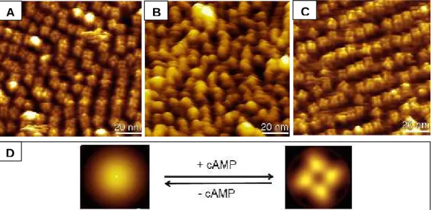
The conformational change induced by cAMP in the full-length channel was visualized by atomic force microscopy, using a mutant with decreased ligand affinity. Such mutant was shown to be functionally active. The conformational change was found to be reversible and dependent on cAMP concentration. In the presence of cAMP, the CNB domains were organized in a four-fold arrangement, while in its absence they became highly unorganized and could not be visualized individually.
iii.
Sumário
Os canais iónicos regulados por nucleótidos cíclicos são proteínas membranares que permeiam iões na presença dos nucleótidos cíclicos 3’, 5’-monofostato cíclico de adenosina (cAMP) ou 3’, 5’-monofosfato cíclico de guanosina (cGMP). Estes canais desempenham funções essenciais na visão e no olfacto, bem como no estabelecimento dos ritmos cardíaco e neuronal. A expressão heteróloga destas proteínas membranares eucarióticas, necessária para estudos estruturais, é frequentemente difícil. Esta dificuldade é geralmente superada através do estudo dos seus homólogos procarióticos.
O MlotiK1 é um canal de potássio regulado por nucleótidos cíclicos, proveniente da bactéria Mezorhizobium loti. Este homotetrâmero contém uma região transmembranar e um domínio de ligação a nucleótidos cíclicos citoplasmático. A presente tese aborda investigação recente neste canal de potássio, com ênfase nos determinantes moleculares de selectividade para o ligando e na sua alteração conformacional global.
Os determinantes moleculares de selectividade para o ligando foram explorados a partir de um conjunto abrangente de mutações pontuais no domínio de ligação. A selectividade para o ligando foi invertida através da combinação de três mutações específicas. Enquanto o domínio de ligação original favorece a ligação de cAMP relativamente a cGMP, o mutante triplo obtido favorece a ligação de cGMP sobre cAMP. Esta inversão de selectividade reflecte-se na activação do canal por nucleótidos cíclicos. A estrutura cristalina do domínio de ligação do mutante triplo revelou que a inversão de selectividade está relacionada com a formação de uma extensa rede de ligações de hidrogénio mediadas por moléculas de água no local de ligação. Em contraste com o local de ligação original, apolar, o local de ligação do mutante triplo é hidrofílico e mais fexível, permitindo acomodar um número variável de moléculas de água. Estas moléculas mediam a ligação de cGMP através de uma rede de ligações de hidrogénio.
A alteração conformacional induzida por cAMP no canal foi visualizada utilizando microscopia de força atómica e um mutante com baixa afinidade para cAMP. O referido mutante é funcionalmente activo. A alteração conformacional é reversível e dependente da concentração de cAMP. Na presença de cAMP, os domínios de ligação distribuem-se num arranjo tetragonal, ficando altamente desorganizados na sua ausência, não podendo ser visualizados individualmente.
iv.
List of publications
The results described on this thesis are based on the following publications, which will be referred to in the text as Paper I and Paper II.
Paper I:
Pessoa J, Fonseca F, Furini S and Morais-Cabral J H (2014) “Determinants of ligand selectivity in a cyclic-nucleotide regulated potassium channel” J. Gen. Physiol. 144 (1): 41-54
Paper II:
Mari S A, Pessoa J, Altieri S, Hensen U, Thomas L, Morais-Cabral J H and Müller D J (2011) “Gating of the MlotiK1 potassium channel involves large rearrangements of the cyclic nucleotide-binding domains” Proc. Natl. Acad. Sci. USA 108 (51): 20802-20807
Contents
i. Acknowledgements... iv
ii. Abstract ...vii
iii. Sumário ... viii
iv. List of publications ... ix
v. Abbreviations... xiii
1. Introduction... 1
1.1. Cyclic nucleotides ... 2
1.1.1. Identification of cAMP and cGMP ... 2
1.1.2. Cyclic nucleotide synthesis, degradation and effectors... 3
1.1.3. Generating cyclic nucleotides: adenyl cyclases and guanyl cyclases ... 3
1.1.4. Degrading cyclic nucleotides: Cyclic nucleotide phosphodiesterases ... 4
1.1.5. Proteins containing CNB domains ... 4
1.1.5.1. Protein kinases activated by cAMP or cGMP ... 5
1.1.5.2. Ion channels... 6
1.1.5.3. Guanine nucleotide exchange factor, Epac ... 7
1.2. Potassium channels ... 7
1.2.1. The beginning of ion channel research... 8
1.2.1.1. Electrical measurements in squid giant axons... 8
1.2.1.2. Early structural insights ... 8
1.2.2. The potassium channel selectivity filter ... 9
1.2.3. Important models of potassium channels: Shaker and KcsA ... 9
1.2.4. Rapid and selective potassium permeation ... 10
1.2.5. Voltage-gated potassium channels... 11
1.2.6. Ligand-gated potassium channels ... 12
1.2.6.1. Inward rectifier potassium channels ... 13
1.2.6.2. Calcium-activated potassium channels ... 13
1.3. Channels activated by cyclic nucleotides... 14
1.3.1. CNG channels... 14
1.3.2. HCN channels ... 15
1.3.3. MlotiK1, a bacterial cyclic nucleotide-regulated potassium channel... 15
1.3.3.1. Structural data... 16
2.1. Introduction...22
2.2. Materials and methods ...23
2.2.1. Expression and purification of ligand-free MlotiK1 CNB domain mutants ....23
2.2.2. Cyclic nucleotide–binding assays ...24
2.2.2.1. High affinity mutants ...26
2.2.2.2. Low affinity mutants ...28
2.2.3. Expression and purification of full-length MlotiK1 channel...29
2.2.4. Liposome reconstitution and radioactive uptake assay ...29
2.2.5. Purification and crystallization of T284S/V288S/A352D mutant CNB domain 30 2.2.6. Molecular dynamics (MD) simulations ...31
2.2.7. Energy calculations...32
2.3. Results ...33
2.3.1. Single mutants...33
2.3.2. Double mutants ...34
2.3.3. A triple mutant with inverted ligand selectivity ...35
2.3.3.1. Binding properties...35
2.3.3.2. Activity of MlotiK1 T284S/V288S/A353D in the full-length channel ...36
2.3.3.3. Crystal structure of MlotiK1 CNB domain T284S/V288S/A352D ...37
2.3.3.4. The binding pocket ...38
2.3.4. Molecular dynamics simulations ...38
2.4. Discussion ...40
3. Visualizing the conformational change by atomic force microscopy (Paper II)...43
3.1. Introduction...44
3.1.1. The atomic force microscope (AFM) precursor: the scanning tunneling microscope (STM)...44
3.1.2. Operating principle of an AFM microscope ...45
3.1.3. The versatility of AFM ...46
3.1.4. Some achievements brought by AFM: from photosynthesis to medicine...47
3.1.5. Two-dimensional crystals: a platform for AFM (and electron crystallography) 48 3.2. Materials and methods ...49
3.2.1. Protein expression and purification ...49
3.2.2. Reconstituting densely packed MlotiK1 membranes ...49
3.2.3. Radioactive flux assay ...50
3.2.4. Atomic force microscopy...50
3.3. Results ... 52
3.3.1. Visualizing the bound state... 52
3.3.2. R348A, a mutant with lower affinity for cAMP ... 53
3.3.3. Visualizing the unbound state... 54
3.3.4. The reversibility of the changes observed ... 56
3.4. Discussion... 56
4. General discussion ... 59
5. Concluding remarks... 65
6. Apendix ... 67
6.1. Exploring the equilibrium between the bound inactive and bound active states of the CNB domain ... 68
6.1.1. 5,5'-dithiobis-(2-nitrobenzoic acid), DTNB ... 68
6.1.2. Estimation of the percentage of saturation ... 69
6.1.3. Data fitting... 69
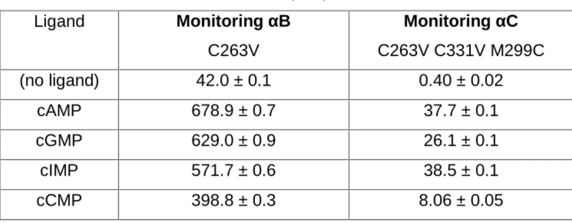
6.1.4. Validating the assay: C263V (αB helix) and C263V C331V M299C (αC helix) 70 6.1.5. The effect of R348 in the bound inactive and bound active distribution ... 72
6.2. A preliminary electron microscopy study of MlotiK1 in NABB particles ... 74
6.2.1. Nanodisc technology ... 74
5.2.1.1. Biological basis ... 75
5.2.1.3. Properties and applications of nanodiscs ... 76
5.2.1.4. Nanoscale apolipoprotein bound bilayer (NABB) particles... 76
6.2.2. Incorporation of MlotiK1 into NABB particles ... 77
6.2.3. EM data and structural model... 77
6.2.4. Analysis of the model obtained... 79
6.3. A fluorescence-based activity assay for potassium channels ... 82
6.3.1. Potassium-binding benzofuran isophthalate (PBFI)... 82
6.3.1.1. Characterization of PBFI binding properties ... 83
6.3.2. Setup of the assay ... 84
6.4. Materials and methods ... 86
6.4.1. DTNB assay... 86
6.4.2. Incorporation of MlotiK1 into NABB particles and EM ... 86
6.4.3. Fluorescence-based activity assay... 87
v. Abbreviations
2D crystal Two-dimensional crystal
AFM Atomic force microscopy
Apo A-1 Apolipoprotein A-1
ATP Adenosine 5’-triphosphate
BK Large conductance calcium-gated potassium channels cAMP Adenosine 3’, 5’-cyclic monophosphate
CAP Catabolite gene activator protein
CCCP Carbonyl cyanide 3-chlorophenyl hydrazone cCMP Cytidine 3’, 5’-cyclic monophosphate
cGMP Guanosine 3’, 5’-cyclic monophosphate
CHAPS [(3-Cholamidopropyl)dimethylammonio]-1-propanesulfonate hydrate cIMP Inosine 3’, 5’-cyclic monophosphate
CNG Cyclic nucleotide-gated
DM n-decyl-β-D-maltopyranoside
DTNB 5,5'-dithiobis-(2-nitrobenzoic acid)
DTT Dithiothreitol
EM Electron microscopy
Epac Exchange protein directly activated by cAMP
GDP Guanosine 5'-diphosphate
GEF Guanine nucleotide exchange factor GPCR G-protein-coupled receptor
GTP Guanosine 5'-triphosphate
HCN Hyperpolarization-activated cyclic nucleotide-modulated HDL High-density lipoprotein
KD Dissociation constant
MD Molecular dynamics
MSP Membrane scaffold protein
MW Molecular weight
NABB Nanoscale apolipoprotein-bound bilayer particle ndHDL Nascent discoidal high-density lipoprotein
NMG N-methyl-β-D-glucamine
PBC Phosphate-binding cassete
PBFI Potassium-binding benzofuran isophtalate
PDB Protein data bank
PDE Cyclic nucleotide phosphodiesterase
PKA cAMP-dependent protein kinases
PKG cGMP-dependent protein kinases
POPC 1-palmitoyl-2-oleoyl-sn-glycero-3-phosphocholine
SD Standard deviation
SDS-PAGE Sodium dodecylsulphate polyacrylamide gel electrophoresis SK Small conductance calcium-gated potassium channels
STM Scanning tunneling microscopy
1.1. Cyclic nucleotides
The cyclic nucleotides adenosine 3’, 5’-cyclic monophosphate (cAMP) and guanosine 3’,5’-cyclic monophosphate (cGMP) are universal signalling molecules, found in all organisms. They are involved in many physiological processes, including vision, olfaction, sugar metabolism and neuronal function.
1.1.1. Identification of cAMP and cGMP
cAMP was first identified as a “heat stable factor” produced in membrane fractions of dog or cat liver homogenates, upon addition of the hormones epinephrine or glucagon. Adenosine 5’-triphosphate (ATP) and magnesium were required and the “heat stable factor” could not be produced in the corresponding soluble fractions under the same conditions. When added to the supernatant, the “heat stable factor” induced glycogen breakdown by stimulating the liver phosphorylase activity (Berthet et al., 1957). The factor was later purified and identified as an adenine ribonucleotide (Sutherland and Rall, 1958). Its structure was determined (Lipkin, 1959) and it was named “adenosine 3’,5’-phosphoric acid”, cyclic 3,5-AMP (cAMP). cAMP was shown to be quantitatively synthesized from ATP and produced not only in the liver, but also in the heart, skeletal muscle and brain (Rall and Sutherland, 1958). The enzyme catalyzing this reaction requires magnesium and was named adenyl cyclase. Its distribution, preparation and properties were described (Sutherland et al., 1962) and pyrophosphate was shown to be another reaction product (Rall and Sutherland, 1962). Importantly, the discovery of cAMP led to the introduction of the concept of secondary messenger. The hormone (primary messenger) activated the membrane-bound adenyl cyclase, increasing the production of cAMP (secondary messenger) from ATP.
cGMP was first isolated from rat urine (Ashman et al., 1963) and animal tissue (Goldberg et al., 1969) (Ishikawa et al., 1969). Unlike cAMP, cGMP biosynthesis could not be initiated by epinephrine or glucagon, showing that it should result from another metabolic pathway (Hardman and Sutherland, 1969). It was shown that cGMP was produced from guanosine 5’-triphosphate (GTP) by a guanyl cyclase (White and Aurbach, 1969), which is largely soluble, has a preference for manganese instead of magnesium
1.1.2. Cyclic nucleotide synthesis, degradation and effectors
cAMP and cGMP are produced by adenyl and guanyl cyclases, respectively, and degraded by cyclic nucleotide phosphodiesterases (PDEs) (Rehmann et al., 2007). Physiological responses elicited by cyclic nucleotides are mostly mediated by cAMP-dependent protein kinases (PKA) (Walsh et al., 1968) (Kuo and Greengard, 1969a) (Kuo and Greengard, 1969b) (Miyamoto et al., 1969) and cGMP-dependent protein kinases (PKG) (Kuo and Greengard, 1970). These two homologous protein families are recognized as the main effectors of cAMP and cGMP-mediated responses (Lincoln and Corbin, 1977).
Other effectors are the bacterial Catabolite gene Activator Protein (CAP) (Emmer et al., 1970), which promotes gene transcription in the presence of cAMP (Zubay et al., 1970), ion channels, which are mostly studied in vision (Cook et al., 1987; Kaupp et al., 1989) and olfaction (Nakamura and Gold, 1987), a microbial cAMP receptor expressed on the surface of the eukaryotic microorganism Dictyostelium discoideum (slime mold) (Klein et al., 1988) and a protein family with Guanine nucleotide Exchange Factor (GEF) activity called Exchange Protein directly Activated by cAMP (Epac) (de Rooij et al., 1998; Kawasaki et al., 1998).
1.1.3. Generating cyclic nucleotides: adenyl cyclases and guanyl cyclases
In mammals, there are at least ten groups of adenyl cyclases (also known as adenylyl cyclases or adenylate cyclases), divided into one soluble and nine membrane-bound subgroups (Taussig and Gilman, 1995). The membrane membrane-bound adenyl cyclases are homologous, containing two catalytic domains that form the catalytic site and are attached to two membrane-embedded domains (Hanoune and Defer, 2001). The whole unit converts ATP into cAMP. Adenyl cyclase activity is controlled by a membrane-bound G-protein-coupled receptor (GPCR) and a heterotrimeric G protein (formed by α, β and γ subunits). Adenyl cyclase activity can be triggered by several molecules, including hormones and neurotransmiters, which bind to their specific GPCR. Upon activation by the GPCR, the guanosine 5'-diphosphate (GDP) in the G-protein α subunit is exchanged by guanosine 5'-triphosphate (GTP). The β and γ subunits are dissociated form it and regulate downstream processes (Taussig and Gilman, 1995), including activation of adenyl cyclase. During this process, the extracellular signal is amplified: one activated GPCR can activate several adenyl cyclases and each adenyl cyclase can synthesize many cAMP molecules. Besides G proteins, adenyl cyclases are regulated by protein
kinases, forskolin (a plant diterpene) and the calcium-binding protein calmodulin (Hanoune and Defer, 2001). Several human diseases are related to defective or excessive cAMP biosynthesis (Sutherland, 1972) (Costache et al., 2013).
There are two groups of guanyl cyclases (also known as guanylyl cyclases or guanylate cyclases): membrane-associated and soluble. Both have one catalytic domain per subunit (Tesmer, 2008). Membrane-associated guanyl cyclases are homodimers containing an extracellular binding domain, whose best known ligands are natriuretic peptides (peptide hormones), a transmembrane region with one or more segments and an intracellular catalytic domain (Wedel and Garbers, 2001) (Rehmann et al., 2007). Soluble guanyl cyclases are heterodimers formed by a α and a β subunit, existing two and three kinds of α and a β subunits, respectively (Friebe and Koesling, 2003). They contain a heme group and are activated by nitric oxide (Rehmann et al., 2007). Like adenyl cyclases, guanyl cyclases also amplify the extracellular signal during its transduction process.
1.1.4. Degrading cyclic nucleotides: Cyclic nucleotide phosphodiesterases
The intracellular concentrations of cAMP and cGMP are regulated mainly by the activities of adenyl/guanyl cyclase and cyclic nucleotide phosphodiesterases (PDEs). PDEs catalyze the conversion of cAMP or cGMP to AMP or GMP, respectively (Beavo et al., 1994). PDEs form a diverse protein family of dimers and multimeric complexes. Their function is achieved by a conserved catalytic domain usually located at the C-terminus. PDEs can be unspecific or specific for cAMP or cGMP (Manganiello et al., 1995). In some PDEs, a N-terminal GAF domain has been identified, whose function is cyclic nucleotide binding (Zoraghi et al., 2004). There are GAF domains specific for cAMP and for cGMP (Gross-Langenhoff et al., 2006). In cGMP-specific GAF domains, ligand binding is favoured through a conserved aspartate. GAF domains are not restricted to PDEs (Aravind and Ponting, 1997) and can bind other small molecules besides cyclic nucleotides (Zoraghi et al., 2004).
1.1.5. Proteins containing CNB domains
which this low molecular weight compound achieves its diversity of effects” (Kuo and Greengard, 1969b).
When the sequences of CAP, PKA and PKG were determined, a common structural motif was found: the cyclic nucleotide-binding (CNB) domain (Shabb and Corbin, 1992). CNB domains are recognized as the main common link among proteins regulated by cyclic nucleotides. The first structure of a CNB domain was obtained when E. coli CAP was crystallized in complex with cAMP (McKay and Steitz, 1981; McKay et al., 1982).
1.1.5.1. Protein kinases activated by cAMP or cGMP
PKAs are divided into several groups (Canaves and Taylor, 2002), which may have different subcellular locations (Theurkauf and Vallee, 1982). Eukaryotic PKAs are heterotetramers composed of two identical regulatory (where cAMP binds) and two identical catalytic subunits, existing four types of each one (Johnson et al., 2001). Each regulatory subunit has two tandem CNB domains (Titani et al., 1984; Diller et al., 2001). The regulatory subunits dimerize through a narrow region and a catalytic subunit is attached to each one of the regulatory subunits (Zhao et al., 1998). In the absence of cAMP, PKA is inhibited through high-affinity binding of the regulatory subunits to the catalytic subunits (Hofmann, 1980). In the presence of cAMP, two ligand molecules bind cooperatively to each regulatory subunit, disrupting the interaction with catalytic subunits and activating the kinase (Johnson et al., 2001) (Anand et al., 2002). cAMP-binding and interaction with a catalytic subunit are achieved through separate regions (Corbin et al., 1978; Herberg et al., 1994). PKA effects are mostly related to stimulation of glycolysis and glycogen breakdown in muscle and liver (Nelson, 2005).
There are at least two types of PKG: PKG1, found mostly in vascular smooth muscle cells, endothelium and platelets and PKG2, found mostly in the intestine, kidneys and brain (Vaandrager and de Jonge, 1996). PKG1 enzymes are homodimers, where each monomer contains one regulatory domain with two CNB domains, plus a C-terminal catalytic domain with kinase activity (Francis et al., 2010), which forms interactions within each monomer (Wolfe et al., 1989). The regulatory domain also forms the dimerization interface. In the absence of cGMP, the enzyme is inhibited by internal contacts, as in PKA (Francis et al., 2010). PKG1 mediates the vasodilation effect of nitric oxide and is, as a consequence, involved in vascular smooth muscle relaxation. Other effects include decrease in endothelial permeability, inhibition of platelet aggregation (Vaandrager and de
Jonge, 1996) and activation of potassium channels regulated by calcium and voltage (Alioua et al., 1998).
1.1.5.2. Ion channels
Ion channels regulated by cyclic nucleotides are best studied in the vision and in the olfaction mechanisms. Cyclic nucleotide gated (CNG) channels are usually heterotetramers, containing one C-terminal CNB domain per monomer. Their specificity for cAMP or cGMP varies (Kaupp and Seifert, 2002). Both visual and olfactory ion channels are specific for cations and permeate calcium (mostly) and also sodium and potassium (Menini, 1995). Unlike other ligand-gated ion channels, they do not desensitize in the continued presence of ligand (Kaupp and Seifert, 2002). They are also regulated by calcium/calmodulin (Trudeau and Zagotta, 2003), pH and phosphorylation (Kaupp and Seifert, 2002).
Light is sensed through closure of cGMP-gated ion channels and consequent termination of neurotransmitter release into neighbouring cells. In the dark, these channels are open. Rod cells in the retina have large amounts of the GPCR rhodopsin, which contains constitutively bound retinal, a photosensitive derivative of vitamin A. Light is sensed through isomerization of retinal, activating rhodopsin. Activated rhodopsin will relay the light signal through the G-protein transducin, exchanging its GDP to GTP and activating it. The α-subunit of activated transducin will activate a PDE, resulting in a decrease in cGMP concentration and consequent closure of cGMP-gated calcium channels, decreasing their current and the release of neurotransmitter. Conversely, the decrease in intracellular calcium concentration stimulates guanyl cyclase activity, enhancing cGMP synthesis by a feedback mechanism (Hille, 1992) (Matulef and Zagotta, 2003). A similar mechanism exists in cones, the cells in the retina responsible for colour vision; however, they are less sensitive and adapt to a wider range of light intensities, possibly because of differences in calcium homeostasis. Mutations in genes encoding these channels can lead to blindness or loss of colour discrimination (Kaupp and Seifert, 2002).
Binding of an odorant to its GPCR in an olfactory neuron stimulates adenyl cyclase, increasing the intracellular concentration of cAMP. cAMP will open calcium channels and the consequent increase in intracellular calcium concentration will open
vast array of odorant receptors. Mice lacking functional cAMP-sensitive olfactory channels exhibited no detectable response to odorants (Kaupp and Seifert, 2002).
1.1.5.3. Guanine nucleotide exchange factor, Epac
Epac is a monomer and contains a regulatory region with a CNB domain, plus a C-terminal catalytic region that mediates GEF activity. GEF activity consists in the transfer of GTP to the GTPase proteins Rap1 and Rap2, which cycle between GDP-bound (inactive) and GTP-bound (active) forms (Gloerich and Bos, 2010). In the absence of cAMP, Epac is inactive, since access of the catalytic domain to Rap is blocked by interactions with the regulatory regions in Epac (Rehmann et al., 2006). cAMP binding causes the CNB domain to swing away from its blocking position, allowing access to Rap proteins (Rehmann et al., 2008).
In mammalian cells, there are two isoforms of Epac: Epac1 and Epac2 (Gloerich and Bos, 2010). Epac2 contains an additional CNB domain at the N-terminus whose binding affinity is lower (de Rooij et al., 2000). This additional CNB domain has a non-canonical sequence and different phosphate-binding cassete (PBC) architecture (Rehmann et al., 2003). Its function may not be cAMP-binding. Repac, the other member of the family, lacks a CNB domain and is a constitutive activator of Rap proteins (de Rooij et al., 2000). Both Epac1 and Epac2 are abundant in the central nervous system and have neuronal functions (Gloerich and Bos, 2010). Epac1 is also abundant in blood vessels, kidney or adipose tissue and is involved in cardiac function, vascular permeability and inflammation (Fukuhara et al., 2005; Gloerich and Bos, 2010). Epac2 is also found in the adrenal gland and pancreas and is involved in insulin secretion (Shibasaki et al., 2007; Gloerich and Bos, 2010).
1.2. Potassium channels
Potassium channels are most likely the fundamental controllers of the cellular electrical homeostasis (Torres et al., 2007). Their diversity is of great importance to understand the variety of electrical responses of cells when subjected to stimuli.
1.2.1. The beginning of ion channel research
The ion channel research field has its foundations in the study of electrical activity of squid giant axons. The unusually large dimensions of these cells allowed measuring their electrical properties by using microelectrodes. Such electrical properties were found to be dependent on ion channels.
1.2.1.1. Electrical measurements in squid giant axons
Squid giant axons were prepared by removing fibers and branches under an optical microscope, after washing with sea water. Prepared axons usually had 6 to 8 centimetres in length and a uniform diameter between 530 and 580 µm. Recordings were done in elongated chambers with circulating sea water. Axons whose electrical properties stabilized within one hour were used in the following 6 to 8 hours, as their electrical properties were constant during such period (Cole and Curtis, 1939).
This system was used for measuring the electrical currents that resulted of applying voltage to giant axons. Such currents resulted mainly of changes in sodium and potassium permeabilities. Upon stimuli, the conductance changed differently and reversibly for each ion. Based on these recordings, an empirical kinetic model was presented to describe sodium and potassium permeability changes upon stimuli, in squid giant axons (Hodgkin and Huxley, 1952b, a, c). Such model became an explanation for their electrical excitability.
1.2.1.2. Early structural insights
Electrical recordings using inhibitory compounds in squid giant axons (Armstrong, 1971) or frog nerve fibers (Armstrong and Hille, 1972) suggested that potassium permeation should be achieved by permeation pores. Such pores should have two distinct regions: a cavity wide enough to accommodate an ion or an inhibitor molecule and a narrower inner region that could accommodate only the ion.
Perfusion of squid giant axons with proteases indicated that sodium and potassium permeation should be carried out by distinct channels and that those should be proteins
channels should contain at least three potassium binding sites occupied by a minimum of two ions at a time (Hille and Schwarz, 1978) and their narrowest region should be a circle of oxygen atoms forming a 3 Å wide pore (Hille, 1973).
1.2.2. The potassium channel selectivity filter
Each potassium channel sequence contains a highly conserved segment called potassium channel signature sequence, which forms the narrow region responsible for selective potassium permeation, known as the selectivity filter (MacKinnon, 2003). Mutations in this segment can severely affect ion discrimination (Heginbotham et al., 1994).
Potassium ions permeate through these channels down their concentration gradient at rates that can be close to 106 ions per second, corresponding to the translocation of one ion every 0.1 millisseconds (Hille, 1992).
1.2.3. Important models of potassium channels: Shaker and KcsA
The Shaker potassium channel (Pongs et al., 1988) cloned from Drosophila melanogaster, the fruit fly, was found to be a tetramer (MacKinnon, 1991). Its external entryway was described as a shallow conical vestibule formed by loops, with the selectivity filter in the apex (Ranganathan et al., 1996). The channel function is affected by pore blockers, which can be trapped inside its cavity by the channel gate (Holmgren et al., 1997). This mechanism is called “gating” and is associated with changes in volume and accessibility in the cavity (Liu et al., 1997).
The KcsA channel from Streptomyces lividans, a filamentous soil bacterium, was shown to be selective for potassium and predicted to have two transmembrane helices per subunit (Schrempf et al., 1995). Purified KcsA is a remarkably stable tetramer in solution (Heginbotham et al., 1997) and was the first ion channel whose structure was determined by X-ray crstallography (Doyle et al., 1998). The structure showed a tetrameric assembly, where each subunit contains two long α-helices that cross the membrane and surround a shorter α-helix. The pore is located at the center of the four subunits (Fig. 1.1 A). The selectivity filter is formed by a loop located between the shorter and the C-terminal α-helices (Fig. 1.1 B). The selectivity filter contains four identical potassium binding sites, where each potassium ion is coordinated by eight oxygen atoms from main chain carbonyl groups and from side-chain hydroxyl groups (Fig. 1.1 C).
Although crystal structures show one ion bound to each of the four potassium binding sites, in fact, only two of those sites are occupied by ions at the same time (Zhou and MacKinnon, 2003). Each pair of ions is separated by one water molecule, alternating rapidly between two configurations, which are potassium-water-potassium-water and water-potassium-water-potassium. When a potassium ion enters in one side of the line, it displaces another potassium ion in the other side, resulting in potassium translocation (Morais-Cabral et al., 2001).
Figure 1.1: Crystal structure of the KcsA potassium channel in ribbon representation. Potassium ions and water molecules are represented as purple and red spheres, respectively. (A) Extracellular view, neighboring subunits are represented in different colors. (B) Side view, only two subunits are shown for clarity and the selectivity filter is indicated by oval. (c) Zoom over selectivity filter, where polar contacts between potassium ions and water molecules or carbonyl groups from the selectivity filter are indicated by black dashed lines. Protein data bank (PDB) code: 1K4C (Zhou et al., 2001).
1.2.4. Rapid and selective potassium permeation
The pore structure present in KcsA is conserved in eukaryotic potassium channels (MacKinnon et al., 1998). Evolutionary optimization of the potassium channel selectivity filter resulted in very high rates of potassium permeation and very high selectivity (MacKinnon, 2003). The very high permeation rate is explained by the weak binding of the ions to the selectivity filter. It has been postulated that such binding is weak for two reasons: 1) repulsion between two consecutive ions separated by one water molecule; 2) the presence of two potassium ions changes the conformation of the selectivity filter, causing an energetic penalty that reduces the total binding energy (MacKinnon, 2003).
of α-helices (Roux and MacKinnon, 1999). It has also been postulated that high ion selectivity is achieved because the selectivity filter has several “tailor-made” binding sites that perfectly mimic the hydration shell of potassium (MacKinnon, 2003). The presence of potassium should impose a large energy barrier to other ions, as lithium or sodium, to bind to the selectivity filter (Thompson et al., 2009). Potassium, or another permeable cation, is required for structural stability of the selectivity filter and overall stability of the tetramer (Krishnan et al., 2005). Its removal causes pore dilation, loss of ion selectivity and ultimately loss of function (Loboda et al., 2001). Potassium channel diversity is due to the different mechanisms for gating (opening and closing of the pore) and regulation, which can be voltage or a ligand (MacKinnon, 2003).
The main families of potassium channels will be discussed below. The members of these families are homo or heterotetramers.
1.2.5. Voltage-gated potassium channels
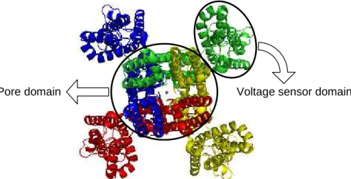
When the membrane electrical field is altered, voltage-gated potassium channels detect the alteration. They become activated, letting potassium ions cross the membrane and restoring the electrical field to its resting state. In these channels, each monomer has six transmembrane segments divided into two domains (Fig. 1.2): the first four helices form the voltage sensor domain and the last two helices form the pore domain, identical to the one seen in KcsA (Fig. 1.1). Gating is directly controlled by the transmembrane electrical field, which is sensed in real time by a voltage sensor, located in the voltage sensor domain. The fourth helix in each voltage-sensor domain is an essential element in voltage sensing. It contains four to eight positively charged residues, mostly arginines, every third position (Tombola et al., 2006). The Shaker channel is a voltage-gated potassium channel and a useful model to explore the voltage sensing mechanism. During voltage sensing, positive charges in the fourth helix accessible on one side of the membrane change their accessibility to the other side by translation and most likely also rotation of the fourth helix (Yellen, 2002; Tombola et al., 2006).
In the structures of Shaker channels (Long et al., 2005; Long et al., 2007), the fourth helix adopts a 310 helix conformation over seven to ten residues. 310 helices are formed when main chain hydrogen bonds are established between residues separated by two other residues, while in -helices the separation is of three residues. Therefore, 310 helices are more tightly wound and less stable than canonical -helices. These helices are relatively rare and most are short (usually with three or four residues). Longer 310 helices are even rarer, since they have specific packing requirements, rarely seen over
long extensions (Vieira-Pires and Morais-Cabral, 2010). As mentioned above, voltage sensing should involve translation and most likely, also rotation of the fourth helix across the membrane. A 310helix conformation would place the positively charged residues all in the same side and the switch to a -helical conformation would provide the rotation mentioned above. Such transition is possible and has been reversibly observed in isolated peptides upon changes in solvent polarity (Karle et al., 1994; Bellanda et al., 2007). Therefore, it has been proposed that voltage sensing might involve transition between the two conformations in part of the fourth helix (Vieira-Pires and Morais-Cabral, 2010). It is still unclear how voltage sensing is coupled to channel gating; however, several studies indicate that the linker between both transmembrane domains and the C-terminal portion of the C-terminal helix is involved (Tombola et al., 2006).
Figure 1.2: Crystal structure of the Shaker potassium channel in a ribbon representation (extracellular view). Each subunit is represented in a different color. The pore is indicated by a potassium ion represented by a sphere. The pore domain and the voltage sensor domain are indicated. PDB code: 2R9R (Long et al., 2007).
1.2.6. Ligand-gated potassium channels
There are two main families of potassium channels regulated by ligands: inward rectifier and calcium-gated, which will be described below.
1.2.6.1. Inward rectifier potassium channels
Inward rectifier potassium channels conduct potassium more efficiently towards the intracellular side (inward) than in the opposite direction (outward), due to pore blocking in the intracellular side by magnesium or polyamines (Hibino et al., 2010). Such blocking is mediated by their regulatory cytoplasmic domain formed by the N- and C-termini from each subunit, which provides a favorable environment for binding of polyamines (Nishida and MacKinnon, 2002). These channels lack a voltage sensor domain. They contain only the pore domain (Tao et al., 2009) and the overall structure is similar to KcsA. Their activity is regulated by a vast array of ligands, mostly ions, phospholipids or proteins, like GPCRs. Their functions include controlling the resting membrane voltage of cells, insulin release and neurotransmitter action (Hibino et al., 2010).
1.2.6.2. Calcium-activated potassium channels
This family is subdivided into large conductance (maxi-K or BK) and small conductance (SK) channels (McCoy and Nimigean, 2012). While both are activated by intracellular calcium, BK channels are also activated by voltage (Rothberg, 2012) (Adelman et al., 2012). Both types have six transmembrane helices, but BK channels have an additional N-terminal helix, which is part of the voltage sensor and also involved in oligomerization (Rothberg, 2012).
In SK channels, calcium regulation is mediated by calmodulin, which binds to the C-terminus of each subunit (Adelman et al., 2012). In BK channels, calcium is sensed by a cytoplasmic C-terminal regulator of conductance of potassium (RCK) domain (Rothberg, 2012), which has a regulatory function in various potassium channels and transporters (Roosild et al., 2009). BK channels are essential to control action potentials and neurotransmitter release, as well as in the control of smooth muscle contraction, triggered by calcium release from the endoplasmic reticulum (Rothberg, 2012). SK channels are expressed in the central nervous system and their activity has rapid effects in excitability and synaptic transmission, as well as in long term changes that affect memory and learning (Adelman et al., 2012).
1.3. Channels activated by cyclic nucleotides
Cyclic nucleotide activated ion channels are divided into two main families: cyclic nucleotide-gated (CNG) and hyperpolarization-activated cyclic nucleotide-modulated (HCN) channels. Both are homo or heterotetrameric, containing six putative α-helices and a cytoplasmic C-terminal CNB domain (Craven and Zagotta, 2006). Despite CNG, HCN channels are also regulated by voltage (Kaupp and Seifert, 2001). CNG channels are non-selective cation channels, permeating mostly sodium and calcium (Kaupp and Seifert, 2002). On the contrary, HCN channels are about 4-fold more permeant to potassium than sodium. The CNG and HCN families are believed to be similar in structure and mechanism; however, they play different physiological roles (Craven and Zagotta, 2006).
1.3.1. CNG channels
Although CNG channels are best studied in vertebrate photoreceptors and olfactory neurons, they are not restricted to these tissues. Another important function of CNG channels is mobility and chemoattraction in sperm cells. They are also found in heart, kidney, pancreas, adrenal gland and colon (Kaupp and Seifert, 2002; Matulef and Zagotta, 2003). Their importance in the central nervous system is being increasingly recognized, since they are probably involved in several processes, including neuronal excitability, neurotransmission and pain perception (Podda and Grassi, 2013). CNG channels have also been studied in plants (Zelman et al., 2012).
In CNG channels, the four N-terminal helices form a domain with homology to the voltage sensor domain of voltage-gated channels; however, these channels are not sensitive to voltage. It has been proposed that although the fourth helix contains some of the positive charges required for voltage sensing, some of the necessary counter charges are missing (Bellanda et al., 2007). Consistently with this hypothesis, the fourth helix of a CNG channel (voltage insensitive) was shown to work as a voltage sensor, by transferring it to a voltage-gated channel (Tang and Papazian, 1997). In CNG channels, this helix might also be “locked” in a fixed conformation by hydrophylic residues of its upstream loop (Kaupp and Seifert, 2002). It was proposed that the CNG channels pore opens through a translation and possible rotation of the intracellular ends of the C-terminal helices, increasing its internal diameter (Flynn and Zagotta, 2001).
1.3.2. HCN channels
In mammals, HCN channels are expressed in the heart and nervous system, where they regulate heart rate and neuronal excitability. Due to such function, they are also called pacemaker channels (Kaupp and Seifert, 2001). Strikingly, and unlike other voltage gated channels, which are activated by membrane depolarization, membrane hyperpolarization activates HCN channels. Nevertheless, the voltage sensing mechanism in HCN channels is probably similar to those of voltage-gated channels (Bell et al., 2004). HCN channel activation is also regulated by cyclic nucleotides, especially cAMP.
Rhythmic contraction of the cardiac muscle results of a cycle of membrane depolarization and hyperpolarization mediated by at least four different types of ion channels, including HCN channels (Kaupp and Seifert, 2001). Some neurotransmitters accelerate the heartbeat rate through an increase in cAMP concentration, favouring activation of HCN channels (Craven and Zagotta, 2006). In neurons, these channels work through a similar, but faster mechanism, and therefore the neuronal action potentials are faster (Craven and Zagotta, 2006). In the nervous system, they control neuronal excitability, synaptic transmission, among other functions (Benarroch, 2013). They are associated with learning and memory and misregulation may be involved in chronic pain and epilepsy (Lewis and Chetkovich, 2011). HCN channels have been explored as potential drug targets (Postea and Biel, 2011).
1.3.3. MlotiK1, a bacterial cyclic nucleotide-regulated potassium channel
The physiological and medical relevance of both CNG and HCN ion channels are important reasons for their study. Functionally, they have been well characterized through electrical recordings. However, structural studies require expression and purification of significantly large amounts of protein. For these eukaryotic membrane proteins, this is frequently not feasible and their prokaryotic homologues, such as MlotiK1, have been a valuable alternative. MlotiK1 is a cyclic nucleotide regulated potassium channel from Mesorhizobium loti, a symbiotic bacterium involved in nitrogen fixation (Kaneko et al., 2000). Both the full-length channel and its isolated CNB domain can be expressed and purified with good yield and purity. Protein folding is facilitated in the presence of a cyclic nucleotide, since ligand depletion during purification causes extensive protein aggregation (Nimigean et al., 2004).
1.3.3.1. Structural data
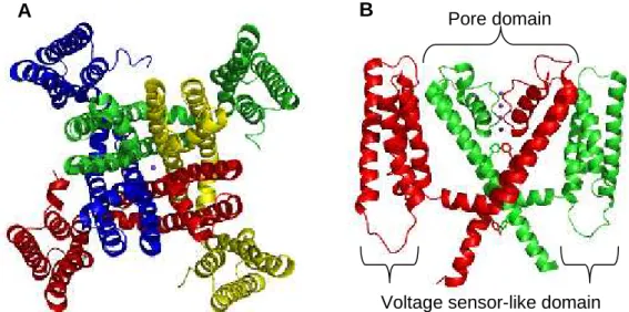
MlotiK1 is a homotetramer and its sequence predicts a transmembrane topology containing six α-helices and a cytoplasmic C-terminal CNB domain. Electron microscopy (EM) of single particles in the bound state shows that the CNB domains are organized with a four-fold symmetry, without any obvious interactions between them (Chiu et al., 2007). The structure of its transmembrane region has been determined at 3.1 Å by X-ray crystallography (Clayton et al., 2008), showing an arrangement similar to those of voltage-gated potassium channels. In each subunit, the four N-terminal α-helices form a voltage sensor-like domain and the two C-terminal α-helices form the pore domain (Fig. 1.3 A). In the voltage sensor-like domain, the fourth helix contains an unusually long 310 helix, with eleven residues (Clayton et al., 2008). This helix does not have positively charged residues at the necessary positions for voltage sensing (Clayton et al., 2008) and therefore, it is unlikely to sense the transmembrane electrical field. Although it has structural homology to voltage-sensor domains, its role, if any, is unclear. Electron crystallography suggests that, in the membrane, the position of the voltage sensor-like domain relative to the pore domain is highly variable, adopting multiple orientations (Clayton et al., 2009). The pore domain has the typical structure found in potassium channels; however, it contains two bulky residues, F203 and Y215, which partially occlude the pore (Fig. 1.3 B) (Clayton et al., 2008). Although the crystals were grown in the presence of cAMP, the structure shows a closed state. The CNB domains were highly disorded and could not be resolved.
Figure 1.3: Crystal structure of the MlotiK1 potassium channel transmembrane region, in a ribbon representation. (A) Extracellular view, where each subunit is represented in a different color. The pore is indicated by a potassium ion represented by a sphere. (B) Side-view, only two subunits are shown for clarity. In the pore domain, the selectivity filter is indicated by four potassium ions (represented by spheres) and F203 and Y215 are represented in stick. PDB code: 3BEH (Clayton et al., 2008).
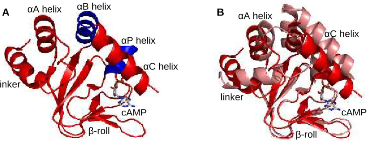
There are several crystal structures of the isolated CNB domain: four structures in complex with cAMP, two structures in complex with cGMP and six apo structures (Clayton et al., 2004; Altieri et al., 2008). There are also nuclear magnetic resonance (NMR) structures: one in complex with cAMP (Schunke et al., 2009) and an apo structure (Schunke et al., 2011). Both show that the isolated CNB domain is a monomer in solution. The structure of MlotiK1 CNB domain in complex with cAMP consists of an antiparalel -roll and five helices, comprising a helical linker to the transmbrane region and four -helices,A, B, C and P (Fig. 1.4 A). Superposition of bound and unbound structures of the CNB domain shows that the β-roll is essentially unchaged in both states, except in the β4-β5 hairpin loop. The linker that connects the CNB domain to the transmembrane region and the other helices are in different positions. Importantly, C helix covers the binding pocket as a lid in the bound state and is deviated in the unbound state (Fig. 1.4 B). The different positions of C helix in different crystal structures of the unbound state suggest that this helix is highly mobile in the absence of ligand (Clayton et al., 2004; Altieri et al., 2008); however NMR data indicates that the CNB domain is rigid in the unbound state (Schunke et al., 2011).
A
B
Pore domainFigure 1.4: Crystal structures of MlotiK1 CNB domain in ribbon representations, with bound cAMP represented in stick. (A) Structure in complex with cAMP colored in red or blue (for αB and αP helices). (B) Superposition of cAMP-bound (red) and unbound (light red) structures through the β-roll. PDB codes: 1VP6 (bound) and 1U12 (unbound) (Clayton et al., 2004).
1.3.3.2. Functional data
MlotiK1 has remarkably slow permeation kinetics (Clayton et al., 2004); (Silverman and Heginbotham, 2007), partly due to the occlusion of the pore by the bulky residues F203 and Y215 (Clayton et al., 2008). It has not been possible to study MlotiK1 function through electrical recordings, most likely because of its slow permeation kinetics. Alternatively, a radioactive flux assay (Nimigean, 2006) has been used to functionally characterize this potassium channel. MlotiK1 is selective for potassium over sodium or lithium, but accurate estimates of the selectivity ratios could not be determined (Nimigean et al., 2004). MlotiK1 mediated potassium transport is enhanced by a cyclic nucleotide, but not totally dependent of it (Silverman and Heginbotham, 2007). The maximum stimulatory effect of cAMP is only about 2-fold (Clayton et al., 2004; Silverman and Heginbotham, 2007). The channel can also be activated by cGMP, but the necessary concentration for half of maximum activity is approximately 10-fold higher than for cAMP (Nimigean et al., 2004; Altieri et al., 2008). The determinants that define this 10-fold selectivity for cAMP versus cGMP are not completely known; however, CNB domain structures in complex with cAMP or cGMP provide some insights. Due to the different
αC helix αA helix linker β-roll cAMP αP helix αB helix
A
B
cAMP β-roll αA helix linker αC helixresidue in the PBC, S308, establishes hydrogen bonds with cGMP, but not with cAMP. Mutating S308 to a valine decreases the affinity for cGMP by 3-fold, without significant effect in cAMP (Nimigean and Pagel, 2007). In CNG channels, an aspartate in αC helix was proposed to be important for the discrimination between cAMP and cGMP (Varnum et al., 1995). In MlotiK1, the equivalent residue is an alanine, which is not resolved in the crystal structures (Clayton et al., 2004; Altieri et al., 2008) and is disordered in the NMR structures (Schunke et al., 2009; Schunke et al., 2011). Mutating it to an aspartate increases affinity for cGMP by 3-fold, without detectable effect in cAMP (Nimigean and Pagel, 2007).
Cyclic nucleotide binding in MlotiK1 is not cooperative (Cukkemane et al., 2007) and is well coupled to channel opening (Nimigean and Pagel, 2007). Upon cAMP binding, the MlotiK1 CNB domain conformational change occurs through an induced-fit mechanism (Peuker et al., 2013) that affects virtually all residues in the CNB domain (Schunke et al., 2007; Schunke et al., 2010; Cukkemane et al., 2012). In the C helix, the R348 residue establishes important interactions with the ligand (Clayton et al., 2004; Altieri et al., 2008; Schunke et al., 2009). Mutating it to an alanine significantly decreases binding affinity (Cukkemane et al., 2007).
MlotiK1 cannot be included either into the CNG or HCN families. It is selective for potassium, like HCN channels; however, it is most likely not voltage-gated. It should therefore be simply considered as separate family of prokaryotic potassium channels regulated by cyclic nucleotides (Nimigean et al., 2004).
1.3.3.3. State of the art and objectives
In contrast to the well characterized conformational change occurring in the CNB domain upon ligand binding/unbinding, the structural data available for MlotiK1 has shown its transmembrane region only in the closed state. It would be of interest to gain insights into the structural differences between the the open and closed states. Additionally, the molecular mechanism that determines ligand selectivity in CNB domains is still poorly understood and the MlotiK1 CNB domain is a good model to investigate it.
The main objectives of the present project are:
- To define the molecular determinants of ligand selectivity in MlotiK1 CNB domain.
- To visualize the cAMP-induced conformational change in the full length channel.
In the present section, a study on the molecular determinants of ligand selectivity in the MlotiK1 channel is described.
2.1. Introduction
The function of cyclic nucleotide dependent effectors is partly determined by the ligand selectivity properties of the CNB domain. These properties have not been completely characterized. One of the difficulties in the study of ligand selectivity is to distinguish the effects related to binding of nucleotide from the effects that are related to efficiency of ligand in fuctional activation. In this study, this difficulty has been overcome by studying the isolated CNB domain. The amino acids of the CNB domain binding pocket that specifically affect binding of either cAMP or cGMP are critical for ligand selectivity. Using site-directed mutagenesis and a fluorescence-based binding assay, we have screened a comprehensive set of point mutations in the binding pocket residues known to interact with the nucleotide base. From the single mutants tested, we generated a triple mutant, which displayed inverted ligand selectivity, so that the mutant has higher affinity for cGMP than for cAMP. This triple mutant was structurally and functionally characterized.
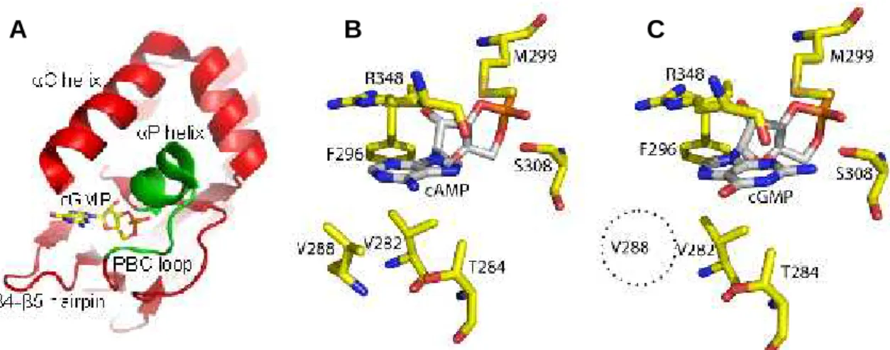
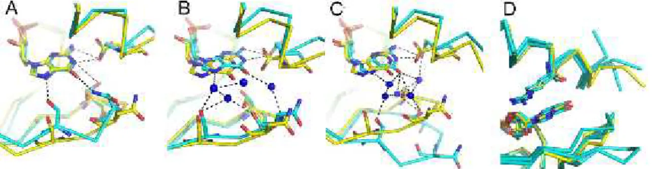
The binding pocket of CNB domains consists of a shallow cavity formed by side chains from several structural motifs, which are the β4-β5 hairpin, the αP helix, the PBC region and a lid, which in MlotiK1 is formed by αC helix (Fig. 2.1 A). Inspection of crystal structures of the MlotiK1 CNB domain binding pocket in complex with cAMP or cGMP shows the residues that interact with the ligand base. V282 and T284 in the β4-β5 hairpin, R348 in the lid of the αC helix, M299 in αP helix and F296 and S308 in the PBC. Comparing the structures in complex with cAMP and in complex with cGMP, the most obvious difference is in another residue in the β4-β5 hairpin: V288, which establishes van der Waals interactions with cAMP (Fig. 2.1 B), but not with cGMP (Fig. 2.1 C), making it a promising candidate in our search for determinants of ligand selectivity. Nevertheless, the other residues in the binding pocket were also explored, except F296, since it is highly conserved and therefore is probably not involved in ligand discrimination.
Figure 2.1: Cyclic nucleotide binding to the MlotiK1 CNB domain. (A) The CNB domain structural elements (in red and green) that form the ligand binding pocket are indicated. cGMP is shown as stick. (B) and (C) Residues interacting directly with ligand bases in the MlotiK1 CNB domain crystal structures bound to (B) cAMP (PDB code: 1VP6) and (C) cGMP (PDB code: 3CL1). Dotted circle indicates that V288 is not one of the residues interacting with cGMP.
2.2. Materials and methods
2.2.1. Expression and purification of ligand-free MlotiK1 CNB domain mutants
MlotiK1 CNB domain mutants were expressed and purified as described previously (Clayton et al., 2004). Phosphate-buffered saline (PBS) buffer containing 2 mM dithiothreitol (DTT) (purification buffer) was used throughout purification. Size-exclusion chromatography was performed in a Superdex 75 column (GE Healthcare). For preparation of apo-domain, freshly purified protein was incubated overnight with cAMP agarose beads (Sigma-Aldrich), and bound protein was extensively washed with purification buffer. cAMP-free protein was eluted by unfolding with purification buffer that included 3 M guanidinium chloride (unfolding buffer). cAMP removal was monitored by determining the OD260/OD280 ratio, as described previously (Peuker et al., 2013). Typical values varied between 0.65 and 0.90 for nucleotide-free CNB domain. Protein was first diluted to 0.3 mg/ml with unfolding buffer and then diluted threefold into refolding buffer (100 mM NaCl, 10 mM sodium phosphate, pH 7.0, 5 mM glutathione [reduced], 0.5 mM glutathione [oxidized], 0.5 mM L-arginine and 10 mM EDTA) (Cukkemane et al., 2007) to a final concentration of 0.1 mg/ml protein and 1 M guanidinium chloride. This solution was
gently stirred at 4°C for 3 h, and then the protein was concentrated to ~0.3 mg/ml. The refolded protein was further dialyzed against refolding buffer to lower guanidinium chloride concentration to ~50 mM. Dialyzed protein was concentrated and loaded onto a Superdex 75 size-exclusion column to remove misfolded protein (Peuker et al., 2013) (folded protein elutes at 12.0–12.5 ml, and misfolded protein elutes at 16.0–17.0 ml). Finally, refolded apo-protein was dialyzed against 10 mM HEPES buffer, pH 7.5, and 100 mM NaCl before binding assays.
2.2.2. Cyclic nucleotide–binding assays
Dissociation constants (KD) were determined at room temperature in 10 mM HEPES pH 7.5, and 100 mM NaCl, with a fluorescence assay that uses the fluorescent nucleotide analogue 8-NBD cAMP (Biolog) (Cukkemane et al., 2007; Altieri et al., 2008). Cyclic nucleotides (cAMP and cGMP [both from Sigma-Aldrich]), were purchased as acids or as sodium salts. For each mutant, the KDof the fluorescent analogue was determined by titrating 50 nM 8-NBD cAMP with increasing protein concentrations and collecting fluorescence emission spectra (excitation at 471 nm) in a spectrofluorometer (Fluoromax-4; Horiba Scientific). For each protein concentration tested, emission at 536 nm was normalized as described previously (Altieri et al., 2008), and data were fitted with the following equation: K P K P K L K P K L y 4 1 1 2 1 1 2
,
where y is the normalized fluorescence intensity, K is the dissociation constant of 8-NBD cAMP, P is protein concentration, and L is 8-NBD cAMP concentration. For determination of the dissociation equilibrium constants of cAMP or cGMP, the above assay was repeated in the presence of a competing fixed concentration of cAMP or cGMP in each sample. For determination of the competing concentration, the protein concentration that resulted in 70–80 % of the maximum signal was selected and titrated with increased ligand concentrations. The ligand concentration that decreased the signal to 40–50 % was then chosen for the competition assay. Data were fitted with the following equation:
0 3 cos 3 2 1 3 3 cos 3 2 1 2 2 y a c b a K a c b a L G y
,
where:P
L
L
K
K
a
1
2
1
2
1
1
2
1
2
2
L
P
K
L
P
K
K
K
b
2
3 3 3 2 2 1 27 9 2 arccos b a P K K ab a c ,
where y is the fluorescence intensity, K1 is the dissociation constant of 8-NBD cAMP, K2 is the dissociation constant of cAMP or cGMP, L1 is the 8-NBD cAMP concentration, P is total protein concentration, L2 is total concentration of cAMP or cGMP,
G is signal gain, and y0 is signal offset. For mutants displaying 8-NBD cAMP dissociation
constants higher than 5 μM, a modified assay was used (Altieri et al., 2008). The concentrations of the protein and 8-NBD cAMP were fixed at approximately the KDvalue and two times the KD value, respectively, and titrated with increasing cAMP or cGMP concentrations. Data were fitted with the previous equation but with a varying cyclic nucleotide concentration.
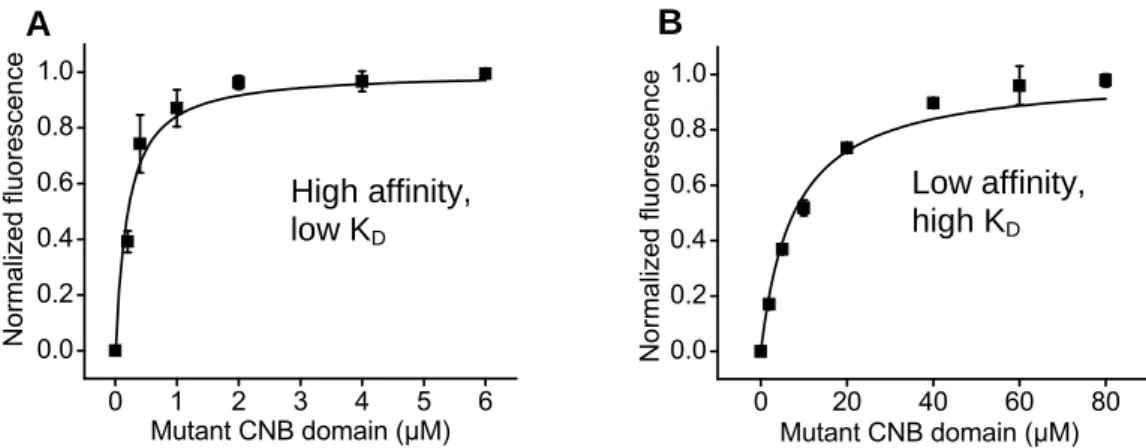
The effect of point mutations on ligand binding was screened using the isolated CNB domain. The approach was a binding assay that uses the fluorescent cAMP derivative 8-NBD cAMP (Biolog). It is well known the the local environment has important effects on fluorescent molecules (Lakowicz, 2006). The intrinsic fluorescence of 8-NBD cAMP is enhanced upon protein binding (Kraemer et al., 2001). For determination of the dissociation constant (KD) values of each CNB domain mutant for cAMP and cGMP, the first step was a protein titration at a fixed concentration of 8-NBD cAMP, usually 50 nM. Affinity for 8-NBD cAMP varied among the mutants screened, resulting on different ranges of protein concentrations required for saturation. If saturation occurs at relatively low (1-2 micromolar) protein concentrations, the mutant has high affinity for the cAMP analog (Fig. 2.2 A). If saturation requires higher protein concentration (tens of micromolar), the mutant has low affinity for the analogue (Fig. 2.2 B). We used different approaches for each of these two groups, as described below.
0 1 2 3 4 5 6 0.0 0.2 0.4 0.6 0.8 1.0 No competitor N or m aliz ed flu or es ce nc e Mutant CNB domain (µM) 0 20 40 60 80 0.0 0.2 0.4 0.6 0.8 1.0 No competitor Norm al iz ed fluores cenc e Mutant CNB domain (µM)
Figure 2.2: Titrations of MlotiK1 CNB domain with 50 nM 8-NBD cAMP (A) T284S and (B) R348A mutants. Determined KDs were 212 ± 22 nM and 7673 ± 818 nM for T284S and R348A, respectively.
2.2.2.1. High affinity mutants
For high affinity mutants, KDs for cAMP and cGMP were determined by repetition of the protein titration with 8-NBD cAMP in the presence of a fixed concentration of cAMP or cGMP. Using the T284S mutant as an example, the procedure used was the following. To determine the concentration of cAMP or cGMP to add to the protein titration curve, first, the fluorescence titration curve is analyzed to identify the protein concentration that resulted in approximately 70-80 % of the maximum signal (indicated by arrow in Fig. 2.3 A). Second, the protein concentration is fixed at that value and mixed with the fluorescent analogue. Incresing concentrations of cAMP or cGMP are added (Fig. 2.3 B), so that fluorescence decreases. The ligand concentration that results in a decrease of the signal to 40-50 % (indicated by arrows in Fig. 2.3 B) is chosen for the assay. The final titration involves adding increasing amounts of protein to a mix of fluorescent cyclic nucleotide analogue with cyclic nucleotide at the concentration determined in the previous step. The competition effect is evident when the binding curves in the absence and presence of cAMP or cGMP are superposed, as saturation is reached at higher concentrations of protein in the presence of non-fluorescent ligand (Fig. 2.3 C). The KDfor 8-NBD cAMP is determined (Fig. 2.3 D) and its value is used to calculate the KDs for cAMP (Fig. 2.3 E) and cGMP (Fig. 2.3 F), taking into account ligand concentrations.
High affinity, low KD
Low affinity, high KD