2019
DEPARTAMENTO DE BIOLOGIA VEGETAL
Study of non-conventional Aspergilli and Penicillia
Ochratoxin A producers
Teresa Ventura Vale Fernandes Dias
Mestrado em Microbiologia Aplicada
Dissertação orientada por:
Prof. Dr. Nelson Lima
Prof.ª Dr.ª Margarida Barata
This Dissertation was fully performed at Universidade do
Minho under the direct supervision of Professor Nelson
Lima.
Professor Margarida Barata was the internal supervisor designated in the scope of the Master in
Applied Microbiology of the Faculty of Sciences of the University of Lisbon.
III
Acknowledgments
This work is the conclusion of several years of hard work and dedication that would not have been possible without the help of family, friends, colleagues and professors. For all that they taught me and the continuous support to always be better, my deepest gratitude.
First and foremost, to Carlos, my partner in life. There are no words to thank him enough for all that he does for me, for the comprehension and for not letting me “lose my mind”. There are also no words to my parents. Nothing would be possible without all their support in all my decisions, specially in the past few years. Their care and love made me who I am. To Carolina and Daniel, for not only being my siblings, but also my friends. This journey was far more special with them by my side.
I would like to address a special thanks to Professor Nelson Lima for the opportunity, guidance and patient. Also, for all the conversations to make me think further, the knowledge and for teaching me “how science is made”. I hope I have the opportunity to keep on listen to the many interesting stories that he always has to tell.
A special thanks to Professor Margarida Barata for giving me the opportunity to experience the mycology world. Without her support and for always being available this work would not have been possible.
I would like to thank all my colleagues from the Applied Mycology Group for always making me feel welcome, for the friendship and help in the last year. I am especially grateful to Célia and Carla, for all that they done for me. Their daily support, advices and guidance when my many doubts arose. Without their help and availability this work would be much more complicated. It was a pleasure to work by their side.
To Professor Olga to let me in in the world of Microbiology, which had an important impact in my life, for her friendship and for the constant preoccupation. Also, to Eduarda for guiding me in the first steps of this world and for making me be focussed and disciplined in my work. To them, my deepest appreciation. My thanks also goes to all my friends from “lab 1.35”. May we keep on having dinners until we are old.
Also, to Carolina, Nuno, Inês and Sofia. All of them contribute in their special way to this work, and their friendship means a lot to me. To Bárbara for the mutual support, help and for always having a wise word to say. Hope to keep on sharing my life and my work with them.
Lastly, I would like to dedicate this work to Rita. If she was still here, I know that she would be proud. I will keep on smiling throw life for her.
IV
Abstract
The continuous growth of the population is pushing the available natural resources to the limit, posing a threat to the balance of the planet. This increase in population means that more food is needed. Consequently, food safety is, nowadays, a matter of interest to the scientific community and world leaders. However, problems related to food/feed contamination have been frequently reported, including those related to fungi. Fungi are diverse, ubiquitous and their role and impact in food security is now an urgent concern. One thing that makes fungi so dangerous is their ability to resist the treatments during the food making process and their ability to produce harmful secondary metabolites like mycotoxins. Mycotoxins are fungal secondary metabolites that can cause severe health issues in either humans or domesticated animals when ingested, inhaled and/or absorbed. From all the mycotoxins reported, some are more studied due to the higher health risks. Ochratoxin A (OTA) is among those and is present in several food/feed products. Aspergillus and Penicillium species are commonly associated with food spoilage and both genera contain species that can produce this toxin. The major OTA producers are P. verrucosum, P. nordicum and A. carbonarius, but more than 20 species of Aspergillus can produce this toxin. However, recent studies reported the presence of OTA in food matrices where known OTA producers are not present. This triggered the scientists involved to try to find the origin of OTA. Based on previous evidence other species like P. crustosum and A. fumigatus are now being considered. Therefore, the main goal of this work was to search for potential OTA producers among P. crustosum and A. fumigatus strains, with different geographic origins, and try to find potential genetic differences at the sub-species level.
A set of 28 Penicillium crustosum strains and 7 Aspergillus fumigatus strains, kindly supplied by Micoteca da Universidade do Minho (MUM), and 16 Penicillium crustosum strains, by Colección Chilena de Cultivos Tipo (CCCT), were studied. The Penicillium isolates are from four different countries: 16 from Italy, 16 from Chile, 6 from Portugal and 4 from Tunisia. The Aspergillus strains are all from Portugal, Spain or with unknown origin. Mycotoxin production was analysed by HPLC-FLD and results were compared with a standard sample. In addition, genes associated with OTA production [two ochratoxin polyketide synthase (Penicillium and Aspergillus related), ochratoxin non-ribosomal peptide synthetase and an ochratoxin transport protein] were tested. RAPD-PCR fingerprinting [M13 and (GACA)4] and beta-tubulin gene (BenA) sequencing were used to perform a wide molecular
characterisation.
Under the studied conditions, and with a HPLC-FLD detection limit of 7.6 ng/ml, preliminary results showed that OTA was not detected for all studied strains. However, regarding the genes associated with OTA production, there were 4 positive strains of P. crustosum for the 3 genes. Genetic differences, based on RAPD fingerprints, between P. crustosum isolates were found allowing the clustering of strains from the same geographic region, except for isolates from Europe. The low number of A. fumigatus strains did not allowed to draw conclusions, although they also presented genetic differences. Sequencing with BenA did not revealed any SNPs. Nevertheless, further studies with a broader array of conditions needs to be considered.
V
Resumo
O crescimento exponencial da população mundial está a levar ao limite os recursos naturais disponíveis representando um risco para o equilíbrio do planeta. O aumento da população tem como consequência o aumento da necessidade de mais alimentos. Consequentemente, a segurança alimentar é, hoje em dia, uma preocupação para a comunidade científica e para os líderes mundiais. Contudo, problemas relacionados com comida e rações animais contaminadas têm sido frequentemente reportados, incluindo aqueles com fungos. Os fungos são um grupo de organismos eucarióticos, diversos, ubíquos e têm um grande impacto na segurança alimentar.
A capacidade de os fungos resistirem aos processos da indústria alimentar, como tratamentos térmicos, e a sua capacidade de produzirem metabolitos secundários prejudiciais, torna-os perigosos contaminantes. Um exemplo desses metabolitos produzidos são as micotoxinas. As micotoxinas quando ingeridas, inaladas e/ou absorvidas podem causar problemas severos de saúde a pessoas e animais. A preocupação com este assunto iniciou-se aquando da morte de cerca de 10000 perus no reino unido, que se deveu a uma contaminação com aflatoxinas. Aspergillus e Penicillium são dois grupos de fungos filamentosos que têm um grande impacto na contaminação alimentar particularmente pela capacidade de produzirem micotoxinas. As micotoxinas mais associadas a estes géneros são aflatoxinas, ocratoxina A, patulina e citrinina. A maior preocupação com estas toxinas é a sua toxicidade aguda e/ou crónica, que pode levar à morte ou a outras patologias. A maioria das micotoxinas são estáveis, resistentes ao calor e podem permanecer mesmo em produtos tratados (por exemplo, produtos pasteurizados). De todas as micotoxinas, a ocratoxina A (OTA) é uma das mais estudadas, devido à sua presença em vários produtos alimentares e também pelos problemas de saúde que pode causar.
A OTA foi descoberta em 1965 no Aspergillus ochraceus. É considerada neurotóxica, nefrotóxica, carcinogénica, hepatotóxica e teratogénica, para diversas espécies. Está classificada com o grupo 2B - possivelmente carcinogénica para humanos. Está presente em diversos produtos alimentares como cereais, vinho, queijo e café. De entre os cereais, o centeio, o trigo e a cevada são os que apresentam os maiores níveis de contaminação. Isto representa um problema pois estes cereais são muito usados no fabrico de farinhas e rações destinadas à alimentação de animais. Consequentemente, os animais domésticos são os mais suscetíveis a ter reações adversas devido à presença da toxina, uma vez que estão expostas a ela por mais tempo e com maior frequência. Os maiores produtores de OTA são P. verrucosum, P. nordicum e A. carbonarius, no entanto cerca de mais 20 espécies de Aspergillus são capazes de a sintetizar. A identificação e a quantificação da OTA são comummente feitas por cromatografia líquida de alta eficiência com um detetor de fluorescência (HPLC-FLD). Além disso há genes que estão associados com a ocratoxina A e que podem ser usados como possíveis marcadores genéticos da capacidade de um fungo a produzir. Recentemente, um estudo identificou a presença de OTA em diversas amostras de queijos Italianos. Contudo, não foi possível identificar nenhum dos conhecidos produtores da toxina. Assim, surgiu a dúvida de qual seria a origem da OTA. Baseados em estudos anteriores, P. crustosum e A. fumigatus estão a ser considerados como possíveis novos produtores. Estes fungos são ubíquos e são diversas vezes encontrados em produtos alimentares. São produtores de toxinas, mas apenas foram descritos uma (A. fumigatus) ou duas (P. crustosum) vezes como produtores de OTA.
O objetivo principal deste trabalho é procurar potenciais produtores de OTA entre um conjunto de estirpes de P. crustosum e de A. fumigatus (com diferentes origens geográficas) e tentar encontrar potenciais diferenças ao nível da subespécie.
Um conjunto de 28 estirpes de P. crustosum e 7 estirpes de A. fumigatus fornecidas pela Micoteca da Universidade do Minho (MUM), e 16 estirpes de P. crustosum, fornecidas pela Colección Chilena de
VI Cultivos Tipo (CCCT), foram estudadas. Os isolados de Penicillium provieram de quatro países e matrizes diferentes: 16 foram isolados de queijos italianos, 16 de merkén do Chile, 6 de diferentes origens em Portugal e 4 de maçãs da Tunísia. As estirpes de Aspergillus são, na sua maioria, de Portugal ou Espanha, no entanto algumas têm origem desconhecida.
A produção de ocratoxina A foi avaliada por HPLC-FLD e os resultados foram comparados com um padrão de concentração conhecida. As estirpes de P. crustosum foram cultivadas em meio enriquecido com sal de forma a terem um stress externo que conduza à produção de micotoxinas. Genes relacionados com a produção de OTA (PKS, NRPS e um transportador) foram também procurados. Adicionalmente foi feito um estudo genético complementar. Apesar de todas as estirpes terem origem em coleções de culturas, o que à partida valida a sua identificação, o gene da beta tubulina foi amplificado. Uma vez que o estudo envolve uma panóplia de estirpes da mesma espécie, principalmente em P. crustosum, com origens geográficas diferentes, há uma possibilidade de nessas estirpes terem ocorrido processos de especiação e haver espécies crípticas por revelar. Espécies crípticas são espécies que são morfologicamente idênticas, no entanto apresentam diferenças genéticas. Para complementar este estudo foram feitas análises de perfis genómicos (fingerprinting) usando pequenos primers: M13 e (GACA)4.
Nas condições testadas, tendo a HPLC um limite de deteção de 7,6 ng/ml, os resultados preliminares mostraram que não se detetou produção de OTA para nenhuma das 51 estirpes. Os cromatogramas obtidos de cada estirpe foram comparados com o cromatograma padrão, onde foi possível observar um pico aos 13 min. No entanto, em relação aos genes relacionados com a biossíntese de OTA os resultados foram mais promissores, exceto para o gene relacionado com a produção de OTA em Aspergillus (Acpks). Para esse gene apenas o controlo positivo foi amplificado e não em nenhuma das estirpes em estudo. Os restantes genes estavam relacionados com Penicillium [otapks (≈500 bp); otanps (≈700bp) e otatra (≈420 bp)]. Para estes genes, 8% estirpes amplificaram todos os genes (sendo todas do Chile), 16% amplificaram somente otapks; 10% otapks e otatra, 8% otanps e otatra; 45% apenas otatra; e 14% não amplificaram nenhum gene. O facto de haver espécies a amplificarem os três genes testados é um indicador que estas podem ter a capacidade de produzir OTA. Além disso a grande diversidade de padrões verificados mostra que as estirpes são distintas entre si. O estudo com os primers de fingerprinting de onde se obtiveram perfis genéticos e com os quais foram construídos dendrogramas mostraram a mesma distinção. Diferentes padrões foram obtidos podendo individualizar cada indivíduo. Além disso, fazendo uma análise aos dendrogramas foi possível verificar que as estirpes chilenas formam um cluster assim como as estirpes da Tunísia. Isto pode demonstrar que a distância geográfica poderá criar processos de especiação. Por outro lado, as estirpes Italianas e Portuguesas não formam nenhum cluster. Estes dois países encontram-se na Europa onde não há muitas restrições de fronteiras. Em relação a A. fumigatus, o baixo número de estirpes, e a falta de informação de algumas delas, não permitiu a formação de clusters, apenas se verificando que também apresentam perfis diferentes.
Este foi um estudo preliminar em que não foi possível a quantificação de OTA nem em P. crustosum nem em A. fumigatus. No entanto, os resultados dos genes biossintéticos mostram que algumas das estirpes podem ter a capacidade de a produzir. Também foi possível verificar que RAPD é uma boa técnica de tipificação e que espécies afastadas geograficamente apresentam perfis genéticos diferentes. Não obstante, estudos com métodos adicionais e com diferentes condições precisam ser considerados.
Palavras chave: Segurança Alimentar; Ocratoxina A; P. crustosum; A. fumigatus; RAPD; Genes biossintéticos.
VII
List of Contents
Acknowledgments ... III
Abstract ... IV
Resumo ... V
List of Figures ... IX
List of Tables ... X
List of Abbreviations ... XI
1. Introduction ... 1
1.1. Ochratoxin A (OTA) ... 1
1.2. OTA biosynthetic genes ... 2
1.3. Penicillium crustosum ... 3
1.4. Aspergillus fumigatus ... 4
1.5. Identification of filamentous fungi ... 5
1.6. Typing techniques ... 6
1.7. Objectives ... 6
2. Materials and Methods ... 7
2.1. Fungal strains ... 7
2.2. DNA extraction ... 7
2.3. Molecular identification ... 8
2.4. Ochratoxin-A (OTA) extraction and quantitative analysis ... 8
2.5. Ochratoxin-A (OTA) biosynthetic genes ... 9
2.6. RAPD-PCR fingerprinting ... 10
3. Results ... 11
3.1. Phylogenetic Analysis ... 11
3.2. Ochratoxin-A (OTA) quantitative analysis ... 13
3.3. OTA biosynthetic genes ... 13
3.4. RAPD-PCR fingerprinting ... 16
3.4.1 Aspergillus fumigatus ... 16
VIII
4. Discussion ... 20
5. Conclusions and Future Perspectives ... 27
6. References ... 28
7. Attachments ... 37
7.1. Attachment I ... 37
IX
List of Figures
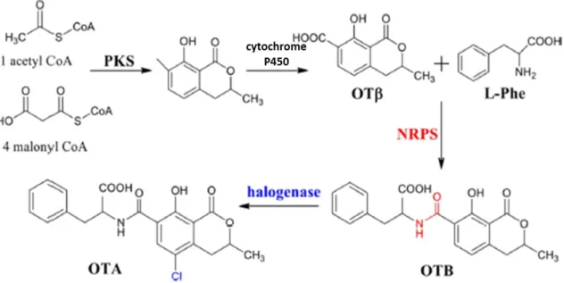
Figure 1.1. - Representation of OTA biosynthetic pathway. OTA biosynthesis begins with biotransformation of malonyl-CoA and acetyl-CoA by a PKS enzyme. Then it is catalysed to ochratoxin β (OTβ) by a cytochrome P450 monooxygenase, that when linked to l-phenylalanin by NRPS, forms ochratoxin B (OTB). Finally, this compound is chlorinated, by a halogenase, to produce OTA (adapted from Wang et al., 2018). ... 3 Figure 1.2 - Macro- and micromorphology of P. crustosum (MUM 16.11), after 7 days growth in MEA at 25 ºC. (Images kindly provided by Célia Soares, curator of Micoteca da Universidade do Minho) .. 4 Figure 1.3 - Macro- and micromorphology of A. fumigatus (MUM 14.30), after 3 days growth in MEA at 37 ºC. (Images kindly provided by Célia Soares, curator of Micoteca da Universidade do Minho) ... 4 Figure 2.1 - Map of the origin of the P. crustosum strains in the world. Chile it is painted in blue, Italy in purple, Portugal in green and Tunisia in pink. ... 7 Figure 3.1 - Evolutionary relationships of A. fumigatus strains (BenA). The evolutionary history was inferred by using the Maximum Likelihood method and Kimura 2-parameter model (Kimura 1980). The tree with the highest log likelihood (-763.88) is shown. The tree is drawn to scale, with branch lengths measured in the number of substitutions per site. This analysis involved 10 nucleotide sequences. All positions containing gaps and missing data were eliminated. There were a total of 378 positions in the final dataset. Evolutionary analyses were conducted in MEGA X (Kumar et al., 2018). ... 11 Figure 3.2 - Evolutionary relationships of P. crustosum strains (BenA). The evolutionary history was inferred by using the Maximum Likelihood method and Kimura 2-parameter model (Kimura, 1980). The tree with the highest log likelihood (-555.71) is shown. The tree is drawn to scale, with branch lengths measured in the number of substitutions per site. This analysis involved 47 nucleotide sequences. All positions containing gaps and missing data were eliminated. There was a total of 298 positions in the final dataset. Evolutionary analyses were conducted in MEGA X (Kumar et al., 2018). ... 12 Figure 3.3 - Chromatogram of an OTA standard (118 ng/mL) with retention time at 13 minutes. ... 13 Figure 3.4 - Fingerprint profiles of the A. fumigatus strains. Amplification with M13 (left) and (GACA)4
(right). ... 16 Figure 3.5 - Joined dendrogram of M13 and (GACA)4 fingerprinting of A. fumigatus strains, generated
in BioNumerics using the average of all experiments method. ... 17 Figure 3.6 - Fingerprint profiles of the Chilean strains (GACA)4. ... 18
Figure 3.7 - Fingerprint profiles of the Tunisian (M13). ... 18 Figure 3.8 - Joined dendrogram of M13 and (GACA)4 fingerprinting of P. crustosum strains, generated
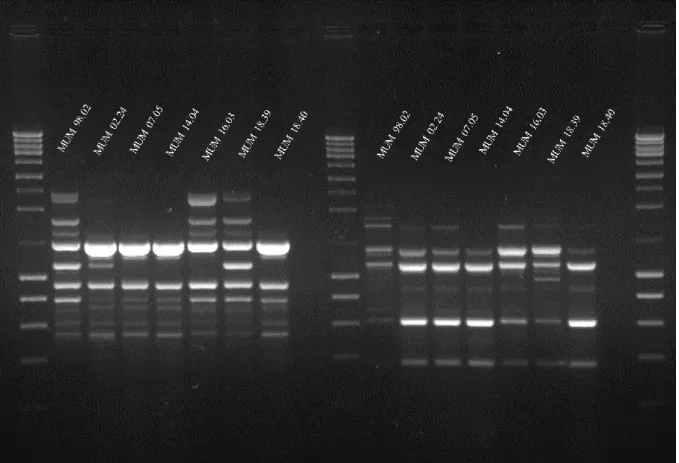
in BioNumerics using the average of all experiments method. Portuguese strains are represented in green, Italian’s in purple, Tunisian’s in pink and Chilean’s in blue ... 19 Figure 7.1 - Representation of PCR amplification of OTA byossinthetic genes: otapks (line 1-5), otanps (line 6-10) and otatra (line 11-15). Amplicons were visualized on 1% agarose gels. MUM 16.08 was used as positive control for all genes. The expected band size, for each gene, are in evidence. For otapks CCCT 18.132 and MUM 16.68, are positive even though that the Chilean strain evidence a non specific band. For this gene is also possible to see non specific amplification in MUM 14.30. For otanps there are also 2 positive strains (CCCT 18.82 and MUM 16.105). Lastly, for otatra all the strains are positive, but only MUM 17.70 doesn’t evidence a non specific band. ... 38
X
List of Tables
Table 3.1 - Results of the PCR amplification of OTA related genes (otapks, otanps, otatra and Acpks). ... 14 Table 7.1 - List of studied strains, their origin and the isolation substrate. ... 37
XI
List of Abbreviations
AFLP – Amplified Fragment Length Polymorphism
BenA – β-tubulin
BLAST – Basic Local Alignment Search Tool
CaM – Calmodulin
CCCT – Colección Chilena de Cultivos Tipo CTAB – Cetyltrimethylammonium bromide DNA – Deoxyribonucleic acid
dsDNA – Double stranded DNA
EFSA – European Food Safety Authority
ELISA – Enzyme-Linked Immunosorbent Assay FAO – Food and Agriculture Organization GC – Gas Chromatography
GRAS – Generally Regarded As Safe
HPLC-FLD – High Performance Liquid Chromatography with Fluorescence Detection HPTLC – High Performance Thin-Layer Chromatography
IARC – International Agency for Research on Cancer ITS – Internal Transcribed Spacer
IUPAC – International Union of Pure and Applied Chemistry LOD – Limit of Detection
LOQ – Limit of Quantification
MALDI-TOF MS – Matrix-Assisted Laser Desorption Ionization-Time-Of-Flight Mass Spectrometry MEA – Malt Extract Agar
MGYP – Malt Extract Glucose Yeast Extract Peptone Medium MLST – Multilocus Sequence Typing
MS – Mass Spectrometry
MUM – Micoteca da Universidade do Minho
NGMLST – Next Generation Multilocus Sequence Typing NGS - Next Generation Sequencing
NRPS – Non-Ribosomal Peptide Synthases OTA – Ochratoxin A
OTB – Ochratoxin B OTα – Ochratoxin α OTβ – Ochratoxin β
PCR – Polymerase Chain Reaction PFGE – Pulse Field Gel Electrophoresis PKS – Polyketide Synthases
RAPD-PCR – Random Amplification of Polymorphic DNA RNA – Ribonucleic acid
RPB2 – RNA polymerase II second largest subunit rRNA – Ribosomal ribonucleic acid
SNPs – Single Nucleotide Polymorphisms TAE – Tris acetate EDTA
UPGMA – Unweighted Pair Group Method Using Arithmetic Average WGST – Whole Genome Sequence Typing
1
1. Introduction
The world population is continuously growing posing a threat to the balance of the planet. Recent projections show that in 2050 the world population is going to reach 9.8 billions (United Nations, 2019). Ensuring the health and safety of all of them will be a priority to world leaders. One of the main concerns is food availability. To meet this demand, substantial increases in food production will be required as well as the need for the safety of the food produced. It is also important to ensure that the food is nutritious, affordable and uses the least resources possible.
In the food chain, the consumers (humans or animals) are the priority. Problems related to food/feed contamination harmful to their health have been increasing (Steinwider et al., 2019). Food spoilage can occur due to microbiological, physical or chemical changes. Microbial spoilage is a major concern since it can cause severe health issues.
The kingdom Fungi encompasses a variety of eukaryotic organisms either unicellular (yeasts) or multicellular (filamentous fungi) that are absorptive chemoorganoheterotrophs. Estimates propose that there are 2.2 to 3.8 million species of fungal species in the world with only 120000 being validly described (Hawksworth and Lücking, 2017). Fungi are diverse, ubiquitous and their role and impact in food security is now an urgent concern. This impact is due to the capacity to resist under the conditions, like extreme heat, used in food production (Biango-Daniels et al., 2019).
Another concern related to fungi is their capacity to produce mycotoxins. Mycotoxins are fungal secondary metabolites that can cause severe health issues in either humans or domesticated animals when ingested, inhaled and/or absorbed. The awareness for this issue started in 1960 when an unknown disease killed more than 100000 turkeys in the United Kingdom. This disease was called “Turkey X disease” (Blount, 1961) and only later it was found to be caused by aflatoxins contamination.
Aspergillus and Penicillium are filamentous fungi of high importance in food contamination, particularly from mycotoxin production. Major mycotoxins associated with aspergilli and penicillia are: aflatoxins [A. flavus and A. parasiticus (Klich, 2007)], ochratoxin A (OTA) [A. carbonarius, P. verrucosum and P. nordicum (Cabañes et al., 2010; Varga et al., 2015)], patulin [P. expansum (Frisvad, 2018)] and citrinin [P. expansum and P. citrinun (Ostry et al., 2013)]. The main concern of these naturally occurring toxins is their acute and/or chronic toxicity, which can cause death and/or deleterious effects. Most of the known mycotoxins are relatively stable, heat resistant and are expected to remain in the heat-treated product.
1.1. Ochratoxin A (OTA)
Ochratoxin A (OTA) is one of the most important toxins. It was first discovered in Aspergillus ochraceus by Van der Merve et al. (1965) who also established its structure. It is considered, among other possible effects, neurotoxic (Paradells et al., 2014), nephrotoxic (Zhao et al., 2016), carcinogenic (Pfohl-Leszkowicz and Manderville, 2007), hepatotoxic and teratogenic (Wu et al., 2018) in various species. The International Agency for Research on Cancer (IARC) classified OTA as a possible human carcinogen (group 2B) (IARC, 1993). This mycotoxin is present in a wide diversity of food and feed products, such as cereals, wine, coffee, dried fruits, beer and in feeds for animals (Joshi et al., 2017). Among cereals, rye, wheat and barley are the ones with the highest levels of contamination (Marin et al., 2013). This represents a serious problem because these cereals are extensively used to make flours, and other derivatives that are the basis for animal feeds. Due to this, livestock are the animals more susceptible to have adverse reactions due to the presence of OTA, as they may be exposed to the toxin for a long period of time.
2 As aforementioned, the major producers of OTA are P. verrucosum, P. nordicum and Aspergillus carbonarius. Many other species within Aspergillus genus can be OTA producers (Varga et al., 2015), such as A. niger, A. steynii, A. westerdijkiae and A. alliaceus. P. verrucosum is the main source of OTA contamination in cereals and their products in cold and temperate climates. In contrast, P. nordicum is usually recovered from dry-cured meat products and cheese and it may be the cause of OTA contamination in these foods (Cabañes et al., 2010). Recently, another Penicillium joined the group of the known OTA producers – Penicillium thymicola, isolated from Canadian cheddar cheese (Nguyen et al., 2016).
OTA was considered, for many years, the cause of the Balkan endemic nephropathy. This disease is characterised by chronic renal disease frequently associated with upper urothelial cancer. This data is plausible because the disease only affects residents from farming villages in the Balkans, that are exposed to relatively high concentrations of OTA. More recent studies demonstrate that OTA is not the cause for the disease but can influence the metabolism of other carcinogens (Stiborová et al., 2015).
To prevent outbreaks of the ochratoxigenic fungi, the Codex Allimentarius (FAO), created Codes of Practice for the prevention and reduction of OTA contamination in cereals (2000), wine (2007), coffee (2009) and cocoa (2013). Mycotoxin production in crops can occur at various points in the food chain: at pre-harvest, harvest, drying, and storage so these codes contemplate all phases. Based on many studies and after assembling of a panel, the European Food Safety Authority (EFSA), established a tolerable weekly intake for OTA of 120 ng/kg body weight (EFSA, 2010).
OTA quantification is commonly performed by high-performance liquid chromatography with fluorescence detection (HPLC-FLD) (Abrunhosa et al., 2014; Mishra et al., 2016; Bonerba et al., 2017). Immunoaffinity columns can be used to obtain more pure and concentrated extracts (Zhang et al., 2018). Other techniques may be used like enzyme-linked immunosorbent assay (ELISA), high-performance thin-layer chromatography (HPTLC), gas chromatography (GC) and mass spectrometry (MS) (Flajs et al., 2009; Zhang et al., 2018; Zhang et al., 2019). Due to the high cost of the majority of these methods HPLC is still the most commonly used technique.
1.2. OTA biosynthetic genes
Knowing and understanding the genetic pathway involved in the biosynthesis of mycotoxins is of the utmost importance, as it could provide tools for diagnostic methods and prevention strategies, for example. In recent years, several works have been conducted to understand the metabolic pathway and the biosynthetic genes involved in OTA production. However, unlike other mycotoxins, this process is still not entirely known.
OTA is a pentaketide composed of a dihydroisocoumarin linked via amide bond to the amino acid phenylalanine (IUPAC, 1992). Polyketide synthases (PKSs) and non-ribosomal peptide synthases (NRPSs) are multimodular enzymes that have roles in the production of fungal secondary metabolites (Nielsen et al., 2017) and OTA is not an exception. The PKS and NRPS were identified in the greatest number of species such as Aspergillus ochraceus (O’Callaghan et al., 2003), Aspergillus carbonarius (Gallo et al., 2012; Gallo et al., 2014), Penicillium verrucosum (O’Callaghan et al., 2013), among others. Besides these two synthases, a cytochrome P450, a halogenase and a bZIP transcription factor (except in Penicillium verrucosum) were also found in a great number of species: Aspergillus westerdijkiae (Chakrabortti et al., 2016, Gil-Serna et al., 2018), Aspergillus steynii (Gil-Serna et al., 2015, Gil-Serna et al., 2018), Aspergillus niger (Gil-Serna et al., 2018), Aspergillus carbonarious (Gil-Serna et al., 2018), Penicillium nordicum (Geisen et al., 2018, Gil-Serna et al., 2018) and Penicillium verrucosum (Geisen et al., 2018). Thus, the most recent studies propose that OTA biosynthesis begins with the
3 biotransformation of malonyl-CoA and acetyl-CoA by a PKS enzyme. Then it is catalysed to ochratoxin β (OTβ) by a cytochrome P450 monooxygenase, that when linked to l-phenylalanine, by NRPS, forms ochratoxin B (OTB). Finally, this compound is chlorinated, by a halogenase, to produce OTA (Wang et al., 2018) (Fig. 1.1). The belief that ochratoxin α, initially proposed by Huff and Hamilton (1979) and others (Harris and Mantle, 2001), was an intermediate in OTA biosynthesis was not corroborated, which indicates that this compound is only a product of OTA hydrolysis.
1.3. Penicillium crustosum
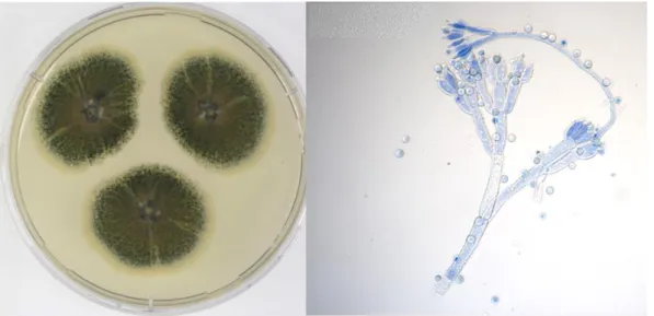
P. crustosum is a ubiquitous fungus, found all over the globe. The fungus presents, typically, low colonies with the surface appearing powdery due to the heavy conidial production (Fig. 1.2). Colonies tend to have a dull green colour on the obverse and on the reverse side yellow to orange-brown (Pitt and Hocking, 2009).
This fungus is usually found associated with food/feed (Wigmann et al., 2016; Decontardi et al., 2017; Greeff-Laubscher et al., 2018). It is, for example, a normal contaminant in cheese (Hymery et al., 2014).
P. crustosum is a consistent producer of several mycotoxins such as roquefortine C, penitrem A–F, thomitrem A and E (Rundberget et al., 2004). However, in 2006, Vega and collaborators identified a P. crustosum strain, isolated from a berry in Mexico, that was able to produce OTA (Vega et al., 2006). Though, the amount of OTA produced was below the allowed by law. Furthermore, recent preliminary studies (personal communication) discovered that several different strains of P. crustosum in specific conditions can produce OTA. These results are very concerning because this fungus may be contaminating several different foods, which represents a safety hazard.
Figure 1.1. - Representation of OTA biosynthetic pathway. OTA biosynthesis begins with biotransformation of malonyl-CoA and acetyl-CoA by a PKS enzyme. Then it is catalysed to ochratoxin β (OTβ) by a cytochrome P450 monooxygenase, that when linked to l-phenylalanin by NRPS, forms ochratoxin B (OTB). Finally, this compound is chlorinated, by a halogenase, to produce OTA (adapted from Wang et al., 2018).
4
1.4. Aspergillus fumigatus
A. fumigatus is also a ubiquitous fungus, highly associated with human health due to the capacity to invade human tissue causing aspergillosis, especially in immunocompromised patients (Lee et al., 2019). This fungus presents low, blue-green colonies, with a velvety texture (Fig. 1.3) (Pitt and Hocking, 2009).
The prime habitat for A. fumigatus is decaying vegetation, however it can be found in food or feed samples (Shapaval et al., 2013; Greco et al., 2014). On this matrix it represents a danger to the consumers’ safety since it can resist high temperatures. This fungus can produce several toxins like gliotoxin, verruculogen, fumagillin and helvolic acid (Boudra and Morgavi, 2005). In 1997, Albarca and collaborators reported an A. fumigatus strain (isolated from food-related materials) that was able to produce OTA (Albarca et al., 1997). In the same study, A. versicolor was also identified as an OTA producer, yet none of the strains was described as an OTA producer after that.
Figure 1.3 - Macro- and micromorphology of A. fumigatus (MUM 14.30), after 3 days growth in MEA at 37 ºC. (Images kindly provided by Célia Soares, curator of Micoteca da Universidade do Minho)
Figure 1.2 – Macro- and micromorphology of P. crustosum (MUM 16.11), after 7 days growth in MEA at 25 ºC. (Images kindly provided by Célia Soares, curator of Micoteca da Universidade do Minho)
5
1.5. Identification of filamentous fungi
The identification of foodborne fungi is very important because with a name comes a lot of knowledge (i.e. resistance under extreme heat, low/higher water activity, ability to produce mycotoxins). An effective and rapid identification can prevent food outbreaks which are a crucial feature in the food industry.
The identification of fungi is traditionally made by classical microbiological techniques. These methods use morphologic criteria and microscopic features, that are grounded on phenotypic characters. Though, fungi have a variety of different forms (anamorphic and teleomorphic) which leads to giving different names to the same species (Hawksworth, 2011). For that reason, the identification based on conventional techniques is rather subjective, time-consuming and prone to errors.
The development of molecular biology allowed to do rapid and sensitive identification of filamentous fungi on food/feed samples (Rico-Munoz et al., 2019). Still using traditional techniques should not be excluded. So, an integration of a phenotypic approach together with a genotypic approach, in a multiple step method is the most effective way to identify foodborne fungi (Simões et al., 2013; Decontardi et al., 2018).
When identifying bacteria, in most of the cases, a well know gene, 16S rRNA, is used as the universal marker since a long time (Fox et al., 1977). Meanwhile, regarding fungi, choosing a universal barcode DNA marker for identification has not been so easy. Only recently, the nuclear ribosomal internal transcribed spacer (ITS) region was chosen for this purpose (Schoch et al., 2012). Ribosomes are responsible for synthetize proteins, and because of this they are highly conserved (Rorbach et al., 2017), thus an excellent target for identification. Albeit ITS is a good DNA marker, is not always the best marker to discriminate to the species level some groups, like Aspergillus or Penicillium. This might be due to the genetic differences that the ITS region presents (Kiss, 2012). For this, secondary markers were chosen for the identification of these groups. For these groups 3 options were chosen: calmodulin (CaM), β-tubulin (BenA) or the RNA polymerase II second largest subunit (RPB2), being CaM more suitable for Aspergillus and BenA for Penicillium (Samson et al., 2014; Visagie et al., 2014).
In the Eumycota kingdom there is a high prevalence of morphologically defined species, with similar phenotypes, i.e. cryptic species. These species, as has been said, are morphologically indistinguishable but have different nucleic acid characters (Perrone and Susca, 2017). The study of a broad array of strains from different species, resorting to PCR based methods, allows the discovery of these “sister species”. To identify and define species boundaries is important to understand processes related to adaptation and speciation. For this, it is crucial to have a strong database that gathers all the information, in this case, gene sequences, that will allow the comparison and recognition of inconsistencies if present (Federhen, 2014). Over the years, several cryptic species have been acknowledged (Cruse et al., 2002; Perrone et al., 2011; Lackner et al., 2019). The ability to classify these species is important in foodborne fungi once similar species can produce different secondary metabolites such as mycotoxins. This causes concern since species with the status of generally regarded as safe (GRAS) may have “sister species” that produce harmful components that can cause health issues to the consumers (Schuster et al., 2002). Notwithstanding this, resolving cryptic species may not be easy. Due to all the evolutionary processes that all livings beings are exposed to, one DNA marker may not be enough to resolve the relationship between closely related fungi. Balasundaram and collaborators (2015), using species from the genus Serpula, demonstrate that to reveal cryptic species with high confidence it may be needed 5 independent loci (different genetic markers). They also showed that ITS was not the best marker to disclose so close related species.
6
1.6. Typing techniques
Typing techniques allow to identify strains and discriminate them to subspecies level. When food security is at stake using these techniques are important to find different individuals within the same species, that might pose different risks, or to try to detect the origin of the contamination.
The Matrix-Assisted Laser Desorption Ionization-Time-Of-Flight Mass Spectrometry (MALDI-TOF MS) technique can be used to type. A soft ionisation results in minimum fragmentation of different biomolecules, such as peptides, proteins, sugars or polymers (Lima and Santos, 2017). This technique has been used for detection of different food-borne fungi (Santos et al., 2016; Elbehiry et al., 2017; Lauterbach et al., 2017).
Amplified fragment length polymorphism (AFLP) is a genotyping technique that uses restriction endonucleases digestion to obtain fragments from a particular genome (Umesha and Manukumar, 2018). Kure et al. (2003) could precise which Penicillium strains and in which stage of the production chain the contamination of cheese was occurring. Pawlik et al. (2012) used AFLP to identify Pleurotus strains to take the most advantage of their potential for the food industry.
RAPD-PCR fingerprinting (random amplification of polymorphic DNA) has also been used to study fungi. It uses short arbitrary primers that produce distinct banding patterns (Umesha and Manukumar, 2018). The congruence of a group of P. nordicum strains, isolated from cured meats, was studied (Bogs et al., 2006). Decontardi et al. (2018) also used this technique to study the clustering of Penicillia and Aspergilli isolated from Italian grana cheese.
A plethora of other techniques can be used, such as microsatellite analysis, multilocus sequence typing (MLST), pulse field gel electrophoresis (PFGE), whole genome sequence typing (WGST), Next Generation Multilocus Sequence Typing (NGMLST), among others (Umesha and Manukumar, 2018; Rico-Munoz et al., 2019). It is the responsibility of the investigator, based on previous knowledge and the resources available, to choose the most adequate techniques that allow a rapid and effective identification.
1.7. Objectives
So, if the major Aspergilli and Penicillia producers of OTA are well recognized, other species have been currently assigned as potential OTA producers. Taking this into consideration, the present work will try to answer a fundamental question: Are other Aspergilli and Penicillia species able to produce OTA? For the reasons mentioned above, the focus will be Penicillium crustosum and Aspergillus
fumigatus. Several isolates from different backgrounds will be analysed for the potential of producing
this mycotoxin, either by quantification or search of OTA related genes. Apart from that, a typing study of all the isolates will try to address potential genetic differences at the sub-species level.
7
2. Materials and Methods
2.1. Fungal strains
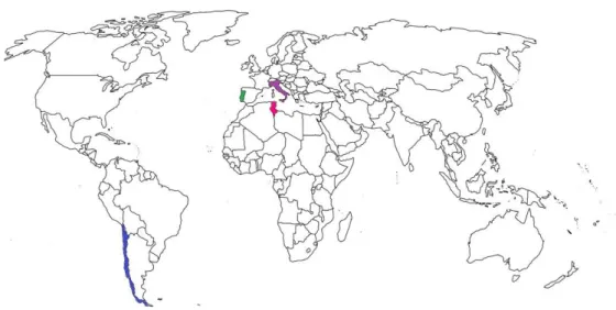
A set of 28 Penicillium crustosum strains and 7 Aspergillus fumigatus strains, kindly supplied by Micoteca da Universidade do Minho (MUM), and 16 Penicillium crustosum strains, by Colección Chilena de Cultivos Tipo (CCCT), were studied. The Penicillium isolates have different backgrounds (Fig. 2.1): 16 were isolated from Italian cheeses (Decontardi et al., 2018), 16 from Chilean Capsicum pepper and in its derivatives such as Merkén (traditional Chilean chili pepper powder), 4 from Tunisian apples, and 6 from different substrates from Portugal. The Aspergillus strains were all from different substrates from Portugal, Spain or unknown origin (Attachment I – Table 7.1).
2.2. DNA extraction
All strains were grown for seven days on Malt Extract Agar [(MEA), agar 20 g/L, glucose 20 g/L, 20 g/L malt extract, 1 g/L peptone)] at 25 ºC in the dark. Spores were collected and preserved in soft agar (4%) pending on further inoculations. With the help of a sterile loop, these suspensions were used to inoculate 50 mL Falcon tubes containing 25 mL of Malt Extract Glucose Yeast Extract Peptone Medium [(MGYP, malt extract 3 g/L, glucose 10 g/L, yeast extract 3 g/L, peptone 5g/L)]. Samples were incubated at room temperature for 4-6 days in an orbital shaker. Fungal biomasses were filtrated and, subsequently, stored at -20 ºC.
For DNA extraction, 200 mg of biomass were placed in a 1.5 mL tube filled with 670 mg of glass beads (425-600 µm). Mechanical lysis was performed using a FastPrep-24TM 5G Instrument (MP
Biomedicals, Santa Ana, California, USA) with 1 mL of CTAB (2%) for 30 s at a velocity of 6.0 m/s. Samples were centrifuged at 14000 × g for 8 min at room temperature and 800 µL of the supernatant were transferred to a new 2 mL tube. Polysaccharides and proteins were precipitated by adding 1 mL of cold sodium acetate (3 M, pH 5.5). Samples were gently mixed by inversion, placed at -20 °C for 10 min and centrifuged at 14000 x g for 10 min at room temperature. Then, 1 mL of the supernatant was transferred to a new tube along with 1 mL of isopropanol at room temperature. Samples were again gently mixed by inversion and incubated 1 h at room temperature. To precipitate the nucleic acid pellet, samples were centrifuged ate 14000 x g for 10 min. The pellet was washed twice with cold 70% ethanol, centrifuged at 6000 x g for 7 min and dried at 40 ºC for 5 min using a SpeedVac Concentrator (Thermo
Figure 2.1 - Map of the origin of the P. crustosum strains in the world. Chile is painted in blue, Italy in purple, Portugal in green and Tunisia in pink.
8 Scientific, Waltham, Massachusetts, USA). Pellets were suspended on 100 µL of sterile ultra-pure water, and incubated in a water bath, at 56 ºC for at least 3 h. DNA samples were stored at -20 ºC. The DNA quality was assessed by quantification of total DNA using NanoDropTM 1000 spectrophotometer
(Thermo Scientific, Waltham, Massachusetts, USA) and by electrophoresis agarose gel 1% (w/v) for 30 min at 80 V. SYBR® Safe DNA Gel Stain (Invitrogen, Waltham, Massachusetts, USA) was used as a
staining element and NZYDNA ladder III (NZYTech, Lisbon, Portugal) was used as a DNA molecular weight marker.
2.3. Molecular identification
Sequence analysis of the β-tubulin gene (BenA) was used to reconfirm and enlarge the knowledge of the previous identification of the fungal strains based on the ITS analysis. The extracted DNA was amplified using the primers Bt2a GGTAACCAAATCGGTGCTGCTTTC-3´) and Bt2b (5´-ACCCTCAGTGTAGTGACCCTTGGC-3´) design by Glass and Donaldson (1995). PCR was carried in a 25 µL mixture containing 12.5 µL of NZYTaq 2× Green Master Mix (NZYTech, Lisbon, Portugal), 0.25 µM of each primer, 25 ng of DNA and ultra-pure water until the final volume. The PCR program was performed in C1000TM Thermo Cycler (Bio-Rad, Hercules, California, USA) and consisted in an
initial denaturing step of 5 min at 95 ºC; 35 cycles of 60 s at 95 ºC, 45 s at 56 ºC, and 90 s at 72 ºC; and a final extension of 10 min at 72 ºC.
PCR products were visualized in ChemiDoc after electrophoresis of 30 min at 80 V, in a 1% agarose gel in 0.5 × Tris-acetate-EDTA (TAE) buffer. SYBR® Safe DNA Gel Stain (Invitrogen, Waltham,
Massachusetts, USA) was used as a staining element and NZYDNA ladder III (NZYTech, Lisbon, Portugal) was used as a DNA molecular weight marker.
Amplicons were purified using E.Z.N.A Cycle Pure Kit (Omega Bio-tek, Norcross, Georgia, USA) according to the manufacturer’s instructions. The identity of amplicons was confirmed by sequencing at Stab Vida (Madan Parque, Caparica, Portugal).
Gene sequences were provided in AB1 format file. Sequence analysis was carried out using BioEdit Sequence Alignment Editor. The processed sequences were compared with the Gene Bank database sequences using the Basic Local Alignment Search Tool (BLAST https://blast.ncbi.nlm.nih.gov/). The phylogenetic trees (one for Penicillium samples and one for Aspergillus samples) were constructed using Mega X software [Molecular Evolutionary Genetics Analysis across computing platforms (Kumar et al., 2018)]. Sequences were aligned using Clustal W alignment (Thompson et al., 1994). Dendrograms were then deduced using MEGAX tool to find the most suitable model for the data. Kimura 2-parameter model was chosen and Maximum Likelihood statistical method was used with a bootstrap resampling method considering 1000 replicates. Penicillium nordicum ATCC 44219 (KJ834476) and Penicillium discolor IMI 285513 (AY674348) were used as outgroups in the Penicillium dendrogram and P. crustosum IMI 91917 (AY674353) as type strain. Aspergillus felis CBS 130245 (JX021700) and Aspergillus lentulus BPI 863540 (EF669825) were used as outgroups in the Aspergillus dendrogram, and Aspergillus fumigatus IMI 16152 (EF669791) as type strain.
2.4. Ochratoxin A (OTA) extraction and quantitative analysis
All P. crustosum strains were three-point inoculated, starting from the spore soft agar suspension, in MEA plates supplemented with 10% NaCl, for 14 days at 25 ºC. The Aspergillus strains were also three-point inoculated in MEA, without any supplementation, for 7 days at 25 ºC.
9 Procedures to extract OTA from all plates were conducted. The agar medium was cut in small pieces and covered with 20 mL of an extraction solution (2% acetic acid; 20% methanol; 78% acetonitrile) on 50 mL tubes. The solution was left on constant stirring for at least one hour. The tubes were centrifuged for 10 min to precipitate the agar medium. Then the solution was filtered twice: first with a 0.45 µm nylon filter followed by a second step using a 0.2 µm nylon filter. The tubes were kept at 4 ºC.
Quantification of OTA was performed using High Performance Liquid Chromatography instrument comprised by a C18 column (YMC-Pack ODS-A/ 5µm, 12 nm, 250x4.6mm equipped with a Varian 9002 pump (Agilent, Palo Alto, CA, USA), a Varian Prostar 410 autosampler and Jasco FP-920 fluorescence detector (Jasco Europe, Cremella, Italy). Excitation and emission wavelengths were set at 333 and 460 nm, respectively. The mobile phase was a mixture of acetonitrile (49.5%), distillate water (49.5%) and acetic acid (1%) that was filtered using a 0.2 µm filter and degassed using a sonicator.
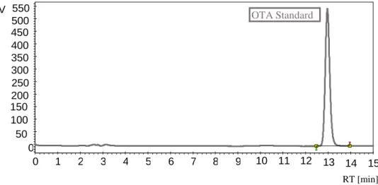
The presence of OTA in samples was identified by comparing the retention time of the peak from the standard and peaks that might be formed in each sample. The limit of detection (LOD) and the limit of quantification (LOQ) were, 7.6 ng/ml and 23.2 ng/ml, respectively.
2.5. Ochratoxin A (OTA) biosynthetic genes
Aiming to explore the bioactive potential, i.e. ability to produce OTA, the molecular screening of OTA related genes was carried out in all the studied isolates.
The primers designed by Geisen et al. (2006) were used to test this ability in Penicillium strains: otapks_for (5´-TACGGCCATCTTGAG CAACGGCACTGCC-3´); otapks_rev (5´-ATGCCTTTCTGGGTCCGATA-3´); otanps_for (5´-AGTCTTCGCTGGGTGCTTCC-3`); otanps_rev (5´-CAGCACTTTTCCCTCCATCTATCC-3`); otatra_for (5´-GGTCGGGCCGATGTTTGATCG-3`); otatra_rev (5´-CCTCGCATCTTGTAAGG AACGC-3`). Penicillium nordicum (MUM 16.08) and Fusarium oxysporum (MUM 18.58) were used as a positive and negative control, respectively.
For the Aspergillus samples, only one set of primers was used: Acpks_for (5´-GCAGCGGGAGTCAATGTAAT-3´); Acpks_rev2 (5´-GCGTCGTACAAAGC CTCTT-3´) (Rossi et al., 2010). Aspergillus carbonarius (MUM 01.08) and Fusarium oxysporum (MUM 18.58) were used as positive and negative control, respectively. In a second phase, Penicillium primers were tested in Aspergillus strains and vice-versa.
PCR reactions for Penicillium primers were performed in a 12.5 µL mixture of 6.25 µL of NZYTaq 2× Green Master Mix (NZYTech, Lisbon, Portugal), 0.20 µM of each primer, 12 ng of DNA and ultra-pure water until the final volume. The PCR program was performed in C1000TM Thermo Cycler
(Bio-Rad, Hercules, California, USA) and consisted in an initial denaturing step of 3 min at 95 ºC; 33 cycles of 30 s at 95 ºC, 40 s at 60 ºC and 60 s at 72 ºC; and a final extension of 10 min at 72 ºC.
PCR reactions for Aspergillus primers were also performed in a 12.5 µL mixture of 6.25 µL of NZYTaq 2× Green Master Mix (NZYTech, Lisbon, Portugal), 0.20 µM of each primer, 12 ng of DNA and ultra-pure water until the final volume. The PCR program was performed in C1000TM Thermo
Cycler (Bio-Rad, Hercules, California, USA) and consisted in an initial denaturing step of 2 min at 94 ºC; 30 cycles of 45 s at 94 ºC, 60 s at 67.1ºC and 60 s at 72 ºC; and a final extension of 7 min at 72 ºC.
PCR products were visualized in ChemiDoc after electrophoresis of 30 min at 80 V, in a 1% agarose gel in 0.5 × Tris acetate EDTA (TAE) buffer. SYBR® Safe DNA Gel Stain (Invitrogen, Waltham, Massachusetts, USA) was used as a staining element and NZYDNA ladder III (NZYTech, Lisbon, Portugal) was used as a DNA molecular weight marker.
10
2.6. RAPD-PCR fingerprinting
To differentiate all the strains from the same species, RAPD-PCR technique was used. The primers M13, 5´-GAGGGTGGCGGTTCT-3´ (Vassart et al., 1987), and (GACA)4 (Ali et al., 1986) were used.
Using Qubit® 2.0 Fluorometer and Qubit dsDNA Broad Range Assay Kit (Invitrogen, Waltham,
Massachusetts, USA), DNA quantification was made, according to manufacturer’s instructions. Qubit device allows to distinguish DNA from RNA which means that the measurements are more precise and accurate. DNA dilutions were conducted so that all samples had the same amount of DNA for the RAPD-PCR technique.
For M13 primer reactions were carried out in a 25 µL mixture of 12.5 µL of NZYTaq 2× Green Master Mix (NZYTech, Lisbon, Portugal), 0.80 µM of primer, 50 ng of DNA and ultra-pure water until the final volume. The PCR program was performed in C1000TM Thermo Cycler (Bio-Rad, Hercules,
California, USA) and consisted in an initial denaturing step of 2 min at 94 ºC; 40 cycles of 20 s at 94 ºC, 60 s at 50 ºC and 20 s at 72 ºC; and a final extension of 6 min at 72 ºC.
For (GACA)4 primer reactions were carried out in a 25 µL mixture of 12.5 µL of NZYTaq 2× Green
Master Mix (NZYTech, Lisbon, Portugal), 0.80 µM of primer, 50 ng of DNA and ultra-pure water until the final volume. The PCR program was performed in C1000TM Thermo Cycler (Bio-Rad, Hercules,
California, USA) and consisted in an initial denaturing step of 2 min at 94 ºC; 40 cycles of 20 s at 94 ºC, 60 s at 48 ºC and 20 s at 72 ºC; and a final extension of 6 min at 72 ºC.
PCR products were visualized in ChemiDoc after electrophoresis of 100 min at 60 V, in a 1.5% agarose gel in 0.5 × Tris acetate EDTA (TAE) buffer. SYBR® Safe DNA Gel Stain (Invitrogen,
Waltham, Massachusetts, USA) was used as a staining element and NZYDNA ladder III (NZYTech, Lisbon, Portugal) was used as a DNA molecular weight marker. Images were saved as TIFF files and processed in BioNumerics software (version 7.6, created by Applied Maths NV. Available from http://www.applied-maths.com). Calculation of similarity between fingerprinting profiles was based on Dice band matching coefficient with 1% band position tolerance. The dendrograms for each primer were deduced from the matrix of similarities by the unweighted pair group method using arithmetic average (UPGMA) algorithm. The co-phenetic correlation was calculated. A dendrogram merging both analyses, i.e. of each primer, was created. Cluster analysis was done using the average of all experiments method.
11
3. Results
3.1. Phylogenetic Analysis
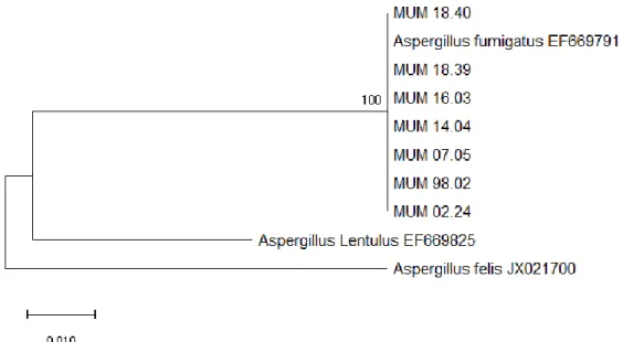
For phylogenetic analysis, the β-tubulin gene of 7 Aspergillus fumigatus and 44 Penicillium crustosum strains were amplified. A search using BLAST (Basic Local Alignment Search Tool) performed on the BenA sequences confirmed the respective identification of each strain, either for A. fumigatus or P. crustosum. Dendrograms were constructed, using the type strains A. fumigatus IMI 16152 (EF669791) and P. crustosum IMI 91917 (AY674353) as references for each. The dendrograms show that all A. fumigatus strains aligned perfectly with the type strain without any nucleotide differences (Fig. 3.1). The bootstrap value of 100% indicates a perfect clustering. This was also verified for the P. crustosum strains (Fig 3.2), also with a high bootstrap value of 99%, and again no nucleotide differences were observed.
Figure 3.1 - Evolutionary relationships of A. fumigatus strains (BenA). The evolutionary history was inferred by using the Maximum Likelihood method and Kimura 2-parameter model (Kimura, 1980). The tree with the highest log likelihood (-763.88) is shown. The tree is drawn to scale, with branch lengths measured in the number of substitutions per site. This analysis involved 10 nucleotide sequences. All positions containing gaps and missing data were eliminated. There were a total of 378 positions in the final dataset. Evolutionary analyses were conducted in MEGA X (Kumar et al., 2018).
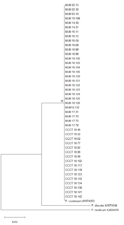
12 Figure 3.2 - Evolutionary relationships of P. crustosum strains (BenA). The evolutionary history was inferred by using the Maximum Likelihood method and Kimura 2-parameter model (Kimura, 1980). The tree with the highest log likelihood (-555.71) is shown. The tree is drawn to scale, with branch lengths measured in the number of substitutions per site. This analysis involved 47 nucleotide sequences. All positions containing gaps and missing data were eliminated. There was a total of 298 positions in the final dataset. Evolutionary analyses were conducted in MEGA X (Kumar et al., 2018).
13
3.2. Ochratoxin-A (OTA) quantitative analysis
To detect the presence of OTA 44 P. crustosum strains and 7 A. fumigatus strains were analysed by HPLC with a fluorescent detector. Alongside a standard sample was included to obtain the retention time of the toxin and for comparison purposes. The retention time was around 13 minutes (Fig. 3.3). However, under the studied conditions, there were not detected OTA producers above the HPLC-FLD detection limit, neither for the Penicillium or the Aspergillus strains.
3.3. OTA biosynthetic genes
The molecular screening of OTA related genes was conducted. This search was a preliminary attempt to find an OTA cassette that each species harbour. Forty-four strains of P. crustosum and seven of A. fumigatus were screened against 3 known OTA genes of P. nordicum (otapks, otanps and otatra) and 1 from A. carbonarius (Acpks). P. nordicum (MUM 16.08) and A. carbonarius (MUM 01.08) were used as positive controls and F. oxysporum (MUM 18.58) as the negative control.
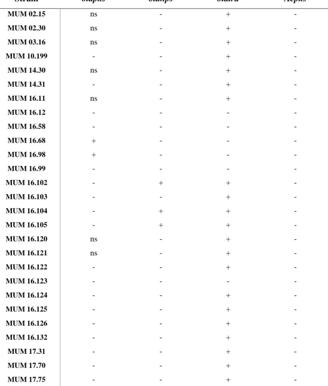
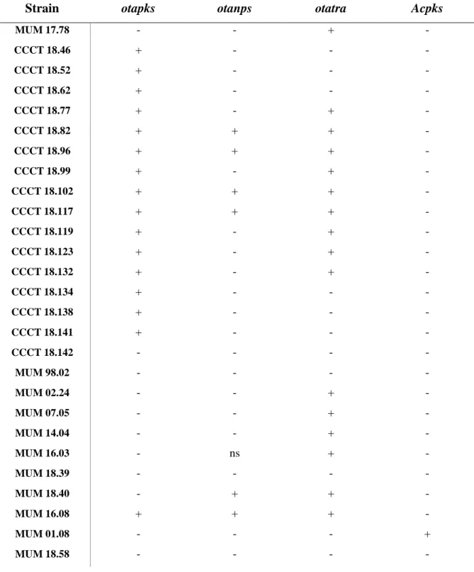
None of the strains was positive for all genes. Regarding Acpks only on the positive control, A. carbonarius (MUM 01.08), was possible to see amplification (Table 3.1).
Considering the other 3 genes studied the results were more complex (Table 3.1). Of the 51 strains tested: around 14% of the strains did not amplify any gene; 16% amplified otapks (≈ 500 bp); 10% otapks and otatra (≈ 420 bp); 8% otanps (≈ 700bp) and otatra; 45% only otatra; and 8% amplified the three genes (see Attachment II, Fig. 7.1, for examples).
The Portuguese strains (MUM 02.15 - MUM 14.31) and the Tunisian strains (MUM 17.31 – MUM 17.78) of P. crustosum were consistent only amplifying otatra. Some of the strains, from Portugal, had nonspecific amplification in otapks however for the study they were considered negative.
Almost all the Italian strains (MUM 16.11 – MUM 16.132) amplified otatra, except for MUM 16.12, 16.58, 16.68, 16.98 and 16.99. On the other side, two of these strains, MUM 16.68 and MUM 16.98, were positive for otapks. Three other strains of Italy, MUM 16.102, 16.104 and 16.105, also have dissimilar results, amplifying otanps besides otatra. In this group 4 strains, MUM 16.12, 16.58, 16.99 and 16.123, were negative for all the genes studied.
Regarding the Chilean strains (CCCT 18.46 - CCCT 18.142) all of them amplified otapks, except for CCCT 18.142, that did not amplify any of the genes. Besides these, this gene was only amplified by the positive control (MUM 16.08) and the two Italian strains aforementioned. The majority of the strains
15 14 13 12 11 10 9 8 7 6 5 4 3 2 1 0 550 500 450 400 350 300 250 200 150 100 50 0 RT [min] OTA Standard V
14 amplified otatra, still 7 did not, which is a large percentage compared to other groups, i.e. Portuguese, Italian and Tunisian strains. Four strains amplifying all the genes, CCCT 18.82, 18.96, 18.102 and 18.117, as the positive control.
Five of the A. fumigatus strains amplified otatra except for MUM 98.02 and 18.39. Only another strain, MUM 18.40, amplified a different gene: otanps. It was registered a nonspecific amplification in MUM 16.03 of otanps, but, once more, not considered pertinent.
Table 3.1 - Results of the PCR amplification of OTA related genes (otapks, otanps, otatra and Acpks). Table 3.1 – Cont.
Strain
otapks
otanps
otatra
Acpks
MUM 02.15 ns - + - MUM 02.30 ns - + - MUM 03.16 ns - + - MUM 10.199 - - + - MUM 14.30 ns - + - MUM 14.31 - - + - MUM 16.11 ns - + - MUM 16.12 - - - - MUM 16.58 - - - - MUM 16.68 + - - - MUM 16.98 + - - - MUM 16.99 - - - - MUM 16.102 - + + - MUM 16.103 - - + - MUM 16.104 - + + - MUM 16.105 - + + - MUM 16.120 ns - + - MUM 16.121 ns - + - MUM 16.122 - - + - MUM 16.123 - - - - MUM 16.124 - - + - MUM 16.125 - - + - MUM 16.126 - - + - MUM 16.132 - - + - MUM 17.31 - - + - MUM 17.70 - - + - MUM 17.75 - - + -
15 Table 3.1 – Cont.
Strain
otapks
otanps
otatra
Acpks
MUM 17.78 - - + - CCCT 18.46 + - - - CCCT 18.52 + - - - CCCT 18.62 + - - - CCCT 18.77 + - + - CCCT 18.82 + + + - CCCT 18.96 + + + - CCCT 18.99 + - + - CCCT 18.102 + + + - CCCT 18.117 + + + - CCCT 18.119 + - + - CCCT 18.123 + - + - CCCT 18.132 + - + - CCCT 18.134 + - - - CCCT 18.138 + - - - CCCT 18.141 + - - - CCCT 18.142 - - - - MUM 98.02 - - - - MUM 02.24 - - + - MUM 07.05 - - + - MUM 14.04 - - + - MUM 16.03 - ns + - MUM 18.39 - - - - MUM 18.40 - + + - MUM 16.08 + + + - MUM 01.08 - - - + MUM 18.58 - - - -
[+] positive (amplifies the expected band size, but can also have non specific amplification); [-] negative (does not amplify ; no bands observed); [ns] non specific amplification (only amplifies unspecific bands)
[+] positive (amplifies the expected band size, but can also have non specific amplification); [-] negative (doesn’t amplify anything); [ns] non specific amplification (only amplifies unspecific bands)
16
3.4. RAPD-PCR fingerprinting
Once there were a lot of strains from just two different species, fingerprinting analyses were conducted to discriminate them. All the strains amplified two primers: M13 and (GACA)4.
Fingerprinting profiles were visualized in agarose gel and analysed using BioNumerics software to construct dendrograms.
3.4.1 Aspergillus fumigatus
Seven A. fumigatus fingerprint profiles of a combined analyse of (GACA)4 and M13 primers were
studied to build a dendrogram (Fig. 3.5). It was not possible to highlight any specific clusters between the different strains. Nor for their matrix of origin (for example air or food) nor their geographic origin (Portugal or Spain). However, differences in fingerprint profiles are noticeable (Fig. 3.4) The strain MUM 98.02 has the most different band pattern in both genes, as MUM 16.03 and MUM 18.39.
17
3.4.2 Penicillium crustosum
Forty-four P. crustosum fingerprint profiles were analysed to build a dendrogram (Fig. 3.8). Genetic differences were found allowing clustering. The Chilean strains (blue) clustered all together as the Tunisian strains (pink). Even though they cluster together, there are some differences in the fingerprint profiles (Figs. 3.6 and 3.7).
The Portuguese strains (green) are scattered between the Italian and the Tunisian strains. They do not show particular similarities between them. All of them are closer together with the Italian strains, apart from MUM 14.30 that is closer with the Tunisian strains.
Finally, the Italian strains (purple) are spread all over the dendrogram. Most of them cluster together with the Portuguese and the Tunisian strains, except for 3 strains (MUM 16.121, 16.122 and 16.124) that cluster at the end of the dendrogram close to the Chilean strains.
Figure 3.5 - Joined dendrogram of M13 and (GACA)4 fingerprinting of A. fumigatus
18 Figure 3.6 - Fingerprint profiles of the Chilean strains (GACA)4.
Figure 3.7 - Fingerprint profiles of the Tunisian strains (M13).
19 100 95 90 85 80 75 70 65 60 55 50 45 40 35 30 25 MUM 02.30 MUM 16.98 MUM 14.31 MUM 16.11 MUM 16.58 MUM 16.68 MUM 10.199 MUM 16.103 MUM 16.104 MUM 16.99 MUM 16.102 MUM 03.16 MUM 16.105 MUM 16.120 MUM 02.15 MUM 16.12 MUM 16.132 MUM 17.31 MUM 17.75 MUM 17.70 MUM 17.78 MUM 14.30 MUM 16.125 MUM 16.126 MUM 16.123 CCCT 18.117 CCCT 18.138 CCCT 18.99 CCCT 18.123 CCCT 18.132 CCCT 18.102 CCCT 18.119 CCCT 18.134 CCCT 18.141 CCCT 18.142 CCCT 18.62 CCCT 18.77 CCCT 18.96 CCCT 18.82 CCCT 18.46 CCCT 18.52 MUM 16.122 MUM 16.124 MUM 16.121
Figure 3.8 - Joined dendrogram of M13 and (GACA)4 fingerprinting of P. crustosum strains, generated in
BioNumerics using the average of all experiments method. Portuguese strains are represented in green, Italian’s in purple, Tunisian’s in pink and Chilean’s in blue
20
4. Discussion
Due to significant economic losses, and negative impact on human and animal health, mycotoxins are a matter of concern to the scientific and political communities. Ochratoxin A has been a highly studied mycotoxin since it can cause various deleterious effects, on humans or animals, when consumed in contaminated products (Paterson and Lima, 2010b). Besides being present in raw materials, OTA is also able to resist some food processes, like making bread, as reported by Milani and Heidari (2016). For this, it is essential to keep control in all phases of food production.
The OTA producing fungi have been established, being Aspergillus or Penicillium species. There are around 27 species of Aspergillus that can produce OTA like: A. steynii, A. ochraceus, A. westerdijkiae from section Circundati, A. carbonarius and A. niger, from section Nigri and A. alliaceus from section Flavi (Perrone and Gallo, 2017). On the other side, there are only three OTA producing Penicillium: P.
nordicum, P. verrucosum and P. thymicola (Nguyen et al., 2016; Perrone and Susca, 2017). Regardless these fungi are the recognize OTA producers by the scientific community additional reports have been published with other species being proposed as having OTA producing ability. P. brevicompactum, P.
crustosum, P. olsonii and P.oxalicum were reported by Vega and collaborators (2006). P. chrysogenum, P. glycyrrhizacola, P. polonicum, isolated from liquorice, were also acknowledged as OTA producers
(Chen et al., 2013). A. fumigatus and A. versicolor are also in the group of potential OTA producers (Abarca et al., 1997).
Recently, Decontardi and colaborators (2017) studied the presence of OTA in Italian cheeses. They found that all the samples of cheese were contaminated with OTA ranging from 1 to 1432 μg/kg. All the cheeses were colonized by fungi with Penicillium being generally dominant, but with the presence of some Aspergillus. However, none of the isolates was identified as any of the major producers of OTA, that is, they were not identified as any Aspergilli OTA producers or any P. verrucosum, P. nordicum or
P. thymicola. These were intriguing results because the presence of OTA was indubitable. The authors
propose that the isolation of the OTA producers Penicillia may have been hampered by a large number of other Penicillium species non-OTA producers. P. crustosum was one of those isolated fungi. Moreover, it’s possible to hypothesise that maybe one of the other isolated species was the source of the OTA contamination. As P. crustosum has already been described in another independent study as a producer, this study focused on this species to try to understand if this fungus could be a threat to food safety as an OTA producer. Concurrently, A. fumigatus also was scrutinized.
A thorough study was conducted on 44 P. crustosum and 7 A. fumigatus strains. Once that all the strains came from culture collections it is expected that the identifications are accurate. For this, no classical methods of identification, to study the morphology of each strain, took place. Withal some species are morphologically indistinguishable, referred to as cryptical species, but are genetically different (Bickford et al., 2007). Therefore, the use of molecular methods is crucial since it does not depend on culture conditions, interpretation from a specialist and could reveal the hidden identity of new species (fixing the names based on DNA barcodes) (Hawksworth, 2015). Once that so many strains of the same species are being studied from different matrices and different parts of the globe, specially P. crustosum strains, there is a possibility that some of them have gone through speciation events.
The authenticity of the identification of each strain was verified by the amplification of the housekeeping BenA gene. This gene was recognized as the best gene, instead of the universal barcode (ITS), to differentiate most of the Penicillium to the species level (Visagie et al., 2014). For Aspergillus, the corresponding gene would be CaM, but BenA is suitable as well (Samson et al., 2014). So, to standardize the present study the same gene was used in both genus. No genetic differences were found