ORIGINAL
ARTICLE
Identification of putative new
Escherichia coli
flagellar
antigens from human origin using serology, PCR-RFlP
and DNA sequencing methods
Authors
Monique Ribeiro Tiba1
Claúdia de Moura2
Marcelo Falsarella Carazzolle3
Domingos da Silva Leite4
1MSc; Dr.; Post-doctorate, Universidade Estadual de Campinas - UNICAMP, São Paulo, Brazil
2MSc; PhD Candidate, UNICAMP, São Paulo, Brazil
3MSc, Dr.; Physicist, UNICAMP, São Paulo, Brazil
4MSc, Dr.; Professor, UNICAMP, São Paulo, Brazil
Submitted on: 08/15/2010 Approved on: 10/21/2010
Correspondence to:
Monique Ribeiro Tiba Rua Visconde de Taunay, 147/41, Vila Itapura, Campinas, SP, Brazil [email protected]
Financial Support:
FAPESP CAPES
We declare no conflict of interest.
ABSTRACT
Escherichia coli has been isolated frequently, showing flagellar antigens that are not recognized by any of the 53 antisera, provided by the most important reference center of E. coli, The International Escherichia and Klebsiella Center (WHO) of the Statens Serum Institute, Copenhagen, Denmark. The objective of this study was to characterize flagellar antigens of E. coli that express non-typeable H antigens. The methods used were serology, PCR-RFLP and DNA sequencing. This characteriza-tion was performed by gene amplificacharacteriza-tion of the fliC (flagellin protein) by polymerase chain reaccharacteriza-tion in all 53 standards E.coli strains for the H antigens and 20 E. coli strains for which the H antigen was untypeable. The amplicons were digested by restriction enzymes, and different restriction en-zyme profiles were observed. Anti-sera were produced in rabbits, for the non-typeable strains, and agglutination tests were carried out. In conclusion,the results showed that although non-typeable and typable H antigens strains had similar flagellar antigens, the two types of strains were distinct in terms of nucleotide sequence, and did not phenotypically react with the standard antiserum, as expected. Thirteen strains had been characterized as likely putative new H antigen using PCR-RFLP techniques, DNA sequencing and/or serology.
Keywords:Escherichia coli; antigens; bacterial; polymerase chain reaction; polymorphism; restric-tion fragment length.
[Braz J Infect Dis 2011;15(2):144-150]©Elsevier Editora Ltda.
INTRODUCTION
Escherichia coli is the predominant faculta-tive member of the normal human intestinal flora. This species also includes different pathovars which are associated with intes-tinal and extraintesintes-tinal diseases in humans
and animals. Some E. coli variants have been
identified as pathogens that encode an array of pathogenic factors harmful for the respec-tive host.1,2 The O polysaccharide and flagellin
are the two major antigens of Gram-negative bacteria, also known respectively as the O and H antigens. Since the early 1940’s, agglutina-tion of these two antigens has served as the
foundation of E. coli serotyping with 187 “O”
and 53 “H” being characterized to date.3
Serology has been used to track strains in epidemiological studies and has allowed
the characterization of pathogenic E. coli
se-rotypes. Two main groups of such frequent serotypes were defined: serotypes from diar-rhoeal disease and serotypes from
extraintes-tinal disease.4 However, several difficulties
have been observed, when the H serotyp-ing of E. coli is applied as routine laboratory standard: (I) the expression of H-antigens can be dependent on various environmental signals and identification of the complete set of serotypes is a time-consuming process and requires the use of 53 specific antisera; and (II) there is a great deal of cross-reactions among E. coli strains.1,2,5
The flagellum (the organelle responsible for motility) consists of repeated subunits of the protein flagellin (fliC). The flagellin proteins are conserved in their terminal domains, where-as, the central domain is variable and carries serotype-specific epitopes.6 Flagellin genes are
suitable for PCR amplification, and variability between the PCR products can subsequently be assessed by restriction analysis (PCR-RFLP) or
DNA sequencing.1,3,7 Molecular biology
serotyping has been the mainstay in the characterization and diagnostic of E. coli, and this technique remains essential for
taxonomic and epidemiological purposes.2,9
The aim of this study was to characterize the H antigens of motile, serologically non-typeable H antigens strains, from various clinical origins (cases of gastroenteritis, bloody diar-rhoea, HUS, urinary tract infection). Rabbit antisera were produced against non-typeable strains. A PCR-restriction fragment length polymorphism (PCR-RFLP) test that de-tects and characterizes fliC was used to build a database of restriction patterns (P-types) and to recognize H-types.1,2 One
non-typeable strain that the H antigen was not recognized by
PCR-RFLP was selected and the fliC gene was sequenced to
compare with those already described in the literature.
MATERIALS AND METHODS
Bacterial strains
The reference strains belonging to various O- and H-antigen groups representing the flagella antigens H1 to
H56 were included in this study,10 and they were obtained
from the E. coli Reference Laboratory, Santiago de
Com-postela, Spain (Table1). Moreover, a total of 20 serologi-cally non-typeable H antigens strains from various clini-cal origins were used in this study (Table 2). The cliniclini-cal
E. coli strains were donated by Dr. Helmut Tschäpe
(Rob-ert Koch Institute, National Reference Centre of
Salmo-nella and other enterics, Wernigerode, Germany) and by
Dr. Jorge Blanco (E. coli Reference Laboratory, Santiago
de Compostela, Spain). All E. coli isolates were stored at
room temperature in nutrient broth (NB) 0.75% agar and preserved in glycerol cultures at -80°C.
Sera, serum absorption, and H-antigen serotyping
To determine the O- and H-antigens, we used antisera
against reference E. coli H-antigens that were obtained
from the E. coli Reference Laboratory, Santiago de
Com-postela, Spain. The application of the E. coli reference
collection and the reference sera produced according to
recommendation of the International Escherichia and
Klebsiella Centre (WHO) was used. Reference E. coli and clinical E. coli strains were serotyped at the Universidade Estadual de Campinas.
Hyperimmune rabbit antisera against non-typeable strains were produced by the Bacterial Antigens
Labo-ratory in Universidade Estadual de Campinas. Using
the clinical E. coli strains and the standard protocol for
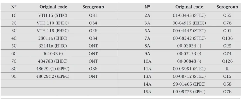
Table 2. E. coli clinical strains carrying serologically non-typeable H-antigens
Nº Original code Serogroup Nº Original code Serogroup
1C VTH 15 (STEC) O81 2A 01-03443 (STEC) O55 2C VTH 110 (EHEC) O84 3A 00-04915 (EHEC) O76 3C VTH 118 (EHEC) O26 5A 00-04447 (STEC) O91 4C 28011a (EHEC) O84 7A 00-08242 (STEC) O136 5C 33141a (EPEC) ONT 8A 00-03034 (-) O25
6C 46103B (-) ONT 9A 00-07153 (-) O74
7C 40478B (EHEC) ONT 10A 00-00848 (-) O126 8C 48629c(1) (EPEC) O86 11A 00-05951 (STEC) R 9C 48629c(2) (EPEC) ONT 13A 00-08712 (STEC) O15
14A 99-01406 (EPEC) O68
15A 00-09775 (EPEC) O76
ONT, undertermined by typing sera; R, rough strains.
(-), negative to virulence factors: eae (enterocyte attaching and effacing), vt1 (verocytotoxin type 1), vt2 (verocytotoxin type 2), bfp ( bundle forming pilus), eaf (EPEC adherence factor).
Table 1. E. coli H-antigens reference strains
O6:H1 O9:H12 O86:H25 086:H36 O156:H47 O3:H2 O18:H13 O38:H26 O42:H37 O16:H48 O53:H3 O23:H15 O58:H27 O69:H38 O6:H49 O50:H4 O46:H16 O148:H28 O110:H39 O8:H51 O4:H5 O15:H17 O138:H29 O41:H40 O11:H52 O120:H6 O17:H18 O86:H30 O137:H41 O148:H53 O1:H7 O32:H19 O73:H31 O70:H42 O161:H54 O105:H8 O126:H20 O114:H32 O140:H43 O4:H55 O30:H9 O146:H21 O60:H33 O3:H44 O139:H56 O108:H10 O158:H23 O160:H34 O125:H45
deriving rabbit antisera.11 The production and
absorp-tion of antisera and tube H-antigen agglutinaabsorp-tion were carried out as described previously by Ewing (1986).
DNA preparation
A single colony was grown in 3.0 mL of Luria-Bertani medium, overnight at 37°C. Genomic DNA was purified by using the “Wizard Genomic DNA Purification Sys-tem Kit” (Promega/EUA). The purified DNA was sus-pended in 100 µL of water and stored at 4°C.
Primers and PCR amplification
The primers used in this study are listed in Table 3. Each PCR was carried out using a 30 µL reaction
mix-ture containing 2 mM MgCl2, each deoxynucleoside
triphosphate at a concentration of 0.2 mM, each primer at a concentration of 10 pmol and 1.5 U of Taq DNA polymerase (Fermentas). PCR conditions included de-naturation for 60s at 94°C, annealing for 60s at 60°C and extension for 120s at 72°C for 30 cycles, in a Ther-mal Cycler (Gene Amp PCR System 9700/Perkin Elmer Corporation, Norwal CT/USA). The amplified DNA product was visualized by standard submarine gel elec-trophoresis using 10 mL of the final reaction mixture on a 1.5% agarose gel in TAE buffer (1.6 M Tris-ED-TA, 0.025 M acetic acid). Amplified DNA fragments of specific sizes were located by UV fluorescence, after staining with ethidium bromide. The 1-kpb DNA lad-der (Fermentas) was used as a standard for determining molecular size of PCR products.
Metaphor (FMC Bioproducts/USA) for 5h at 4.8 V/cm.1
A 100-bp DNA ladder (Fermentas) was used as external and internal fragment size standard. The restriction frag-ments were stained with ethidium bromide and docu-mented by Image Master VDS (Amersham Pharmacia Biotech/ USA. Gel Compar II (Applied Maths/ Belgium) was used to identify RFLP patterns and to establish a
da-tabase for fliC fingerprinting. Fragments were considered
identical if their sizes did not differ by more than 3.5% (allowed error).
DNA manipulation and sequencing
The fliC gene was first PCR amplified, and the PCR
product was inserted into pGEM T-easy kit (Promega/ USA). Analysis of cloned fragments and transformation in DH5α strain were performed using standard
meth-ods.12 fliC PCR products were purified with the enzyme
ExoSAP-IT, according to the instructions of the
manu-facturer (GE Health Care/USA). Subsequently, 5.0 μL of purified PCR product were mixed with 4.0 μL ET
Termi-natorTM mix (GE Health Care/USA), 1.0 μL sequencing
primers T7 (forward) and M13 (reverse). The thermal program consisted of 30 cycles of 20s at 95°C, 15s at 50°C and 1 min at 60°C. The purification of the sequenc-ing products was obtained by mixsequenc-ing 1 μL of ammonium acetate (7.5M) and 27.5 μL absolute ethanol, followed by incubation in the dark for 30 min, and subsequent centrifugation at 3,700 rpm for 75 min at 4°C. Separa-tion of the DNA fragments was obtained in a Megabace 1,000 system (GE Health Care/USA). Voltage and time of injection were 3kV and 120s. Running was performed at 9kV for 100 min at 44°C.
DNA sequence was assembled and edited by us-ing the programs Phred, Phrap, and Consed. BLAST was used for searching databases, including the Gen-Bank. Sequence alignment and comparison were car-ried out using ClustalW. After analysis, an internal
primer pair was constructed: fliC 1C:
AACTAACG-GTACTAACTCTGACA and fliC1Crev:
CCACTAC-CGTCTCAGCTTT to obtain a complete fliC
se-quence, because the entire gene was large and when the DNA sequencer (Megabace 1000 system) was used approximately just 600 pb were obtained.The DNA sequence has been deposited in GenBank under the accession nº GQ423574.
RESULTS
Serotyping
Determination of the O- and H-antigens was performed according to Ewing, 1986, by agglutination with specific hyperimmune rabbit antisera. All H-antigen reference collection and from various clinical origin strains were
Restriction patterns
The PCR-RFLP protocol developed by Fields et al.,7 and
Machado et al.,1 was carried out. The amplified fliC gene
was cleaved with HhaI restriction endonuclease
(Invit-rogen), when fliC(M) primers were used, and RsaI
re-striction endonuclease (Invitrogen), when fliC(F) prim-ers were used. Fifteen microlitprim-ers of each PCR product was digested with restriction endonuclease, according to the manufacture’s instructions. Restriction fragments were separated by electrophoresis on a 2% agarose gel
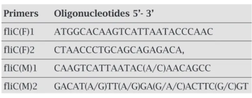
Table 3. Sequence of primers used for PCR amplification
Primers Oligonucleotides 5’- 3’
fliC(F)1 ATGGCACAAGTCATTAATACCCAAC fliC(F)2 CTAACCCTGCAGCAGAGACA, fliC(M)1 CAAGTCATTAATAC(A/C)AACAGCC
serotyped with respect to their H-antigens using the classical agglutination tests.
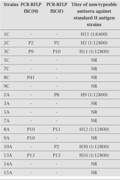
All clinical strains were titrated with all existing 53 antisera initially in 1:100 dilutions and the results of ag-glutination tests were negative, meaning that the clinical strains used in this work, had non-typeable H-antigens. To analyze the flagellar serology of the non-typeable strains, hyperimmune rabbit antisera against the H-antigen were produced. Antibody cross-absorption as-says were carried out, and the H-antigen agglutination tests were performed in tubes. Moreover, the results of serotyping (Table 4) showed that these antisera produced against non-typeable strains shared a specific partial H-antigen factor absent in the reference strains. All
non-typeable E. coli clinical strains were negative to
se-rotyping using reference antisera (53 H-antisera).
fliC-RFLP analysis of E. coli reference strains
To correlate the H-antigen pattern with fliC
polymor-phisms, PCR-amplified fliC fragments were subjected to
RFLP analysis. This analysis was performed three times or more for each strain studied. Patterns were
designat-ed by a letter P, followdesignat-ed by a number (Table 1). All E.
coli reference strains tested gave rise to a PCR product
(varying in size from 0.8 to 2.7 kbp) with the exception
of fliC(F) H17, H25, and H53. The fliC was not
ampli-fied either in the H53 antigen when fliC(M) was used, even under different PCR condition, indicating inad-equate primer homology.
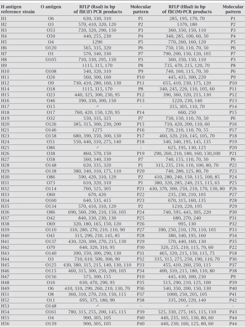
HhaI-fliC gene restriction fragments were observed
in 52 E. coli reference strains. A total of 44 different
patterns were observed after HhaI restriction (Table 5)
and a total of 40 different patterns were observed
af-ter RsaI restriction (Table 5). When RsaI- fliC(F) was
used, a common pattern was observed for the fliC from
the H1, H28, H31 strains (P1), the H2, H30 and H35 strains (P2), the H7, H19 and H27 strains (P7), the H9 and H14 strains (P8), the H11 and H47 strains (P10).
When HhaI-fliC(M) was used, the H3 and H8 strains
(P3), the H6, H10, H19 and H27 strains (P6), the H11 and H47 strains (P9), the H23 and H43 strains (P18), the H28 and H42 strains (P22) had a common pattern.
The fliC genes for H11, H19, H27, H28 and H47
anti-gens were indistinguishable with both restriction en-zymes.
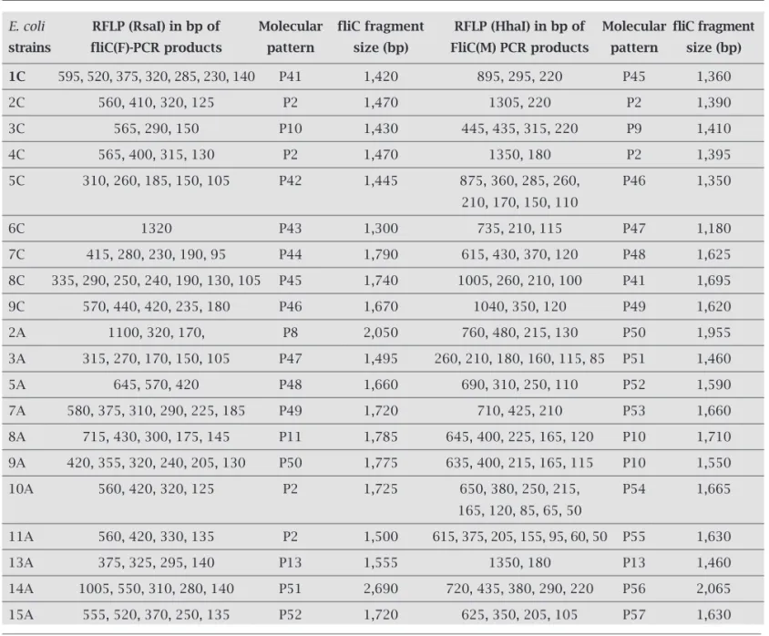
Detection of non-typeable antigen by PCR-RFLP
Since many pathogenic E. coli strains were motile but,
non-typeable by serotyping, the determination of fliC
polymorphism might be a quick altenative for H-antigen typing. The flagellin gene was amplified in all strains studied (Table 6). We detected single bands ranging from 1.1 to 2.6 kbp when fliC(M) was used and sin-gle bands from 1.3 to 2.7 kbp when fliC(F) was used.
When RsaI-flic(F) was used, in eleven non-typeable
strains there were no patterns comparable to those
from E. coli reference strains. Three strains sharing the
P2 pattern, and four strains sharing the P8, P10, P11,
and P13 patterns respectively (Table 5). When HhaI
-fliC(M) was used there were no patterns comparable to those from reference strains in thirteen non-typeable strains. Two strains shared the P2 pattern, two other strains shared the P10 pattern and three strains shar-ing the P9, P13 and P41 patterns respectively (Table 6). Two strains had the same pattern (P2) when both tech-niques were used. This strain was identified as being similar to the H2 antigen. Most of these non-typeable strains revealed unknown RFLP patterns among the H antigens H1 to H56 (Table 6).
Table 4. Results of PCR-RFLP and serotyping of non-typeable E. coli strains
Strains PCR-RFLP PCR-RFLP Titer of non-typeable
fliC(M) fliC(F) antisera against
standard H antigen strains
1C - - H11 (1:6400) 2C P2 P2 H2 (1:12800) 3C P9 P10 H11 (1:12800)
5C - - NR
7C - - NR
8C P41 - NR
9C - - NR
2A - P8 H9 (1:12800)
3A - - NR
5A - - NR
7A - - NR
8A P10 P11 H12 (1:12800)
9A P10 - NR
10A - P2 H30 (1:12800) 13A P13 P13 H16 (1:12800)
14A - - NR
15A - - NR
Table 5. Polymorphisms of fliC(F) and fliC(M) PCR products and their restriction patterns obtained (molecular pattern)
H antigen O antigen RFLP (RsaI) in bp Molecular RFLP (HhaI) in bp Molecular
reference strain of fliC(F) PCR products pattern of fliC(M)-PCR products pattern
H1 O6 630, 330, 310 P1 285, 195, 170, 70 P1
H2 O3 570, 410, 320, 120 P2 1370, 180 P2
H3 O53 720, 320, 290, 150 P3 360, 350, 150, 110 P3 H4 O50 440, 255, 230 P4 340, 285, 100, 60, 50 P4
H5 O4 1290 P5 770, 260, 160, 120 P5
H6 O120 565, 335, 320 P6 750, 150, 110, 70, 50 P6 H7 O1 570, 340, 330 P7 790, 200, 150, 120, 105 P7 H8 O105 710, 330, 295, 150 P3 360, 350, 150, 110 P3 H9 1115, 315, 170 P8 735, 470, 215, 120, 70 P8 H10 O108 540, 320, 310 P9 740, 160, 115, 70, 50 P6 H11 O26 560, 300, 160 P10 445, 435, 300, 220 P9 H12 O9 730, 410, 280, 160, 130 P11 655, 410, 230, 175, 120 P10 H14 O18 1115, 315, 170 P8 340, 245, 220, 110, 105, 60 P11 H15 O23 440, 325, 300, 230, 95 P12 390, 360, 320, 215, 130 P12 H16 O46 390, 330, 300, 150 P13 1220, 230, 140 P13
H17 O15 -a - 355, 305, 110, 70 P14
H18 O17 760, 420, 150, 120, 95 P14 660, 250 P15 H19 O32 550, 335, 325 P7 750, 150, 110, 70, 50 P6 H20 O126 385, 315, 300, 230, 200 P15 710, 420, 200, 110, 60 P16 H21 O146 1275 P16 720, 210, 110, 70, 55 P17 H23 O158 680, 390, 350, 300, 130 P17 460, 320, 210, 145, 105, 70 P18 H24 O51 550, 440, 310, 275, 140 P18 540, 340, 195, 145, 135 P19
H25 O86 -a - 625, 195, 130, 125 P20
H26 O38 860, 570, 150 P19 290, 260, 210, 180, 160, 130,100 P21 H27 O58 560, 340, 330 P7 740, 155, 110, 70, 50 P6 H28 O148 620, 335, 320 P1 315, 235, 210, 110, 100, 80, 70 P22 H29 O138 380, 340, 310, 175, 110 P20 740, 280, 125, 80, 70 P23 H30 O86 590, 420, 310, 120 P2 410, 280, 240, 150, 115, 100, 85 P24 H31 O73 610, 320, 310 P1 380, 320, 285, 240, 215, 115, 65 P25 H32 O114 760, 525, 305 P21 430, 370, 300, 250, 210, 170, 130, 80 P26 H33 O60 670, 420 P22 235, 230, 210, 105 P27 H34 O160 640, 535, 415 P23 670, 315, 160, 135 P28 H35 O134 570, 410, 310, 120 P2 1210, 220, 195 P29 H36 O86 690, 560, 290, 210, 150, 105 P24 740, 595, 445, 305, 220 P30 H37 O42 840, 330, 230, 130 P25 680, 270, 240 P31 H38 O69 320, 180, 165, 150, 120 P26 995, 130 P32 H39 O110 310, 280, 270, 210, 110, 90 P27 390, 250, 210, 170, 110, 105 P33 H40 O41 315, 290, 250, 145, 85 P28 380, 340, 195, 160 P34 H41 O137 430, 320, 300, 270, 215, 130 P29 570, 440, 160, 130 P35 H42 O70 640, 320, 310, 95 P30 320, 235, 210, 115, 70, 60 P22 H43 O140 390, 350, 300, 290, 130 P31 465, 320, 215, 150, 115, 75 P18 H44 O3 710, 610, 500, 300, 90 P32 335, 315, 275, 250, 190, 110, 70 P36 H45 O125 430, 380, 315, 215, 140, 130, 110 P33 455, 410, 260, 250, 115 P37 H46 O115 460, 315, 300, 250, 200, 105 P34 400, 310, 215, 180, 110, 80 P38 H47 O156 575, 300, 155 P10 445, 430, 300, 230 P9 H48 O16 630, 470, 290, 95 P35 515, 290, 210, 125, 100 P39 H49 O6 410, 310, 290, 260, 210, 130, 70 P36 540, 350, 200, 150, 130 P40 H51 O8 360, 310, 270, 210, 150, 115 P37 1000, 250, 205, 105 P41 H52 O11 695, 375, 180, 90 P38 335, 260, 220, 140 P42
H53 O148 -a - -a
-H54 O161 780, 315, 255, 200, 145, 115 P39 525, 330, 275, 165, 115, 110 P43 H55 O4 900, 305, 105 P40 440, 235, 165, 130, 80, 60 P44 H56 O139 900, 305, 105 P40 440, 230, 160, 125, 80, 60 P44
Table 6. fliC gene restriction analysis of non-typeable E.coli strains using RsaI and HhaI
E. coli RFLP (RsaI) in bp of Molecular fliC fragment RFLP (HhaI) in bp of Molecular fliC fragment
strains fliC(F)-PCR products pattern size (bp) FliC(M) PCR products pattern size (bp)
1C 595, 520, 375, 320, 285, 230, 140 P41 1,420 895, 295, 220 P45 1,360 2C 560, 410, 320, 125 P2 1,470 1305, 220 P2 1,390 3C 565, 290, 150 P10 1,430 445, 435, 315, 220 P9 1,410 4C 565, 400, 315, 130 P2 1,470 1350, 180 P2 1,395 5C 310, 260, 185, 150, 105 P42 1,445 875, 360, 285, 260, P46 1,350
210, 170, 150, 110
6C 1320 P43 1,300 735, 210, 115 P47 1,180 7C 415, 280, 230, 190, 95 P44 1,790 615, 430, 370, 120 P48 1,625 8C 335, 290, 250, 240, 190, 130, 105 P45 1,740 1005, 260, 210, 100 P41 1,695 9C 570, 440, 420, 235, 180 P46 1,670 1040, 350, 120 P49 1,620 2A 1100, 320, 170, P8 2,050 760, 480, 215, 130 P50 1,955 3A 315, 270, 170, 150, 105 P47 1,495 260, 210, 180, 160, 115, 85 P51 1,460 5A 645, 570, 420 P48 1,660 690, 310, 250, 110 P52 1,590 7A 580, 375, 310, 290, 225, 185 P49 1,720 710, 425, 210 P53 1,660 8A 715, 430, 300, 175, 145 P11 1,785 645, 400, 225, 165, 120 P10 1,710 9A 420, 355, 320, 240, 205, 130 P50 1,775 635, 400, 215, 165, 115 P10 1,550 10A 560, 420, 320, 125 P2 1,725 650, 380, 250, 215, P54 1,665
165, 120, 85, 65, 50
11A 560, 420, 330, 135 P2 1,500 615, 375, 205, 155, 95, 60, 50 P55 1,630 13A 375, 325, 295, 140 P13 1,555 1350, 180 P13 1,460 14A 1005, 550, 310, 280, 140 P51 2,690 720, 435, 380, 290, 220 P56 2,065 15A 555, 520, 370, 250, 135 P52 1,720 625, 350, 205, 105 P57 1,630
Nucleotide sequence analysis
The full gene sequence was obtained for one strain and T7 and M13 primers based on the pGEMT-easy vector were used. An internal pair of primers based on within sequenced
E. coli fliC gene was also constructed. The non-typeable strain, showed two expected conserved regions in the N-ter-minal and C-terN-ter-minal portions, whereas the central region
was quite variable. The complete nucleotide sequence of fliC
gene has 1,541bp (accession number GQ423574).
DNA alignment was based on the amino acid alignment stored in the database of the National Center for Biotecnol-ogy Information (NCBI). Our sequence for the type strain VTH-15 is 99% identical to those of H21 antigen. Synon-ymous and nonsynonSynon-ymous substitution were observed throug the program BLASTx. The deduced amino acid se-quences of this fliC gene differ in up to one amino acid from those of the H21 type strain.
DISCUSSION
E. coli of specific serotype can be associated with certain clinical syndromes, even though the serological antigens do not correlate with virulence. It has been shown that an-tigenic typing of E. coli is extremely useful in epidemiologi-cal studies.4 Currently, some isolates are generally not very
motile and non-typeable and several difficulties have been
observed, when the H serotyping of E. coli was applied as a
routine laboratory standard.1,2,5
exact identification. An important relationship exists
be-tween E. coli H-antigens H11 and H21.11 We demonstrated
that the antiserum obtained from VTH-15 strain had the fi-nal antiserum dilution of 1:6,400, while nucleotide sequenc-ing demonstrated similarity of 99% to H21 type strain. Re-sults by tests in absorbed antiserum were negative to H11 and H21 antigens. Defining and establishing new H-antigen types will remain a task of the International Escherichia and
Klebsiella Centre (WHO).
Using the fliC PCR-RFLP method several authors
showed that non-motile E. coli strains possess fliC- RFLP
patterns that did not correspond to known H E. coli
anti-gens.7,8 However, non-typeable strains have fliC RFLP
pat-terns that did not correspond to the pattern identified for the H1 to H56 antigens and might therefore represent novel H-antigen types.
In the present study, we have shown that the fliC gene
could be amplified in all non-typeable E. coli strains, and a
considerable polymorphism of the HhaI and RsaI
restric-tions products of the amplified fliC gene could be detected
(Table 6) and used for a flagellar identification system. The diversity of amplification products was examined
with the use of HhaI and RsaI, which demonstrated to be a
feasible and rapid method for identification and subtyping
of H-antigens. For each of the fliC products obtained from
non-typeable strains, a restriction pattern (P-type) was gen-erated. A total of 12 kinds of P-types were determined, when
RsaI (PCR-RFLP RsaI) was used and a total of 13 kinds of
P-types were detected with the use of HhaI.
Nucleotide sequencing of the non-typeable E. coli
(VTH-15) from human clinical isolates is deposited in GenBank as GQ423574. Flagellin genes are identified on the basis of the homology with known flagellin genes. Complete nucleotide
sequencing of fliC gene from non-typeable strain
demon-strated similarity of 99% to those previously published for the H21 type strain. Although most of the H-antigens of
E. coli have been already described at the molecular level3
a few remained to be analyzed, especially the non-typeable strains.
In conclusion, fliC diversity has been showed by using
the PCR-RFLP technique in non-typeable strains. These
pu-tative new H groups in E. coli strains isolated from humans
will be used in the epidemiological and occurrence studies. However, defining and establishing new H antigens type will remain a task of the International Escherichia and Klebsiella
Centre (WHO).
REFERENCES
1. Machado J, Grimont F, Grimont PAD. Identification of
Es-cherichia coli flagellar types by restriction of the amplified fliC
gene. Res Microbiol 2000; 151:535-546.
2. Prager R, Strutz U, Fruth A, Tschäpe H. Subtyping of
patho-genic Escherichia coli strains using flagellar (H) – antigens:
serotyping versus fliC polymorphisms. Int J Med Microbiol
2003; 292:477-486.
3. Wang L, Rothemund D, Curd H, Reeves PR. Species-wide
variation in the Escherichia coli flagellin (H-antigen) gene. J
Bacteriol 2003; 185:2936-2943.
4. Ørskov I, Ørskov F. Escherichia coli serotyping and in man and
animals. Can J Microbiol 1992; 38:699-704.
5. Ratiner YA. Temperature-dependent flagellar antigen phase
variation in Escherichia coli. Res Microbiol 1999; 150:457-463.
6. Schoenhals G, Whitfield C. Comparative analysis of flagellin
sequences from Escherichia coli strains possessing
serologi-cally distinct flagellar filaments with a shared complex surface pattern. J Bacteriol 1993; 175:5395-5402.
7. Fields PI, Blom K, Hughes HJ, Helsel LO, Feng P, Swaminathan B. Molecular characterization of the gene encoding H antigen
in Escherichia coli and development of a PCR-Restriction
frag-ment length polymorphism test for identification of E. coli
O157:H7 and O157:NM. J Clin Microbiol 1997; 35:1066-1070. 8. Amhaz JMK, Andrade A, Bando SY, Tanaka TL, Moreira-Filha
CA, Martinez MB. Molecular typing and phylogenetic analysis
of enteroinvasive Escherichia coli using the fliC gene sequence.
FEMS Microbiol Lett 2004; 235:259-264.
9. Scheutz F, Cheasty T,Woodward D, Smith HR. Designation of O174 and O175 to temporary O groups OX3 and OX7, and six
new E. coli O groups that include Verocytotoxin-producing E.
coli (VTEC): O176, O177, O178, O179, O180 and O181.
AP-MIS 2004; 112:569-584.
10. Ørskov F, Ørskov I, Jann B, Jann K. Serology, chemistry, and
genetics of O and K antigens of Escherichia coli. Bacteriol Rev
1977; 41:667-710.
11. Ewing WH, Edwards, PR. The genus Escherichia.
Identifica-tion of Enterobacteriaceae, Burgess, Minneapolis, 1983. 12. Sambrook J, Fritsch EF, Maniatis T. Molecular cloning: A