Universidade de Aveiro 2017
Departamento de Química
Sofia Maria Soares da
Costa
Impacto cardiovascular da citotoxicidade e do
envelhecimento na agregação de proteínas
Cardiovascular impact of cytotoxicity and aging in
protein aggregation
Universidade de Aveiro 2017
Departamento de Química
Sofia Maria Soares da
Costa
Impacto cardiovascular da citotoxicidade e do
envelhecimento na agregação de proteínas
Cardiovascular impact of cytotoxicity and aging in
protein aggregation
Dissertação apresentada à Universidade de Aveiro para cumprimento dos requisitos necessários à obtenção do grau de Mestre em Bioquímica, realizada sob a orientação científica da Mestre Dulce Fontoura, Mestre em Fisiopatologia Cardiovascular e Especialista pela Universidade de Aveiro, e co-orientação da Professora Doutora Inês Falcão Pires, Professora Auxiliar da Faculdade de Medicina da Universidade do Porto e da Professora Doutora Rita Ferreira, Professora Auxiliar do Departamento de Química da Universidade de Aveiro.
o júri
presidente Prof. Doutor Manuel António Coimbra Rodrigues da Silva
Professor associado com agregação, Universidade de Aveiro
Mestre Dulce Marlene Martins Fontoura
Mestre em Fisiopatologia Cardiovascular, Especialista, Faculdade de Medicina da Universidade do Porto
Doutor José Pedro Quinta Araújo Castro
agradecimentos No final de mais uma etapa académica resta-me agradecer a todos os que de certa forma contribuíram para a realização deste trabalho.
Ao Professor Doutor Adelino Leite Moreira, Diretor do Departamento de Fisiologia e Cirurgia Cardiotorácica do Centro de Investigação Médica da Faculdade de Medicina da Universidade do Porto, agradeço pela oportunidade de realizar a minha tese de mestrado nesta instituição.
À Professora Doutora Inês Falcão Pires agradeço pela oportunidade e pelo privilégio em ter feito parte da sua equipa de investigação. Muito obrigada pelo tempo despendido durante este ano bem como por todas as correções e sugestões para melhorar este trabalho.
À minha orientadora, Dulce Fontoura, Mestre em Fisiopatologia Cardiovascular, agradeço por me ter recebido tão bem e por me ter
acompanhado de forma presente e dedicada no decorrer deste projeto. Muito obrigada por todo o apoio, compreensão, encorajamento e disponibilidade sempre demonstrada durante este ano. Espero ter sido capaz de corresponder a todas as expetativas.
Aos Professores Doutores Rui Vitorino e António Barros agradeço pela colaboração e por todos os ensinamentos que muito contribuíram para o enriquecimento deste trabalho.
À Professora Doutora Rita Ferreira agradeço pelo entusiasmo, pelas sugestões e pela disponibilidade sempre demonstrada para esclarecer qualquer dúvida.
Não podia deixar de agradecer ao Fábio Trindade por toda ajuda e disponibilidade. Sem dúvida que os teus conhecimentos científicos foram imprescindíveis para a realização deste trabalho. É sempre bom trabalhar com pessoas tão profissionais e ao mesmo tempo tão prestáveis.
Agradeço também à Daniela Miranda, Cláudia Mendes e Glória Almeida pela simpatia com que me receberam, por todas as gargalhadas e ensinamentos ao longo deste ano.
À Patrícia Rodrigues, um agradecimento muito especial por todo o apoio desde o início. Obrigada pela tua amizade, pela motivação e por todos os conselhos nas horas mais difíceis. Obrigada por estares sempre disponível para me esclarecer todas as dúvidas e pela tua dedicação a esta tese. Aprendi muito contigo. Gosto muito de ti Tixa.
À Tânia Lima e à Raquel Videira obrigada por serem o meu apoio durante este ano. Obrigada pela amizade sincera, por ouvirem os meus desabafos e acima de tudo pelos bons momentos. Gosto muito de vocês miúdas.
Aos meus amigos do coração obrigada por estarem sempre presentes. Ao João, por estares sempre comigo e pelo apoio incondicional. Por fim, um agradecimento muito especial a toda a minha família. Em particular, à minha irmã por me aturar sempre. Ao meu irmão por relativizar as coisas. Aos meus pais, às pessoas mais importantes da minha vida, obrigada por tornarem tudo isto possível, por terem acreditado sempre em mim e por me terem ensinado a nunca desistir. A vocês dedico este trabalho.
palavras-chave Agregação proteica, sistemas de controlo de qualidade proteica,
envelhecimento, doxorrubicina, cardiotoxicidade, doenças cardiovasculares.
resumo A estrutura nativa das proteínas, decorrente do folding proteico, constitui um pré-requisito para a sua funcionalidade. No entanto, vários fatores promovem o folding incorreto (misfolding) de proteínas, causando a sua agregação.
Recentemente, o misfolding e a agregação de proteínas têm sido associados às doenças cardiovasculares, que representam a principal causa de morte em todo o mundo. De forma a impedir a formação de agregados proteicos
potencialmente tóxicos, os cardiomiócitos desenvolveram sistemas de controlo de qualidade proteica. Contudo, o seu comprometimento potencia a
acumulação de proteínas disfuncionais na forma de agregados. Neste contexto, e tendo em consideração o papel do envelhecimento e da
doxorrubicina (Doxo) no aumento do risco para o desenvolvimento de doenças cardiovasculares, será importante esclarecer a associação entre cada um destes fatores de risco e a agregação proteica. Deste modo, procuramos otimizar uma metodologia de enriquecimento de agregados proteicos provenientes de ventrículo esquerdo (VE) de um modelo animal de envelhecimento e de um modelo animal de cardiotoxicidade induzida pela Doxo, visando identificar as proteínas presentes nestes agregados por GeLC-MS/MS. A técnica de isolamento de frações enriquecidas em agregados proteicos foi otimizada com sucesso em ambos os modelos animais. A análise por GeLC-MS/MS permitiu a identificação de 1279 e 1260 proteínas no VE de ratos WKY novos e velhos, respetivamente. A análise diferencial revelou que 15 e 18 proteínas apresentavam níveis mais elevados nos agregados do grupo novos e velhos, respetivamente. Entre as proteínas presentes em maior quantidade nos ratos envelhecidos destacam-se aquelas relacionadas com a contração cardíaca (miosina-6 e miosina-7), o folding mediado pelas
chaperonas (TRiC) e sistemas proteolíticos (catepsina D). Em relação ao modelo animal de cardiotoxicidade, 274 proteínas foram identificadas no grupo controlo e 267 no grupo Doxo. A análise diferencial revelou que apenas uma proteína, a glicoproteína rica em histidina (fragmento), apresentava níveis mais elevados nos agregados de animais tratados com Doxo. Esta proteína está envolvida na regulação de vários processos biológicos, como a inflamação e a angiogénese, sugerindo um possível papel da mesma na cardiotoxicidade induzida pela Doxo. A identificação destas proteínas bem como o
conhecimento da sua relevância biológica fornece informações valiosas sobre o comprometimento da homeostasia das proteínas, tanto no envelhecimento como em condições de cardiotoxicidade. Desta forma, estudos futuros serão necessários para elucidar sobre o impacto real da agregação destas proteínas no envelhecimento cardíaco e em condições de cardiotoxicidade, bem como potenciais alvos terapêuticos.
keywords Protein aggregation, protein quality control systems, aging, doxorubicin, cardiotoxicity, cardiovascular diseases.
abstract Native structure of proteins, acquired by protein folding, is required for them to function properly. However, several factors promote incorrect protein folding (misfolding), causing their aggregation. Recently, protein misfolding and aggregation have been associated with cardiovascular diseases, the leading cause of death worldwide. In order to avoid the generation of potentially toxic protein aggregates, cardiomyocytes have developed protein quality control systems. However, failure of these systems promotes the accumulation of abnormal protein aggregates. In this context, and taking into account the burden of aging and doxorubicin (Doxo) for cardiovascular diseases
progression, it will be important to clarify the association between these tworisk factors and protein aggregation. Therefore, we aimed to optimize the
methodology for protein aggregates enrichment from left ventricle (LV) of aging and Doxo-induced cardiotoxicity animal models, and also to identify the
proteins presented in these aggregates by GeLC-MS/MS. In both animal models, the technique for isolation of protein aggregates-enriched fractions was successfully optimized. GeLC-MS/MS analysis allowed the identification of 1279 and 1260 proteins in young and aged WKY LV, respectively. Differential protein analysis revealed that 15 and 18 proteins were presented at higher amounts in young and aged groups, respectively. Among proteins with greater amounts in aged rats, we highlighted those related to cardiac contraction (myosin-6 and myosin-7), chaperone-mediated protein folding (TRiC) and proteolytic systems (cathepsin D). Regarding the animal model of
cardiotoxicity, 274 proteins were identified in the control group and 267 in the Doxo group. Differential protein analysis revealed that only one protein, histidine-rich glycoprotein (fragment), was presented in higher amounts in aggregates from Doxo-treated animals. This protein is involved in the regulation of several biological processes, such as inflammation and angiogenesis, suggesting that it can play a role in Doxo-induced cardiotoxicity. The identification of these proteins as well as the knowledge of their biological relevance provides valuable information about protein homeostasis impairment, both in aging and in cardiotoxicity conditions. Therefore, future studies are necessary to elucidate the real impact of the aggregation of these proteins on cardiac aging and cardiotoxicity conditions, as well as potential therapeutic targets.
ABBREVIATIONS: ACN: Acetonitrile
ADP: Adenosine Diphosphate Akt (or PKB): Protein Kinase B AMP: Adenosine Monophosphate. AMPK: AMP-Activated Protein Kinase ATF6: Activating Transcription Factor 6 Atg: Autophagy-Related Genes
ATP: Adenosine Triphosphate Bcl-2: B-cell lymphoma 2 DNA: Deoxyribonucleic Acid Doxo: Doxorubicin
DTT: Dithiothreitol
E1: Ubiquitin-activating enzyme E2: Ubiquitin-conjugating enzyme E3: Ubiquitin ligase
EF: Ejection Fraction
Erk1/2: Extracellular Signal-Regulated Kinase 1/2 FDR: False Discovery Rate
GeLC-MS/MS: SDS-PAGE followed by Liquid Chromatography-Tandem Mass Spectrometry GRAVY: Grand Average of Hydropathy
GRP78: Glucose-Regulated Protein 78 HF: Heart Failure
HRG: Histidine-Rich Glycoprotein HRR: Histidine-Rich Region HSP: Heat Shock Proteins IAA: Iodoacetamide IL-1: Interleukin-1
IRE1: Inositol-Requiring Enzyme-1
LAMP: Lysosome-Associated Membrane Protein LC3: Microtubule-Associated Protein Light Chain 3 LV: Left Ventricle
MAFbx: Muscle Atrophy F-box MAO: Monoamine Oxidase
MAPK: Mitogen-Activated Protein Kinase MS: Mass Spectrometry
mTOR: Mammalian Target of Rapamycin MURF-1: Muscle RING-finger protein-1 MVP: Major Vault Protein
MyBPC: Myosin Binding Protein C
Na+/K+-ATPase: Sodium/Potassium-transporting ATPase NBR1: Neighbor of BRCA1 Gene 1
NEF: Nucleotide-Exchange Factors NIH: National Institutes of Health
P70S6K: Ribosomal Protein S6 Kinase Beta-1 PE: Phosphatidylethanolamine
PERK: Protein Kinase RNA-like Endoplasmic Reticulum Kinase PGC-1α: Peroxisome proliferator-activated receptor-gamma coactivator PI3K: Phosphatidylinositol 3-Kinase
ROS: Reactive Oxygen Species RyR: Ryanodine Receptor Sarcosyl: N-lauroylsarcosinate SDS: Sodium Dodecyl Sulphate
SDS-PAGE: SDS Polyacrylamide Gel Electrophoresis SERCA: Sarcoplasmic Reticulum Ca2+-ATPase sHSP: Small Heat Shock Proteins
SR: Sarcoplasmic Reticulum TCA: Trichloroacetic Acid
TFEB: Transcription Factor EB
TRiC: T-complex protein-1 ring complex ULK1: UNC-51 like kinase 1
UPS: Ubiquitin-Proteasome System WKY: Wistar Kyoto
INDEX
1.
INTRODUCTION ... 2
1.1 Protein misfolding and aggregation ... 4
1.2 Protein quality control systems ... 5
1.2.1 Chaperones ... 5
1.2.2 Ubiquitin-proteasome system ... 7
1.2.3 Autophagy ... 9
1.3 Dysregulation of proteostasis in aging ... 12
1.4 Dysregulation of proteostasis caused by doxorubicin ... 15
1.5 Methodological approaches to study protein aggregates ... 19
2.
AIMS ... 20
3.
MATERIALS AND METHODS ... 21
3.1 Animal model ... 21
3.2 Baseline protocol for protein aggregates extraction and purification ... 21
3.3 In-gel protein digestion ... 22
3.4Protein identification by GeLC-MS/MS ... 22
3.5 Bioinformatic and Statistical analysis ... 23
4.
RESULTS ... 24
4.1 Methodological optimization for the isolation and characterization of the protein aggregates-enriched fractions ... 24
4.2 Optimized protocol for the isolation of protein aggregates-enriched fractions ... 30
4.3 Aging animal model ... 31
4.3.1 Characterization of the protein aggregates-enriched fractions isolated from left ventricle of young and aged rats ... 31
4.3.2 Proteomic analysis after GeLC-MS/MS ... 32
Total protein aggregates-enriched fractions ... 32
Hydropathy analysis ... 33
Biological processes ... 33
Differential protein analysis ... 35
Human diseases associated with aggregated proteins ... 37
4.4 Doxo-induced cardiotoxicity animal model ... 39
4.4.1 Characterization of the protein aggregates-enriched fractions isolated from left ventricle of Control and Doxo-treated rabbits ... 39
4.4.2 Proteomic analysis after GeLC-MS/MS ... 40
Total protein aggregates-enriched fractions ... 40
Hydropathy analysis ... 41
Biological processes ... 41
Differential protein analysis ... 42
Human diseases associated with aggregated proteins ... 43
5. DISCUSSION ... 45
5.1 Methodological optimization for the isolation and characterization of the protein aggregates-enriched fractions ... 45
5.2 Proteomic analysis of protein aggregates-enriched fractions ... 47
Cardiovascular impact of aging in protein aggregation ... 47
Cardiovascular impact of Doxo-induced cardiotoxicity in protein aggregation ... 52
6. CONCLUSIONS AND FUTURE PERSPECTIVES ... 56
1. INTRODUCTION
The process by which proteins acquire their native structure, named as folding, is crucial for proteins to perform their functions in cell and, consequently, essential to all biological processes. On the other hand, unfolding or incorrect folding (misfolding) of proteins and consequent loss of function may have catastrophic effects for cells functioning and trigger protein aggregation, which is per se toxic for the cell (1, 2). At the protein native structure, hydrophobic residues are mainly found in their core. However, when protein misfolds, a structural change exposes the hydrophobic residues, allowing them to interact in a non-specific way with other proteins causing abnormal protein aggregation (1). Moreover, it is known that excessive accumulation of misfolded proteins is related to the manifestation or progression of several cardiovascular diseases (2, 3). Therefore, the existence of protein quality control systems that ensure the correct synthesis and folding of proteins as well as the identification and repair or degradation of proteins with abnormal conformation are crucial for cells survival, namely cardiomyocytes (2). The systems responsible for protein quality control, which include chaperones and proteolytic systems, are essential for proteostasis, which means, the maintenance of proteome homeostasis.
Chaperones are specialized proteins that interact, stabilize or assist other proteins to acquire their functionally active conformation, being absent in the final structure. Beyond participating in proteins folding, these molecules recognize and repair misfolded proteins, by binding to hydrophobic regions, thereby preventing aggregation. Additionally, chaperones are involved in protein transport, assembly of oligomeric proteins and proteolytic degradation (4). However, if chaperone activity becomes compromised, other systems will be initiated in order to restore proteostasis (2). Thus, sarcoplasmic reticulum (SR)-associated protein degradation, proteasomes, calpains system and lysosomal and mitochondrial proteolytic enzymes will work together in recycling or removing protein aggregates at a cellular level (5). Between proteolytic systems, ubiquitin-proteasome system (UPS) and autophagy are the two main pathways for protein degradation.
UPS represent the second line of defense, being responsible for the degradation of misfolded, mutated or any other damaged soluble proteins. Moreover, UPS is responsible for the degradation of normal proteins that are no longer needed by the cells, contributing to the temporal regulation of protein activity. This process involves two main steps: protein ubiquitination and the subsequent degradation mediated by proteasomes. Ubiquitination consists in a post-translational modification in which the binding of a polyubiquitin chain to the target protein occurs through a cascade of enzymatic reactions, promoting its subsequent degradation by the proteasome (6).
Lastly, autophagy allows degradation of dysfunctional organelles, misfolded proteins and protein aggregates (7). Autophagy, more specifically macroautophagy, involves the formation of a double-membrane vesicle around the substrates and its subsequent degradation through lysosomes (8, 9). Thus, chaperones and proteolytic systems ensure an optimal proteostasis and an adequate function of cardiomyocytes, both under physiological and stress conditions.
However, when protein quality control systems are compromised, pathological conditions associated with protein misfolding and aggregation become evident. In fact, abnormal intracellular or extracellular accumulation of protein aggregates has been associated with the pathogenesis of several neurodegenerative diseases, such as Alzheimer’s, Huntington’s and Parkinson’s disease (1). The toxicity of these aggregates has been attributed to diverse factors, such as oxidative stress and mitochondrial dysfunction (10), proteasome inhibition (11) and autophagy impairment (12). Similarly, recent evidence suggests the presence of protein aggregates in cardiovascular diseases (3, 7), the leading cause of death in Europe, representing an important public health problem nowadays. It is estimated that more than 4 million of people die each year due to cardiovascular diseases.There are several risk factors associated with the development of cardiovascular diseases, including hypertension, obesity, aging, antineoplastic agents, among others (13, 14).
Indeed, aging associates to the rising of cardiovascular diseases prevalence, aggravated by the ageing of the population, mostly ascribed to the improvement of medical treatments and consequent increase in the average of life expectancy (15). As part of aging process several changes in cardiac structure and function take place, such is the case of: 1) increased formation of reactive oxygen species (ROS) and oxidative stress; 2) progressive accumulation of damages in deoxyribonucleic acid (DNA), in ribonucleic acid (RNA), in lipids and in proteins (16, 17); 3) aggregation of proteins (18); 4) impaired metabolism; 5) changes in calcium (Ca2+) homeostasis (17); 6) cardiac fibrosis; 7) mitochondrial dysfunction; modifications in UPS and autophagy, mitophagy and apoptosis processes (16, 19), and 9) decreased number of cardiomyocytes compensated by hypertrophy of the remaining cardiomyocytes (20).
Oncological diseases are the second cause of death worldwide. Nowadays, there are different radioactive and chemical treatments aiming to eliminate tumor cells. Over the last few years, enhancement in antineoplastic therapies has contributed to a significant improvement in the prognosis of oncological patients and also to a reduction in mortality of this population (13). Doxorubicin (Doxo), a member of the anthracycline family, is widely used in the treatment of several types of cancers, including leukemia, lymphoma and breast cancer. Besides Doxo effectiveness in tumor treatment, this chemotherapy drug is known to induce cardiovascular side-effects, such as arrhythmia, cardiomyopathy and heart failure (HF), therefore limiting its clinical use (21-23). These cardiotoxic effects are usually progressive and irreversible, causing a negative
impact both in success of oncological treatment and in the life quality of patients (13, 23). At the molecular level, there are many mechanisms proposed to explain Doxo-induced cardiotoxicity, despite not been fully understood. Among the proposed mechanisms it is important to highlight the changes in mitochondrial function, ROS and oxidative stress increase, dysregulation of Ca2+ handling, myofibrillar degradation, DNA damage as well as UPS and autophagy dysfunction and apoptosis (23-26).
For all these reasons, this thesis aims to address the role of protein quality control systems in cardiac tissue, as well as the consequences of its dysregulation in the formation of protein aggregates. At the same time, it will also provide a general perspective of the impact of aging and Doxo in proteostasis dysregulation and protein aggregation associated to the progression of cardiovascular diseases.
1.1 Protein misfolding and aggregation
It is known that partially folded or misfolded proteins are prone to aggregation. Protein aggregates can adopt distinct structural organizations, such as soluble oligomers, amorphous aggregates, amyloid fibrils, inclusion bodies and aggresomes (4, 27, 28). With regard to the soluble oligomers, small aggregates composed by approximately 3-50 monomers (29), they represent intermediate species that can give rise to amorphous aggregates or amyloid fibrils (27). The way how aggregation goes depends on several factors such as temperature, pH (28) and peptide concentration (30).
Amyloid fibrils consist in very well organized β-sheet conformation structures. These insoluble aggregates are rigid, unbranched and deposited in the extracellular or intracellular medium. Up to now, several amyloidogenic proteins were identified, being the main feature of these proteins their structural instability, which can be induced by mutations, post-translational modifications or specific medium conditions (such as pH and temperature) (31). Furthermore, formation of these structures occurs in several organs, namely in the heart, which may lead to the development of many cardiac diseases, such as HF (32).
Additionally, the formation of larger protein aggregates, termed inclusion bodies, can occur. These structures resulted from coalescence of individual aggregates into a single one, and can contain amorphous aggregates and amyloid fibrils. Inclusion bodies can be transported along microtubules to a perinuclear region, where they form a structure denominated aggresome (27). Aggresome formation is thought to represent a cellular protective mechanism, because it allows the sequestration of misfolded proteins and aggregates in a single site, helping their subsequent removal by autophagy (29). However, it remains unclear which are the toxic species to the cells. In
fact, some studies demonstrate that soluble and smaller intermediate species, such as pre-amyloid oligomers, represent the major toxic threat for cells in detriment of larger aggregates, being the latter poorly correlated with the severity of clinical manifestation, for example the Alzheimer’s disease (27, 33).
There are many internal and external conditions that contribute to protein aggregation, namely mutations, environmental stress factors such as temperature, aging and oxidative stress (34). The role of post-translational modifications, which can occur as a consequence of the above-mentioned conditions, onprotein aggregates formation has been increasingly recognized (5). For instance, when amino acids like proline, arginine, threonine or lysine are exposed to high levels of ROS, they became more susceptible to carbonylation. In turn, protein carbonylation is associated with loss of enzymatic activity, loss of ligand binding properties, increased susceptibility to proteolytic activity, aggregation and modification in transcriptional activities (35-37). Post-translational modifications are not only involved in the formation of protein aggregates but also in dysregulation of proteolytic system components. In fact, excessive levels of ROS, like hydrogen peroxide (H2O2) and lipid peroxides, can inhibit or change many proteasome subunits, thereby
compromising protein degradation and promoting aggregation of misfolded protein (5, 38, 39).
1.2 Protein quality control systems 1.2.1 Chaperones
Heart is constantly under stress, even under physiological conditions, thereby requiring a continuous recycling and maintenance of proteins in order to work properly (2). For this reason, cardiomyocytes have developed quality control systems to establish a perfect balance between protein synthesis and degradation. In first place, chaperones play a crucial role in protein homeostasis (40), being present in cytosol, nucleus, mitochondria and SR of cardiomyocyte (41). Besides their participation in protein folding, chaperones regulate protein synthesis, transport, aggregation, disaggregation and degradation (4). They are also involved in intracellular signaling, controlling the conformational changes necessary to activation or deactivation of signaling mediators and their binding to signalosomes (42), a complex intracellular network composed of several components of different signaling pathways (43).
Chaperones are proteins constitutively expressed in cardiomyocytes. Nevertheless, their expression increases under stress conditions in order to maintain protein homeostasis and prevent protein aggregates formation. Insoluble aggregates of non-sarcomeric or sarcomeric misfolded proteins are toxic to the cardiac cell, and may even trigger apoptosis. Increased chaperones
expression has as main goal to avoid accumulation of these misfolded proteins, by promoting their refolding or directing them to be degraded by UPS or autophagy (40).
The chaperone machinery is composed by numerous proteins that cooperate with each other, and are typically known as stress proteins or heat shock proteins (HSP), once its expression is increased under stress conditions. In cardiomyocytes, they are usually divided into three main classes: the general chaperones (HSP70 and HSP90), the small heat shock proteins (sHSP) and the SR chaperones (40). Proteins that belong to HSP70 and HSP90 class, which have an ATPase domain, recognize and bind to exposed hydrophobic regions of the non-native conformation proteins, promoting their folding or refolding through several cycles of ATP binding and hydrolysis (4). HSP70 and HSP90 interact with other proteins, the co-chaperones, which support chaperones to perform their functions, including protein folding (44). For example, the HSP70 reaction cycle is controlled not only by co-chaperone HSP40 but also by nucleotide-exchange factors (NEF). The hydrolysis of ATP into ADP is strongly accelerated by HSP40, thereby triggering the binding between substrate and HSP70. Moreover, HSP40 is capable of directly binding to unfolded polypeptides and recruit HSP70. After ATP hydrolysis, the binding of NEF to HSP70 ATPase domain triggers the catalysis of ADP-ATP exchange, resulting in substrate release. Accordingly, the substrate release allows fast-folding molecules to hide hydrophobic residues, avoiding their aggregation (Figure 1). Proteins unable to acquire their native structure after HSP70 cycle, such as actin and tubulin, can be redirected to chaperonins (HSP60 class). Chaperonins exhibit a structure capable of attaching a single unfolded protein at a time, allowing more stable conditions for a proper protein folding (4, 45). Regarding HSP90, this chaperone has been associated with several important signaling pathways in eukaryotic cells, including cell cycle, telomere maintenance, apoptosis, mitotic signal transduction, vesicle-mediated transport, innate immunity and target protein degradation (4). The evolution and maintenance of these functional networks is thought to depend on the ability of HSP90 to attenuate the effects of structurally destabilizing mutations in the protein complexes, thereby allowing the appearance of new potentialities in protein. Essentially, HSP90 acts in structural maturation and conformational regulation of various signal-transduction molecules, such as kinases and steroid receptors (46), using, like other chaperones, other regulators and co-chaperones to perform these functions (4).
The sHSP constitute a class of chaperones which have smaller molecular weights and function in an ATP-independent manner. sHSP binds to unfolded/misfolded proteins promoting their folding/refolding in an independent manner or transferring damaged protein to ATP-dependent chaperones, enabling protein refolding or degradation (47). Mutations in sHSP genes can result in protein aggregates formation, namely aggresomes, as observed in desmin-related cardiomyopathy which is triggered by a mutation in sHSP α-β-crystallin (3).
SR is the main organelle where protein folding occurs, being associated with the presence of various chaperones, specifically glucose-regulated protein 78 (GRP78), that belongs to HSP70 family. In fact, dysfunctional SR results in accumulation of misfolded or unfolded proteins. SR chaperones mainly interact with three protein complexes, the activating transcription factor 6 (ATF6), the inositol-requiring enzyme-1 (IRE1) and the protein kinase RNA-like ER kinase (PERK), present in membrane of this organelle. Under normal conditions, GRP78 chaperone associates with these complexes, inhibiting their functions. However, under stress conditions, GRP78 binds to misfolded proteins and dissociates from these complexes, activating them. These protein complexes function as transmembrane sensors that are capable of detecting the accumulation of misfolded or unfolded proteins, and consequently promote an increase expression of chaperones present in this organelle, inhibiting protein synthesis and activating misfolded proteins degradation, in order to restore proteostasis (40, 48).
Figure 1: The HSP70 chaperone cycle. General representation of the HSP70 reaction cycle, ATP-dependent, controlled by the HSP40 co-chaperone and nucleotide-exchange factors (NEF).
Adapted from (4).
1.2.2 Ubiquitin-proteasome system
Proteasomal proteolytic pathway is responsible for 90% of intracellular proteins degradation in eukaryotic cells. Therefore, it ensures that not required, misfolded, oxidized and other type of damaged proteins do not accumulate in cells, avoiding their toxic effects. This system exists mainly in two forms, the ATP-ubiquitin independent (20S) and ATP-ubiquitin dependent (26S). The first form is mostly responsible for the degradation of oxidized proteins as resulting from oxidative stress, being this system ubiquitin independent. On the other hand, ATP-ubiquitin dependent involves protein ATP-ubiquitination and its subsequent degradation by 26S
proteasome (49, 50) (Figure 2). Ubiquitination refers to the addition of ubiquitin molecules to a protein substrate through a cascade of enzymatic reactions. This process involves three steps: 1) the ubiquitin-activating enzyme (E1) covalently binds to ubiquitin in an ATP-dependent manner, thereby resulting in its activation; 2) the ubiquitin-conjugating enzyme (E2) transfers ubiquitin molecule from the E1 to itself; 3) the ubiquitin ligase (E3) recognizes the specific protein substrate and transfers ubiquitin molecule from the E2 to a lysine residue present in substrate (Figure 2). The fate of ubiquitinated proteins depends on the length of the ubiquitin chain formed and on the specific sites of lysine residues in ubiquitin used for linkage. When substrates are tagged with four or more ubiquitin molecules (poly-ubiquitination), being attached to each other by their lysine 48, they become targeted for degradation by the 26S proteasome (2, 51).
The 26S proteasome is a multicatalytic complex composed by two 19S regulatory subunits and a 20S proteolytic core. The 19S regulatory subunits, positioned at both ends of the 20S proteolytic core, have as main components chaperones, ATPases and enzymes that remove ubiquitin molecules from substrates (deubiquitinases). Hence, these subunits recognize poly-ubiquitin chains formed in substrates, remove poly-ubiquitin molecules from proteins, promote protein unfolding and its entry into the 20S proteolytic core, in an ATP-dependent manner (41). The 20S core has a cylindrical structure, being constituted by two β-rings and two external α-rings, containing each ring seven unique protein subunits. Three major types of peptidase activities have been identified in 20S proteolytic core: chymotrypsin-like, trypsin-like and caspase-like activities (also designated as peptidylglutamyl-peptide hydrolase), attributed to β5, β2 and β1 subunits, respectively. α-rings avoid the entry of non-specific substrates into the 20S proteolytic core, regulates the exit of peptides products from this core and can directly regulate the assembly and peptidase activities of β-rings (2) (Figure 2). Since UPS is responsible for the majority of protein degradation, loss of its activity may lead to accumulation of dysfunctional proteins and, consequently, cause dysfunction and cellular death.
Figure 2: Ubiquitin-proteasome system (UPS). Proteins degradation by 26S proteasome involves the poly-ubiquitination of substrates through a cascade of enzymatic reactions. E1-Ubiquitin-activating enzyme; E2-Ubiquitin-conjugating enzyme; E3-Ubiquitin ligase.
1.2.3 Autophagy
In eukaryotic cells, autophagy is the second protein degradation pathway, although it is the main cellular pathway responsible for degradation of long-lived proteins and unnecessary or dysfunctional organelles (9). This process is essential for degradation of proteins that deposit in larger aggregates, since they cannot be degraded by proteasome proteolytic cavity. Therefore, autophagy is crucial for cellular homeostasis maintenance, protecting cardiomyocytes function (7, 52). There are three distinct forms of autophagy: macroautophagy, microautophagy and chaperone-mediated autophagy. Macroautophagy is the most prevalent form of autophagy, so it is usually referred as autophagy. Macroautophagy begins with the formation of a double membrane, the phagophore, around cytoplasmic material and organelles. The fusion of the phagophore expanding ends originates a vacuole, called autophagosome. Then, autophagosome external membrane fuses with lysosome, forming the autolysosome. Subsequently, autolysosome content is degraded by lysosomal enzymes and the remaining components are released to the cytoplasm (53). The main difference between macroautophagy and microautophagy is that, in the latter, cytoplasmic content is directly incorporated into lysosomes through lysosomes membrane invaginations. Chaperone-mediated autophagy consists in the recognition of cytosolic proteins by HSP70, which interact with lysosome-associated membrane protein type 2a (LAMP2A) receptor, present on lysosome membrane, allowing protein translocation into the lysosomes where they will be degraded (54, 55).
Under basal conditions, autophagy activation occurs at low levels, but it can rapidly increase in response to various stimuli, such as starvation, thereby promoting cellular survival through the release of energetic substrates by degradation of cellular constituents (8, 9). Autophagy also seems to function in a selective manner in the elimination of dysfunctional organelles, protein aggregates and intracellular pathogens. Selective types of autophagy must ensure efficient recognition and sequestration of the specific substrates within autophagosomes. This process is mediated by proteins like p62 and neighbor of BRCA1 gene 1 (NBR1), which are capable of recognize ubiquitinated proteins in protein aggregates and connect them to the autophagic membranes (7, 56, 57). Nevertheless, excessive and uncontrolled autophagy activation may promote cellular death, through the destruction of proteins and organelles that are essential to the cells (9). Mitophagy is a particular case of selective autophagy responsible for the degradation of dysfunctional mitochondria. Dysfunctional mitochondria produce less ATP, generate increased amounts of ROS and are more prone to trigger apoptosis, thereby contributing to the development of HF (58, 59). In this context, the elimination of these damaged mitochondria, by mitophagy, is crucial for cardiomyocytes to work properly. Mitophagy is a rigorously controlled and very selective process that includes mitochondrial sequestration by autophagosome, which fuses with lysosome for posterior degradation (59). It has been described that PTEN-induced putative
kinase 1 (PINK1) and Parkin proteins play an important role in mitophagy regulation. Under healthy conditions, PINK1 is imported into mitochondria, through a process dependent of mitochondria membrane potential, being rapidly degraded and maintained at low levels. Upon loss of mitochondrial membrane potential, PINK1 accumulates on the mitochondrial surface, leading to the recruitment of E3 ubiquitin ligase Parkin, that ubiquitinates many mitochondrial outer membrane proteins. The ubiquitinated proteins serve as a signal for p62/SQSTM1 protein, which assists to the recruitment of autophagosome to mitochondria, thereby promoting mitophagy (59, 60).
Autophagy-related proteins (Atg), initially identified and characterized in yeast, are essential for the autophagic process since they are needed for the formation of autophagosome. After autophagy induction, these proteins associate following a hierarchical direction, to first mediate the phagophore formation and then its expansion into autophagosome (57). Autophagy is usually determined by microtubule-associated protein light chain 3 (LC3) protein levels. LC3 protein consists in an autophagy marker which is involved in autophagosome formation. Briefly, LC3 protein is converted to its cytosolic form LC3-I, by Atg4 cysteine protease. Then, LC3-I is activated by Atg7, transferred to Atg3 and conjugated with phosphatidylethanolamine (PE), thereby converting in LC3-II, that is recruited to phagophore double membrane, forming the autophagossome. Therefore, the amount of LC3-II is associated with the number of autophagosomes (53, 55). There are many signaling pathways that play an important role in autophagy regulation, namely mammalian target of rapamycin (mTOR), phosphatidylinositol 3-kinase-I/protein kinase B (PI3K-I/Akt), a AMP-activated protein kinase (AMPK) and mitogen-activated protein kinase/extracellular signal-regulated kinase 1/2 (MAPK/Erk1/2) (54, 55), as described in Figure 3.
Figure 3: Autophagy regulation. The kinase mTOR is the main regulator of autophagy. mTOR activation promotes autophagy suppression, whereas mTOR inactivation promotes autophagy induction. mTOR can be positively regulated by PI3K-I/Akt and MAPK/Erk1/2 signaling pathways and negatively by AMPK and p53. mTOR regulates autophagy through several pathways, namely acting in ULK1 complex, in kinase p70S6 and in transcription factor TFEB. On the other hand, AMPK is also capable of regulating autophagy by acting directly in ULK1 complex and in 1. Additionally, Bcl-2, an anti-apoptotic regulator, also regulates autophagy by beclin-1 inhibition. AMPK- AMP activated protein kinase; PI3K-I/Akt- phosphatidylinositol 3-kinase/protein kinase B; MAPK/Erk1/2- mitogen-activated protein kinase/extracellular signal-regulated kinase 1/2; mTOR- mammalian target of rapamycin; ULK1- UNC-51 like kinase 1; TFEB- transcription factor EB; P70S6K- Ribosomal Protein S6 Kinase Beta-1; Bcl-2- B-cell lymphoma 2.
In addition to UPS and autophagy, proteases also integrate the main proteolytic systems present in cardiomyocytes. Calpains are cytoplasmic Ca2+-dependent cysteine proteases which participate in several Ca2+-mediated intracellular processes. Although its physiological function has not yet been fully understood, it is known that calpains are involved in cell migration, apoptosis, autophagy and plasma membrane repair. Nevertheless, m-calpains and µ-calpains, expressed in cardiomyocytes, are responsible for degradation of myofibrillar proteins, particularly troponin, tropomyosin, myosin and titin (61). Suppression of calpain-1 activity, by overexpression of calpastatin (a specific endogenous inhibitor for calpain), resulted in development of dilated cardiomyopathy, which is accompanied by a decrease of ubiquitinated proteins, protein aggregates formation, increase in autophagy and destruction of sarcomeres integrity (62). So, calpain-1 activity is essential in maintenance of cardiac function and regulation of specific sarcomeric proteins (62). Additionally, it is known that dystrophins are degraded by calpain. Degradation of dystrophins impairs the integrity of the sarcolemma, leading to cardiac dysfunction and HF. Therefore, calpain inhibition may be a therapeutic target in the regulation of dystrophin degradation, delaying the progress to HF (61).
1.3 Dysregulation of proteostasis in aging
Aging is characterized by progressive decline of cellular proteostasis, and consequently, by accumulation of misfolded and damaged proteins in different organs and tissues (63). In fact, it has been reported the accumulation of protein aggregates in the heart during its natural aging process. Ayyadevara et al. reported an aging-dependent increase of detergent-insoluble aggregates isolated from mice hearts (18). Since protein aggregation is suppressed by protein quality control systems, it becomes relevant to clarify how these systems change through aging and its implications on protein aggregates formation, and consequently on cardiac aging (Figure 4). Knowledge of the mechanisms that contribute to cardiovascular aging would have important clinical impact in cardiovascular disease prevention, early diagnosis and development of treatments (18).
Figure 4: Overview of molecular changes during aging. Aging promotes among other, damage in DNA, RNA, lipids and proteins, changes in Ca2+ homeostasis, mitochondrial dysfunction, ROS and oxidative stress increase. Aging is also associated with dysregulation in protein quality control systems, namely, chaperones, UPS, autophagy and mitophagy. All these changes may lead to a disruption in protein homeostasis and, consequently, to the increase of protein aggregates formation.
The induction of HSP synthesis, in response to stress factors, is crucial to maintain the proteostasis balance, preventing the formation of protein aggregates potentially toxic to cardiomyocytes (40). Nevertheless, different studies have shown that the induction of several chaperones in cardiac tissue from aged animals was compromised in response to various stress stimuli, such as temperature increase and physical exercise (64, 65). Thus, aging is associated with a decreased ability for chaperone synthesis, under stress conditions. Furthermore, Ayyadevara and colleagues found that HSP70, HSP90α and HSP90β levels were significantly diminished in aggregates from aged hearts, whereas HSP75, a mitochondrial HSP which protects against cardiac
hypertrophy and fibrosis, was absent (18). At the same time, the authors observed an increase of HSP70 in aggregates from young mice, highlighting its protective functions. Regarding sHSP (HSP-β6, HSP-β7 and HSP-β8), they were more abundant in aggregates from aged hearts. Moreover, tropomyosin levels were increased in aggregates of aged hearts, which can indicate that the rise in the sequestration of tropomyosin within aggregates may contribute to a decrease in cardiac contractility during aging (18).
When protein function cannot be restored from misfolded proteins or aggregates, chaperones direct them to the proteolytic systems (Figure 4). The presence of ubiquitinated proteins in aggregates, a feature of different diseases, can be related to inefficient directing of proteins to proteasomes, resistance to degradation or UPS impairment, thereby contributing to the accumulation of these proteins (63). It has been reported that the activities of ubiquitinating enzymes E1, E2 and E3 did not show any consistent change with age, while the accumulation of ubiquitinated and oxidized proteins is evident both in aging and in diseases associated with aging (66).
Dysfunction of UPS, namely proteasome activity in both hypertrophic cardiomyopathy and failing hearts has been reported, suggesting that proteasome functional insufficiency plays a major role in cardiac pathogenesis (39). The decrease of peptidase activity present in proteasome, namely the chymotrypsin-like activity (67), has been reported in aged tissues, specifically in cardiac tissue. Li et al. demonstrated that chymotrypsin-like activity of 20S proteasome was significantly reduced in the cardiac tissue of old rats (25 months) comparative to younger rats (3 months). The decline in 20S proteasome function may be due, in part, to the decrease in the amount of β subunits observed in aged hearts, since these subunits are responsible for the peptidase activities of the proteasome. (68). Furthermore, aging also seems to influence other peptidase activities besides the chymotrypsin-like activity. So, analysis of 20S proteasome purified from rat’s hearts with different ages revealed a significantly decrease in peptidylglutamyl-peptide hydrolase and trypsin-like activities on aging, with a consequent reduction of the ability of the 20S proteasome to degrade protein substrates (69). These results suggest that a decrease in proteasome activity occurs during aging.
When protein aggregation is not controlled by chaperone and proteasome activities, autophagy is promoted (5) (Figure 4). The effect of aging in cardiac autophagy has been reported in some studies, although the results revealed to be inconsistent. Taneike et al. found in homogenates from C57BI/6 mice hearts with 6, 14 or 26 months that LC3-II levels were lower comparing to the LC3-II levels present in mice with 10 weeks, suggesting that autophagy decreases with age (19). On the other hand, Inuzuka et al. observed that LC3-II levels remained constant in both young (3 months) and old (20-24 months) rats, indicating that autophagy does not change during aging (70).
Wohlgemuth et al. studied the impact of aging in expression levels of proteins involved in the formation of autophagosome (beclin-1 and LC3-II) and in the degradation by autolysosomes and lysosomes (procathepsin D and LAMP-1). These authors observed an increase of beclin-1 and procathepsin D expression and a decrease of LAMP-1 expression in hearts from old rats (26 months) comparing with young controls (6 months). In addition, they also verified that LC3-II protein expression was significantly increased in older rats (71). The differences in results between the studies could be due to experimental variables, such as animal model features. Besides, it is important to refer that these studies were based on determination of LC3-II levels, however, the increase in LC3-II does not always represent an induction of autophagy, but it can indicate either an increase in autophagosome formation or a blockade of autophagy pathway. In order to evaluate autophagy induction it is essential the determination of autophagic flux, since it is representative of the whole process, namely from the inclusion of substrate within the autophagosome, its delivery to lysosomes (through fusion of autophagosome with lysosome) and the release of the resulting degradation macromolecules into the cytoplasm (53). Therefore, more studies are needed to characterize autophagic flux changes in cardiomyocytes through aging.
As mentioned before, aging is associated with the increase of dysfunctional mitochondria. Thus, the elimination of damaged mitochondria by mitophagy is important during aging. It has been described that the process of Parkin-mediated mitophagy is impaired in aged hearts (60). In fact, the deletion of Parkin promotes the accumulation of dysfunctional mitochondria in mice hearts with aging (72). Moreover, overexpression of this protein leads to an increase of mitophagy in aged cardiomyocytes, accompanied by a decrease of dysfunctional mitochondria and an improvement in cardiac function associated with aging. Therefore, the activation of mitophagy could be a novel target to attenuate the development of cardiovascular diseases induced by aging (60).
Many studies also shown that changes in lifestyle can efficiently retard aging through improvement of autophagy. For instance, He et al. demonstrated that physical exercise increases heart autophagy (73) and has beneficial effects in prevention of both cardiovascular diseases and age-related diseases (74). Furthermore, caloric restriction is also a potent inducer of autophagy, with cardioprotective effects (71). Although physical exercise and caloric restriction manifest beneficial effects in aging prevention, pharmacological induction of autophagy is often used to prevent aging and several age-related diseases (54). Resveratrol has been investigated as a potential therapeutic strategy, although its beneficial effects in cardiomyocytes seem to be dose dependent. Therefore, it is essential to develop pharmacodynamics studies to better understand their cardioprotective or cardiotoxic effects (75). Rapamycin, an mTOR inhibitor, is also considered promising in aging treatment since it can provide functional benefits in aged hearts (76, 77). Wilkinson et al. have shown that rapamycin-treated rats presented a delay in the development of
many diseases associated to aging, including degenerative changes in myocardium. On the other hand, chronic treatment with this drug caused harmful side-effects that can impair functions in some tissues (77). In conclusion, there is still a long way to go to fully understand the role of protein aggregation in proteostasis deregulation induced by aging.
1.4 Dysregulation of proteostasis caused by doxorubicin
The pathophysiological mechanisms associated with Doxo-induced cardiotoxicity have not been fully elucidated. Nevertheless, previous studies have consistently reported the cumulative-dose related cardiotoxic profile of Doxo, a feature that limits the extent to which it can be used safely. Moreover, its cardiotoxicity is also dependent of the administration schedule, the concomitant use of other cardiotoxic therapies, history of cardiac diseases, age and gender (13, 78). Anthracycline-induced cardiotoxicity presents both acute and chronic effects. Acute cardiotoxicity occurs during or immediately after Doxo treatment, being rare and not dose dependent (79). Doxo-induced cardiotoxicity may become clinically evident later in the course of chemotherapy and within one year after therapy finalization (early-onset chronic cardiotoxicity), or more than one year after its completion (late-onset chronic cardiotoxicity) (13). In the latter, cardiac abnormalities are usually persistent and progressive, eventually triggering cardiac cells death (23, 78). This injury is typically characterized by the development of left ventricular systolic dysfunction, which subsequently progresses towards dilated cardiomyopathy and HF (23, 80).
Over the last few years, it has been demonstrated that Doxo is able to induce changes in chaperones, UPS as well as in autophagy processes (Figure 5). The dysregulation of protein quality control can promote an increase in protein aggregates formation, which may induce toxic effects in cardiomyocytes. As mentioned above, evidences suggest a link between anthracycline-induced cardiotoxicity and changes in chaperone expression (22). For instance, cardiac biopsies from rabbits treated with other anthracycline, daunorubicin (3mg/kg, weekly, 10 weeks), showed an increase in the expression of various chaperones and proteins involved in chaperone-mediated autophagy, as well as an activation of proteolytic machinery (81). Moreover, Doxo-induced cardiotoxicity was also related to SR chaperones dysregulation/inhibition, such as GRP78 chaperone. In fact, Fu et al. showed that Doxo induced an apoptotic response in cardiomyocytes, through the activation of ER mechanotransmembrane sensors, accompanied by the inhibition of GRP78 expression, which in turn potentiates the increase of ER stress (48).
Figure 5: Overview of molecular mechanisms in Doxo-induced cardiotoxicity. Doxo promotes among other, DNA damage, dysregulation of Ca2+ handling, myofibrillar degradation, mitochondrial dysfunction, ROS and oxidative stress increase. This drug is also capable to dysregulate protein quality control systems, namely, chaperones, UPS, autophagy, mitophagy and calpain. All these changes may lead to a disruption in protein homeostasis and, consequently, to the protein aggregates formation.
For this reason, recently, chaperones have been intensively studied as potentially new therapeutic targets for cardiac diseases induced by antineoplastic agents. Based on experiments performed in Doxo-induced cardiotoxicity animal model, in which a decrease of HSP20 expression in cardiac tissue was observed, Fan et al. demonstrated that overexpressing this chaperone triggered an improvement in Doxo-induced deleterious effects, including higher resistance to apoptosis, enhanced cardiac function and increase animal survival after chronic Doxo treatment (82). These authors also showed that the mechanisms underlying these beneficial effects are associated with the preservation of Akt phosphorylation/activity, as well as the decrease in oxidative stress induced by Doxo (82, 83).
The specific effects of Doxo in UPS have been already reported, although some results remain controversial. Briefly, when Doxo is administrated it diffuses through cellular membrane into the cytosol where it interacts with the proteasome. The complex formed is translocated to the nucleus, and there Doxo is released from proteasome which allows its binding to DNA (Figure 5) (84). Protein ubiquitination is generally a requirement for proteins to be degraded by the 26S proteasome, being that the specificity of ubiquitination resides mainly in ubiquitin ligase E3, as already mentioned (50, 51). In H9C2 cardiomyoblasts cell culture and in hearts from tumour-bearing GFP-LC3 mouse model, the treatment with Doxo induced a significant increase of E3
ligases RING-finger protein-1 (MURF-1) and muscle atrophy F-box (MAFbx) expression, which tag by ubiquitination, myofibril proteins in skeletal and cardiac muscle. Moreover, protein ubiquitination was also significantly augmented both in vitro and in vivo models, with proteins tagged with poly-ubiquitin chains displaying the higher range of molecular weights and possibly indicating degradation of proteins involved in cardiac muscle structure, by the 26S proteasome (85). However, Doxo caused a significantly reduction on proteasome chymotrypsin-like activity, indicating that this drug is able to inhibit the proteasome (85). The mechanism by which Doxo alters proteasome activity appears to be regulated by the amount of Doxo molecules binding onto the proteasome (86), namely, proteasome activation at lower concentrations and its inhibition at higher Doxo concentrations (25, 87).
Doxo is also capable of inducing post-translational modifications, such as carbonylation, in some sarcomeric proteins (Figure 5), thereby increasing the probability of these proteins to be degraded by proteasome. Recently, it was demonstrated in hypertensive rats that Doxo-induced oxidative stress induces high levels of carbonylation in sarcomeric proteins, more evidenced in myosin binding protein C (MyBPC). In addition, an increase in MyBPC degradation by proteasome was observed, resulting in a reduction of MyBPC levels in cardiomyocytes, as well as in impairment of the interaction of this protein with actin, thereby affecting cardiac cell contractility (88).
With regard to Doxo effects on cardiac autophagy they are still controversial. For instance, a study demonstrated that Doxo promotes autophagic flux in cardiomyocytes. In fact, treatment with bafylomicin-A, a lysosomal H-ATPase inhibitor that impairs the fusion of autophagosome with lysosome, originated a significant increase in LC3-II levels in cardiomyocytes treated with Doxo compared with their controls. In addition, the increase in autophagy induced by Doxo potentiated cardiomyocytes death, suggesting that autophagy activation contributes to Doxo-induced cardiotoxicity (89). In contrast, several studies appear to contradict these results by reporting that Doxo inhibits autophagic flux in cardiomyocytes rather than induces it (90, 91). This result can be explained by the effect of Doxo on the Akt/mTOR pathway activation which leads to suppression of autophagy. The significantly decrease in proteins involved in this process like beclin-1, LC3 and p62, after Doxo treatment, also corroborate this finding (91). Further, the block of autophagic flux in cardiomyocytes can be a consequence of Doxo’s capacity to inhibit lysosomal acidification, and consequently, lysosome function. In fact, neonatal rat cardiac myocytes treated with Doxo demonstrated an increase in lysosome pH from 4.6 to 5.2 thereby compromising lysosome hydrolases activities and proteolysis process. So, blocking the autophagic flux leads to autolysosome accumulation, promoting an increase of ROS, which contribute to Doxo-induced cardiotoxicity (90). Interestingly, the same authors showed that inhibition of the autophagic flux
induced by Doxo seems to be transitional (90). Also, it was demonstrated that Doxo is capable of impairing cardiac autophagy by suppressing TFEB expression, which regulates the transcription of genes related to autophagy and lysosomal biogenesis and function. In fact, the loss of TFEB in cardiomyocytes led to a reduction in macroautophagy protein expression, inhibition of autophagic flux, impairment in lysosomal cathepsin B activity and activation of cell death. Whereas TFEB overexpression in Doxo-treated cardiomyocytes augments lysosomal proteolysis, decreases ROS overload, attenuates cleaved caspase-3 expression and improves cardiomyocyte viability (92).
Efficient and functional mitochondria networks are essential to myocardial contraction as well as for the cardiomyocytes survival. Doxo triggers a cellular cascade leading to mitochondria damage, that eventually culminates with cell death (Figure 5). Since mitochondrial density is greater than 35% in cardiomyocytes, Doxo-induced mitochondrial dysfunction may cause a severe lack of energy supply to these cells (93). A recent study, using in vitro and in vivo models, indicates that exposure to Doxo promotes a decrease in mitophagy mediated by Parkin protein. This reduction seems to be due to the interaction between cytosolic p53 protein and Parkin protein, which inhibits the translocation of the latter to the dysfunctional mitochondria. Thus, elimination of dysfunctional mitochondria by mitophagy seems to be compromised in cardiotoxicity, leading to cardiac dysfunction (60). However, in H9C2 cardiac myoblasts treated with Doxo, Weiner et al. reported an increase in mitochondrial fragmentation and in mitophagy. These results were accompanied by an increase of protein levels and mitochondrial translocation of Parkin and PINK1 2 hours after Doxo treatment, however only PINK1 protein levels maintained higher during 24 hours. Considering this, the same authors studied the effect of Parkin overexpresssion (via adenovirus), that accelerated Doxo-induced mitochondrial morphology changes and induced mitophagy, which in turn was associated with an increase in cell death. On the other hand, Parkin genes inhibition (via siRNA) prevented Doxo-induced mitophagy and diminished apoptosis markers levels (94).
Regarding these evidences, Doxo has the ability to induce modifications in protein quality control systems, being associated with the formation of protein aggregates, which may contribute to the cardiotoxicity induced by this anthracycline. Since the existing information is still controversial, it is essential to carry out further studies in order to understand the role of Doxo in protein aggregates formation and its relationship with the cardiotoxicity.
1.5 Methodological approaches to study protein aggregates
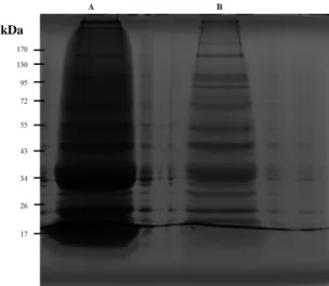
The characterization of protein aggregates can provide valuable insights into the understanding of cardiomyocytes’ remodeling in response to aging and mechanisms underlying cardiotoxicity. Several methodological approaches have been proposed. Bastos et al. (5) highlighted the potential of mass spectrometry (MS) for protein aggregation research once it allows protein identification and characterization from complex mixtures of biological origin. Protein aggregates are not homogeneous in regard to their size, composition and morphology. Such heterogeneity makes the study of protein aggregation challenging (95). The identification of aggregates components is troubled by difficulties in the isolation of protein aggregates, thereby no standard method was yet proposed. The generally approach for the isolation of proteins aggregates are based on the fact that insoluble aggregates, namely amyloids, are resistant to strong detergents. This property allows the isolation of amyloids by centrifugation in the presence of detergent (96). Different detergents have been proposed to isolate protein aggregates, such as sodium dodecyl sulphate (SDS) or sodium N-lauroylsarcosinate (sarcosyl) (18, 97). Some studies demonstrated that amyloids can be isolated from yeast cell lysates by sedimentation in the presence of SDS, showing the role of SDS in the isolation of protein aggregates (96). However, it has been reported that the use of a milder detergent, like sarcosyl, instead SDS that presents a strong denaturant effect and impairs protein’s native conformation, allowed the purification of amyloid aggregates which cannot withstand SDS treatment (97).Furthermore, in cardiac tissue, the use of sarcosyl, was also shown to be efficient in the isolation of protein aggregates-enriched fractions (18). Resolubilization of insoluble aggregates is a crucial step to ensure adequate separation of proteins in SDS polyacrylamide gel electrophoresis (SDS-PAGE). Due the high insolubility of protein aggregates, the use of high percentage of strong detergents to protein aggregates solubilization is essential to achieve maximal protein recovery (98). Gel electrophoresis is commonly used for estimating protein size, identifying proteins and determining sample purity (95). SDS-PAGE is a simple and very frequently used approach to separate and analyze the extracted protein aggregates (95). Also, this separation method allows downstream identification of proteins by MS after in-gel digestion. This method is preferable to the alternative in-solution digestion, because many MS interferents are trapped inside the gel, while peptides can be easily recovered from the polyacrylamide matrix (99, 100). The study of protein aggregates is still a major challenge, taking into account that protein aggregation is a dynamic process that can be influenced by several factors, such as temperature, protein concentration and pH (95). Therefore, development/optimization of methods for isolation of the aggregates and identification of the associated proteins can be essential for understanding the cellular processes underlying the aggregation-driven toxicity.
2. AIMS
The main objectives of the work carried out were to investigate the potential involvement of protein aggregation in the pathophysiological mechanisms underlying the cardiovascular changes associated with aging, as well as to understand the mechanisms by which the protein aggregation promotes Doxo-induced cardiotoxicity. For this purpose, the first goal was to optimize the methodology for protein aggregates enrichment isolated from left ventricle (LV) of an animal model of aging and an animal model of cardiotoxicity. Then, the second goal was to identify proteins from aggregates-enriched fractions by SDS-PAGE followed by liquid chromatography-tandem mass spectrometry (GeLC-MS/MS). The last goal was to understand if the biological processes associated to the proteins present in aggregates differ between young and aged rats LV and between LV from healthy and Doxo-treated animals, as well as their biological implications.
3. MATERIALS AND METHODS
3.1 Animal model
3.1.1 Aging animal model: Left ventricular samples from a rat model of aging previously
established in our laboratory were used for studying protein aggregation. Briefly, young and aged male Wistar Kyoto rats (WKY, n=5 each group) (Charles River, US) were maintained for 25 weeks-old and 20 months-old, respectively, in individually ventilated chambers, in a controlled environment with a 12:12h light/dark cycle at 20 ± 2ºC room temperature. Water and standard diet were provided ad libitum.
3.1.2 Doxo-induced cardiotoxicity model: Left ventricular samples from a rabbit model of
cardiotoxicity previously established in our laboratory were used for studying protein aggregation. Briefly, a well-documented regimen was used for the induction of HF due to doxorubicin toxicity (101). Male New Zealand white rabbits (n=5) received a bolus of doxorubicin (Doxo group, 1mg/Kg, iv) via marginal ear vein, twice a week for 8 weeks, followed by a washout period of 1 week. Control rabbits (n=5) received 0.9% saline (vehicle) in equivolumetric doses over the same period. The animals were housed in stainless steel cages in a controlled environment with a 12:12h light/dark cycle at 20 ± 2ºC. Water and standard diet were provided ad libitum.
At the end of the experiments, rats and rabbits were euthanized under anesthesia and subsequently LV were collected, snap frozen in liquid nitrogen and stored at -80ºC for protein aggregates studies. Experiments conformed to the Guide for the Care and Use of Laboratory Animals published by the National Institutes of Health (NIH Publication no. 85–23, revised 2011) and was approved by the ethics committee of the Faculty of Medicine of University of Porto.
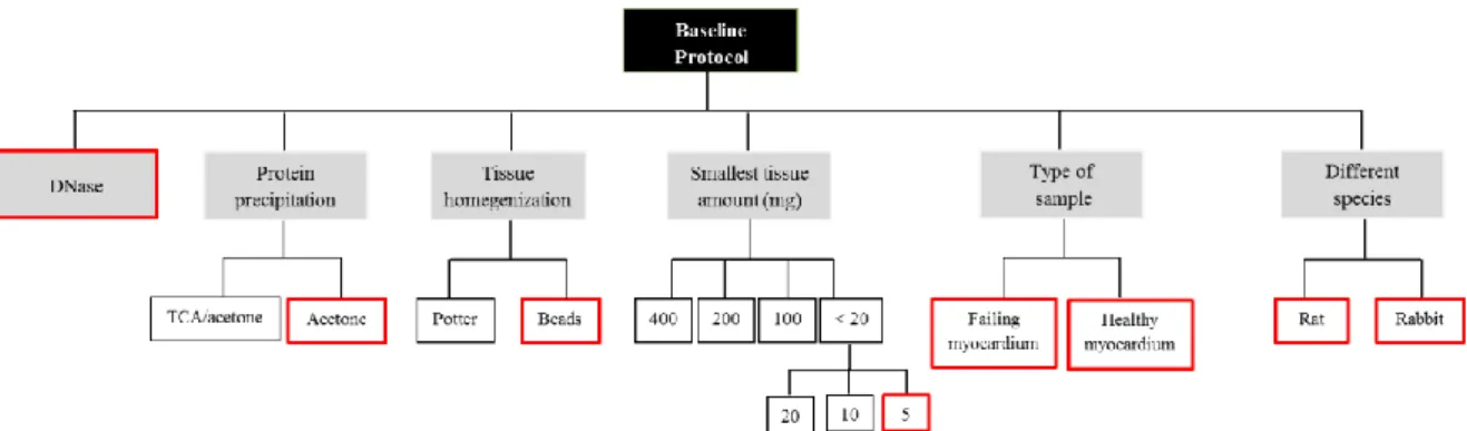
3.2 Baseline protocol for protein aggregates extraction and purification
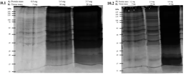
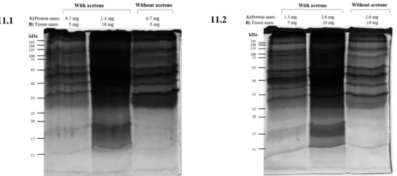
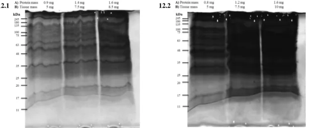
The methodological approach for the isolation of protein aggregates-enriched fractions was based on the Ayyadevara et al. protocol (18). In order to optimize the technique, different conditions were tested: 1) tissue homogenization method (potter or beads); 2) DNase treatment; 3) protein precipitation method (TCA/acetone or acetone); 4) tissue amount; 5) sample type (failing and healthy myocardium); 6) different animal species (rat and rabbit). The optimized protocol is described in “Results” section.