The role of secondary hyperparathyroidism
in left ventricular hypertrophy
of patients under chronic hemodialysis
1Programa de Pós-Graduação em Nefrologia, 2Serviço de Cardiologia, 3Serviço de Nefrologia, Hospital de Clínicas de Porto Alegre, 4Departamento de Medicina Interna, Faculdade de Medicina,
Universidade Federal do Rio Grande do Sul, Porto Alegre, RS, Brasil R.B. Randon1, L.E. Rohde2,
L. Comerlato3, J.P. Ribeiro2,4
and R.C. Manfro1,3,4
Abstract
End-stage renal disease (ESRD) patients frequently develop structural cardiac abnormalities, particularly left ventricular hypertrophy (LVH). The mechanisms involved in these processes are not completely understood. In the present study, we evaluated a possible association between parathyroid hormone (PTH) levels and left ventricular mass (LVM) in patients with ESRD. Stable uremic patients on intermittent hemodialysis treatment were evaluated by standard two-dimensional echocardiography and their sera were analyzed for intact PTH. Forty-one patients (mean age 45 years, range 18 to 61 years), 61% males, who had been on hemodialysis for 3 to 186 months, were evaluated. Patients were stratified into 3 groups according to serum PTH: low levels (<100 pg/ml; group I = 10 patients), intermediate levels (100 to 280 pg/ml; group II = 10 patients) and high levels (>280 pg/ml; group III = 21 patients). A positive statistically significant association between LVM index and PTH was identified (r = 0.34; P = 0.03, Pearson’s correlation coefficient) in the sample as a whole. In sub-group analyses, we did not observe significant associations in the low and intermediate PTH groups; nevertheless, PTH and LVM index were correlated in patients with high PTH levels (r = 0.62; P = 0.003). LVM index was also inversely associated with hemoglobin (r = -0.34; P = 0.03). In multivariate analysis, after adjustment for age, hemoglo-bin, body mass index, and blood pressure, the only independent predictor of LVM index was PTH level. Therefore, PTH is an inde-pendent predictor of LVH in patients undergoing chronic hemodialy-sis. Secondary hyperparathyroidism may contribute to the elevated cardiovascular morbidity associated with LVH in ESRD.
Correspondence
R.C. Manfro Serviço de Nefrologia
Hospital de Clínicas de Porto Alegre Rua Ramiro Barcelos 2350, Sala 2030 90035-903 Porto Alegre, RS Brasil
Fax: +55-51-2101-8001 E-mail: rmanfro@hcpa.ufrgs.br Research partially supported by the Research Incentive Fund (FIPE), Hospital de Clínicas de Porto Alegre. L.E. Rohde is a CNPq investigator. The present address of R.B. Randon is Departamento de Pediatria e Divisão de Nefrologia, Universidade de Caxias do Sul, Caxias do Sul, RS, Brasil.
Received September 22, 2004 Accepted June 3, 2005
Key words
•End-stage renal failure •Parathyroid hormone •Echocardiography
•Left ventricular hypertrophy
Introduction
End-stage renal disease (ESRD) has been associated with changes in the structure and function of the myocardium (1). A signifi-cant percentage of patients who start dialysis
pathogen-esis of ESRD-related LVH, such as hyper-tension, chronic anemia, arteriovenous fis-tulas, concurrent ischemic heart disease, and hypoalbuminemia. These factors have inde-pendent effects, which possibly act syner-gistically leading to LVH and its related morbidity and mortality (4,5).
Recently, parathyroid hormone (PTH) has been identified as an important cardiotoxin in ESRD. Previous studies have supported the view that high PTH serum levels in uremic patients may cause deleterious ef-fects in myocardium metabolism and func-tion (4). The associafunc-tion between PTH lev-els and LVH has been reported by some investigators, with inconsistent results (6-9). The present study was therefore conducted to evaluate the independent association be-tween intact PTH levels and the presence of LVH in end-stage stable uremic patients under chronic hemodialysis, in whom other known risk factors for LVH were not pres-ent.
Material and Methods
Subjects
Forty-one adult ESRD patients receiving renal replacement therapy with hemodialy-sis for at least three months were included. Patients with diabetes mellitus, severe ane-mia (hematocrit <20%), body mass index (BMI) <15 kg/m2, coronary artery disease,
uncontrolled hypertension, primary or sec-ondary cardiomyopathies, known valvular heart disease or pericarditis, a history of heavy alcohol consumption associated with altered liver function tests, parathyroidec-tomy, and connective tissue disease were excluded. All patients who agreed to partici-pate in this protocol signed an informed consent document. The research protocol was approved regarding its ethical and meth-odological aspects by the Ethics Committee in Research Procedures of Hospital de Clínicas de Porto Alegre, Universidade
Fe-deral do Rio Grande do Sul Medical School, accredited by the Brazilian National Re-search Committee, and registered at the Of-fice for Human Research Protection (OHRP-USDHHS) as an Institutional Reviewing Bureau (IRB 00000921).
Two-dimensional echocardiography
Color Doppler echocardiograms were performed by one of the investigators (LER) using the ATL HDI 5000 (ATL Ultrasound, Bothel, WA, USA) ultrasound equipment with a 2.5-3.5 MHz transducer and harmonic imaging. Echocardiograms were performed with the patients within a maximum of 3% above the estimated dry weight (weight when normotensive and free of edema). The echo-cardiographic parameters studied in this pro-tocol were evaluated according to the rec-ommendations of the American Society of Echocardiography. For each measurement, 3-5 consecutive cardiac cycles were ana-lyzed and the average was computed. Left ventricular mass (LVM) was calculated ac-cording to the modified cube formula pro-posed by Devereux et al. (10). LVM index was calculated by dividing LVM by body surface. An LVM index above 100 g/m2 for
women and above 131 g/m2 for men was
considered to indicate LVH (11).
Blood analysis
Other laboratory tests were carried out by standardized clinical laboratory methods and included total alkaline phosphatases, bone-fraction alkaline phosphatase, total calcium, phosphorous, pre- and post-dialy-sis urea, creatinine, alanine aminotransferase, aspartate aminotransferase, albumin, total cholesterol, triglycerides, hematocrit, hemo-globin, serum iron, and serum aluminum. Bone-fraction alkaline phosphatase was measured by the thermal inactive enzyme method (12). Averaged 12-month data of urea fractional clearance and urea reduction rate were used for the biochemical evalua-tion of dialysis adequacy.
Other measurements
Blood pressure was evaluated by averag-ing the measurements obtained before he-modialysis over a period of 6 months pre-ceding the echocardiographic examination. BMI was calculated using the formula: BMI = weight/height2 (kg/m2).
Statistical analysis
Data are reported as means ± SD or as percentages. Differences in echocardiographic, clinical, and laboratory variables between
dif-ferent PTH groups were evaluated by analysis of variance. The Tukey test was used for multiple comparisons. The association between PTH and LVM index and the other variables was evaluated by Pearson’s correlation coeffi-cient. Multiple linear regression analysis was used to evaluate the relation between PTH and LVM index, controlling for variables that may potentially influence LVM index. A two-tailed P value <0.05 was considered to be statisti-cally significant. Statistical analysis was car-ried out using the SPSS Program, version 8.0.
Results
The demographic and anthropometric characteristics of the patients are presented in Table 1. Causes of renal failure were: a) hypertensive nephrosclerosis in 10 (24%) patients, b) polycystic kidney disease in 8 (20%), c) glomerular diseases in 5 (12%), d) obstructive uropathy in 4 (10%), e) Alport syndrome, chronic pyelonephritis, U-shaped kidneys and HIV nephropathy in 1 (2%) patient each, and e) undetermined etiology in 11 (24%) patients.
Laboratory variables are shown in Table 2 for the patients as a whole and stratified by PTH groups. Alkaline phosphatase levels were significantly higher in the high PTH
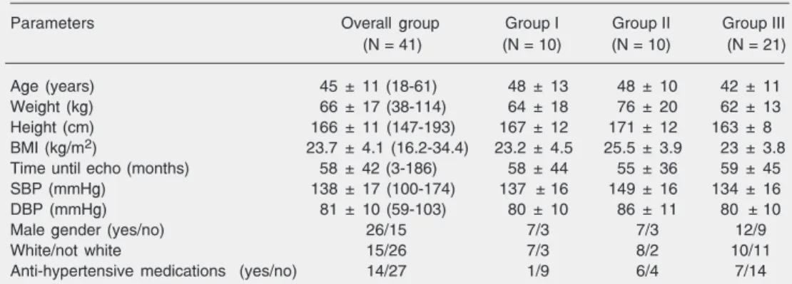
Table 1. Clinical characteristics of the patients as a whole and divided into groups according to parathyroid hormone levels.
Parameters Overall group Group I Group II Group III
(N = 41) (N = 10) (N = 10) (N = 21)
Age (years) 45 ± 11 (18-61) 48 ± 13 48 ± 10 42 ± 11
Weight (kg) 66 ± 17 (38-114) 64 ± 18 76 ± 20 62 ± 13
Height (cm) 166 ± 11 (147-193) 167 ± 12 171 ± 12 163 ± 8
BMI (kg/m2) 23.7 ± 4.1 (16.2-34.4) 23.2 ± 4.5 25.5 ± 3.9 23 ± 3.8
Time until echo (months) 58 ± 42 (3-186) 58 ± 44 55 ± 36 59 ± 45
SBP (mmHg) 138 ± 17 (100-174) 137 ± 16 149 ± 16 134 ± 16
DBP (mmHg) 81 ± 10 (59-103) 80 ± 10 86 ± 11 80 ± 10
Male gender (yes/no) 26/15 7/3 7/3 12/9
White/not white 15/26 7/3 8/2 10/11
Anti-hypertensive medications (yes/no) 14/27 1/9 6/4 7/14
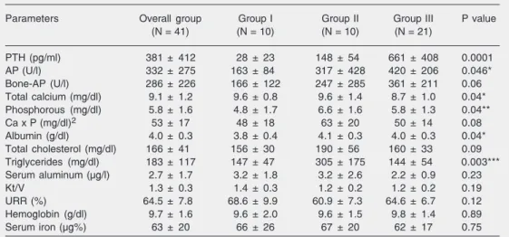
Table 2. Laboratory measurements.
Parameters Overall group Group I Group II Group III P value
(N = 41) (N = 10) (N = 10) (N = 21)
PTH (pg/ml) 381 ± 412 28 ± 23 148 ± 54 661 ± 408 0.0001
AP (U/l) 332 ± 275 163 ± 84 317 ± 428 420 ± 206 0.046*
Bone-AP (U/l) 286 ± 226 166 ± 122 247 ± 285 361 ± 211 0.06
Total calcium (mg/dl) 9.1 ± 1.2 9.6 ± 0.8 9.6 ± 1.4 8.7 ± 1.0 0.04*
Phosphorous (mg/dl) 5.8 ± 1.6 4.8 ± 1.7 6.6 ± 1.6 5.8 ± 1.3 0.04**
Ca x P (mg/dl)2 53 ± 17 48 ± 18 63 ± 20 50 ± 14 0.08
Albumin (g/dl) 4.0 ± 0.3 3.8 ± 0.4 4.1 ± 0.3 4.0 ± 0.3 0.04*
Total cholesterol (mg/dl) 166 ± 41 156 ± 30 190 ± 56 160 ± 33 0.09
Triglycerides (mg/dl) 183 ± 117 147 ± 47 305 ± 175 144 ± 54 0.003***
Serum aluminum (µg/l) 2.7 ± 1.7 3.2 ± 1.8 3.2 ± 2.6 2.2 ± 0.9 0.23
Kt/V 1.3 ± 0.3 1.4 ± 0.3 1.2 ± 0.2 1.2 ± 0.2 0.19
URR (%) 64.5 ± 7.8 68.6 ± 9.9 60.9 ± 7.3 64.6 ± 6.7 0.12
Hemoglobin (g/dl) 9.7 ± 1.6 9.6 ± 2.0 9.6 ± 1.5 9.8 ± 1.4 0.89
Serum iron (µg%) 63 ± 20 66 ± 26 67 ± 20 62 ± 17 0.75
Data are reported as means ± SD. Groups I, II, III = low, intermediate and high serum PTH levels, respectively. PTH = parathyroid hormone; AP = total alkaline phosphatases; Bone-AP = bone fraction of alkaline phos-phatase; Ca x P = calcium x phosphorous product; Kt/V = urea fractional clearance; URR = urea reduction rate. ANOVA *between groups I and III; **between groups I and II; ***between groups I and II, and groups II and III.
Table 3. Echocardiographic parameters.
Parameters All patients Group I Group II Group III
(N = 41) (N = 10) (N = 10) (N = 21)
Diastolic septal thickness wall (cm) 1.2 ± 0.2 1.3 ± 0.3 1.2 ± 0.2 1.2 ± 0.2
Diastolic LV diameter (cm) 4.8 ± 0.7 4.7 ± 0.8 5.2 ± 0.5 4.7 ± 0.8
Diastolic posterior wall thickness (cm) 1.0 ± 0.2 1.0 ± 0.1 1.1 ± 0.1 1.0 ± 0.2
Systolic LV diameter (cm) 3.0 ± 0.6 3.0 ± 0.8 3.2 ± 0.5 2.9 ± 0.5
LVM (g) 204 ± 68 194 ± 50 231 ± 45 196 ± 81
LVM index (g/m2) 119 ± 41 117 ± 39 124 ± 29 118 ± 48
LV ejection fraction (%) 68 ± 9 68 ± 10 68 ± 7 68 ± 10
Data are reported as means ± SD. Groups I, II, III = low, intermediate and high serum PTH levels, respectively. LV = left ventricle; LVM = left ventricular mass. There were no significant differences between groups (ANOVA).
group compared to the low PTH group (P < 0.05) and a similar trend was observed for bone-specific alkaline phosphatase (P = 0.06). Serum total calcium levels were sig-nificantly lower in the high PTH group, while serum phosphorous, calcium-phosphorous product, triglycerides, total cholesterol, and albumin were higher in the intermediate PTH group. Other laboratory variables did not differ among groups (Table 2).
Echocardiographic variables were not significantly different among groups. Table 3 shows the echocardiographic data for the patients as a whole and according to PTH levels. Overall, left ventricle dimensions and
systolic function were within normal values, except for a slightly increased diastolic sep-tal wall thickness.
Table 4. Person’s correlation coefficients between parathyroid hormone and clinical parameters.
Parameters All patients Group I Group II Group III
(N = 41) (N = 10) (N = 10) (N = 21)
r P r P r P r P
Time until echo 0.36 0.02 -0.06 0.87 0.14 0.69 0.63 0.002
AP (U/l) 0.55 0.001 -0.20 0.58 0.08 0.82 0.72 0.001
Bone-AP (U/l) 0.58 0.001 -0.16 0.66 0.07 0.85 0.69 0.001
Groups I, II, III = low, intermediate and high serum PTH levels, respectively. Time until echo = hemodialysis time until echocardiography; AP = alkaline phosphatase.
Table 5. Clinical predictors of left ventricular mass index in multiple regression analysis.
Variables Overall group Group I + Group II Group III
(N = 41) (N = 20) (N = 21)
ß P value ß P value ß P value
PTH 0.03 0.02 0.84 0.44 0.07 0.01
Hemoglobin -6.51 0.11 -6.18 0.18 -2.78 0.71
SBP 0.34 0.35 -0.35 0.45 0.70 0.27
Age 1.08 0.10 1.04 0.22 0.72 0.48
BMI -3.3 0.08 -4.21 0.09 -3.09 0.29
R2: 0.31 R2: 0.38 R2: 0.47
Groups I, II, III = low, intermediate and high serum PTH levels, respectively. ß = ß coefficient; PTH = parathyroid hormone; SBP = systolic blood pressure; BMI = body mass index.
Figure 1. Dispersion diagram demonstrating the correlation be-tween PTH levels and LVM index for all patients and for the patients divided into groups according to PTH levels. Groups I, II, III = low, intermediate and high serum PTH levels, respectively. LVM = left ventricular mass; PTH = parathy-roid hormone. r = 0.34 (P = 0.03) for all patients and r = 0.62 (P = 0.003) for high PTH group (Pearson’s correlation coeffi-cient).
a significant correlation between PTH and LVM index was observed in all patients (r = 0.34; P = 0.03). The correlation was more prominent in the group of patients who pre-sented the most pronounced degree of hy-perparathyroidism (high PTH group; r = 0.62; P = 0.003).
No significant correlations were observed between LVM index and anthropometric or clinical variables. However, a negative cor-relation was observed between LVM index and hemoglobin levels (r = -0.34; P = 0.03).
Trends were also noted for associations be-tween LVM index and alkaline phospha-tases (r = 0.27; P = 0.08).
Multivariate analysis was performed in-cluding age, BMI, systolic blood pressure, hemoglobin, and PTH as independent vari-ables and LVM index as dependent variable. Models were created for all 41 patients (model 1), aggregating the low and intermediate PTH groups (model 2) and in analyses restricted to the high PTH group (model 3). In this analysis (Table 5), LVM index was significantly and
LVM index (g/m
2) 300
Group III Group II Group I
0 400 800 1200 1600 2000
PTH (pg/ml) 250
200
150
100
50
0 ❇
❇
❇ ❇ ❇ ❇ ❇ ❇ ❇
❇
independently associated with PTH, both in model 1 (P = 0.02) and model 3 (P = 0.01).
Discussion
Several factors contribute to the devel-opment of LVH in patients on intermittent hemodialysis treatment, including chronic hypervolemic state, anemia, elevated blood pressure, and arteriovenous fistula (3). It has been suggested that secondary hyperpara-thyroidism may also play a role in the car-diovascular disease of end-stage uremia (2,7), although previous studies evaluating the role of PTH in LVH in ESRD patients have led to contradictory results (6-9).
In the present study, a well-selected sample of patients was examined. Our data indicate that LVH is positively correlated with PTH levels, particularly in patients with significant secondary hyperparathyroidism. However, this correlation does not hold true for patients with intermediate or low PTH levels. These findings were unchanged after multiple regression adjustment for other pa-rameters involved in LVH. Interestingly, in-termediate PTH levels are considered ad-equate for the bone remodeling processes in uremic patients (13).
Our results agree with those of Stack and Saran (14) who reported that PTH levels above 157 pg/ml correlated independently with the development of LVH. Neverthe-less, contradictory findings about the role of secondary hyperparathyroidism in cardiac changes have been reported in chronic ure-mic patients. London et al. (6) presented contradictory results concerning the correla-tion between PTH levels and LVM index, describing a significant negative associa-tion. The reason for this discrepancy is not clear, but may be related in part to some methodological aspects. Firstly, PTH values were relatively higher in our patients with great variability in the group of more severe hyperparathyroidism. Secondly, London et al. (6) used a PTH radioimmunoassay
tech-nique detecting the terminal C-fragment. This technique measures predominantly inactive PTH metabolites whose excretion is reduced in renal failure (15). These metabolites have biological effects on target tissues including the heart that differ from those of intact PTH.
Anemia is considered to be an important factor in the pathogenesis of LVH, contrib-uting to increased cardiovascular mortality in dialysis patients (16). In the overall group, we observed that hemoglobin was negatively associated with LVM index. However, in multivariate analysis, the correlation of PTH with LVM index was independent of anemia and several other potential confounding fac-tors. Moreover, in patients on dialysis, the presence of anemia is combined with chronic volume overload and it has been shown that the use of human recombinant erythropoi-etin can attenuate LVH by correcting ane-mia (17).
High blood pressure is also strongly as-sociated with LVH development (16,18,19). Foley et al. (18) showed that hypertension in chronic dialysis is associated with concen-tric hypertrophy of the left venconcen-tricle. Hyper-tensive hemodialysis patients have LVM in-dices that are significantly higher than their normotensive counterparts. However, LVM indices are similar to those of non-uremic hypertensive patients, demonstrating that inadequate blood pressure control is an im-portant factor for the development of LVH (20). In order to avoid such an interaction, we selected for the present study only nor-motensive or well-controlled hypertensive patients. Among the 19 patients using anti-hypertensive medications, 14 were taking 1 drug and 5 were taking two drugs for blood pressure control (data not shown). Systolic blood pressure, which was included in the multivariate analysis, did not affect the asso-ciation of PHT with LVH.
that its chronic elevation in ESRD patients adversely affects myocardial metabolism and function (4). LVH in uremic patients is not only characterized by an increased myocar-dial fiber mass but also by myocarmyocar-dial inter-stitial fibrosis (21). The mechanisms by which PTH induces LVH have not been completely elucidated. Studies have shown increased cytosolic calcium and/or protein kinase C activation. Expression of cardiac proto-oncogene may be enhanced, which in turn may lead to altered expression of sev-eral genes involved on cardiac structure and action and ultimately stimulate the transla-tion of contractile and non-contractile car-diac muscle proteins leading to LVH (22). These studies also suggest that PTH has a permissive role for fibroblast activation and myocardial fibrosis. Thus, it has been ob-served that elevated PTH levels in ESRD cause irreversible interstitial fibrosis with collagen deposition (23). Following para-thyroidectomy in animals with chronic renal failure, a reduction of collagen deposition in the myocardium is consistently observed (22). Interactions between PTH levels and cardiac abnormalities specifically related to LVH and left ventricular diastolic dysfunc-tion were shown in patients with primary hyperparathyroidism as well (24-26). Such abnormalities were independent of plasma calcium levels and hypertension and also regressed following parathyroid mass
re-duction (24-27).
Since numerous factors are involved in the genesis of LVH in chronic hemodialysis patients, one should not expect that isolated interventions on specific targets will lead to complete correction of this condition. Con-trol of these factors, however, is vital and may significantly reduce LVH related to ESRD. This can be observed in patients after successful renal transplantation (28). More-over, it has been demonstrated that parathy-roidectomy exerts a significant role in de-creasing LVM index among dialysis patients with tertiary hyperparathyroidism (29).
Although several comorbidities associ-ated with chronic renal failure may contri-bute to the development of LVH, the find-ings of the present study suggest that sec-ondary hyperparathyroidism plays an im-portant and independent role in this process. Based on the current state of our knowledge, patients with early renal failure should have their PTH levels monitored and kept within adequate levels, by the judicious use of rou-tine interventions, including dietary protein restriction, the prescription of phosphate binders and the proper use of vitamin D analogs. It also seems adequate to periodi-cally evaluate left ventricular morphologi-cal parameters and cardiac geometry by echo-cardiography, especially in those patients with multiple risk factors for LVH develop-ment.
References
1. Parfrey PS, Harnett JD & Barre PE (1991). The natural history of
myocardial disease in dialysis patients. Journal of the American
Society of Nephrology, 2: 2-12.
2. Foley RN, Parfrey PS, Harnett JD et al. (1995). Clinical and echo-cardiographic disease in patients starting end-stage renal disease
therapy. Kidney International, 47: 186-192.
3. Harnett JD, Parfrey PS, Griffiths SM et al. (1988). Left ventricular
hypertrophy in end-stage renal disease. Nephron, 48: 107-115.
4. Massry SG & Smogorzewski M (1996). The heart in uremia.
Semi-nars in Nephrology, 16: 214-221.
5. Foley RN & Parfrey PS (1997). Cardiac disease in chronic uremia:
clinical outcome and risk factors. Advances in Renal Replacement
Therapy, 4: 234-248.
6. London GM, De Vernejoul MC, Fabiani F et al. (1997). Secondary hyperparathyroidism and cardiac hypertrophy in hemodialysis
pa-tients. Kidney International, 32: 900-907.
7. Amann K & Ritz E (1997). Cardiac disease in chronic uremia:
pathophysiology. Advances in Renal Replacement Therapy, 4:
212-224.
8. Avram MM, Sreedhara R, Avram DK et al. (1996). Enrollment para-thyroid hormone level is a new marker of survival in hemodialysis
and peritoneal dialysis therapy for uremia. American Journal of
Kidney Diseases, 28: 924-930.
9. Strozecki P, Adamowicz A, Nartowicz E et al. (2001). Parathormone, calcium, phosphorus and left ventricular structure and function in
10. Devereux RB, Alonso DR, Lutas EM et al. (1986). Echocardio-graphic assessment of left ventricular hypertrophy: comparison to
necropsy findings. American Journal of Cardiology, 57: 450-458.
11. Levy D, Savage DD, Garrison RJ et al. (1987). Echocardiographic criteria for left ventricular hypertrophy: the Framingham Heart Study.
American Journal of Cardiology,59: 956-960.
12. Moss DW & Whitby LG (1975). A simplified heat-inativation curves
of alkaline phosphatase isoenzymes in serum. Clinica Chimica Acta,
61: 63-71.
13. Rostand SG & Drüeke TB (1999). Parathyroid hormone, vitamin D,
and cardiovascular disease in chronic renal failure. Kidney
Interna-tional, 56: 383-392.
14. Stack AG & Saran R (2002). Clinical correlates and mortality impact of left ventricular hypertrophy among new ESRD patients in the
United States. American Journal of Kidney Diseases, 40:
1202-1210.
15. Endres DB, Villanueva R, Sharp Jr CF et al. (1989). Measurements
of parathyroid hormone. Endocrinology and Metabolism Clinics of
North America, 18: 611-629.
16. Harnett JD, Kent GM, Barre PE et al. (1994). Risk factors for the development of left ventricular hypertrophy in a prospectively
fol-lowed cohort of dialysis patients. Journal of the American Society of
Nephrology, 4: 1486-1490.
17. MacDougall IC, Lewis NP, Saunders MJ et al. (1990). Long-term cardiorespiratory effects on amelioration of renal anaemia by
erythropoetin. Lancet, 335: 489-493.
18. Foley RN, Parfrey PS, Harnett JD et al. (1996). Impact of hyperten-sion on cardiomyopathy, morbidity and mortality in end-stage renal
disease. Kidney International,49: 1379-1385.
19. London GM (2002). Left ventricular alterations and end stage renal
disease. Nephrology, Dialysis, Transplantation, 18 (Suppl 1): 29-36.
20. Cuspidi C, Lonati L, Sampieri L et al. (2000). Impact of blood
pressure control on prevalence of left ventricular hypertrophy in
treated hypertensive patients. Cardiology, 93: 149-154.
21. Mall G, Huther W, Schneider J et al. (1990). Diffuse
intermyocardio-cytic fibrosis in uraemic patients. Nephrology, Dialysis,
Transplanta-tion, 5: 39-44.
22. Mall G, Rambausek M, Neumeister A et al. (1988). Myocardial interstitial fibrosis in experimental uremia - implication for cardiac
compliance. Kidney International, 33: 804-811.
23. Amann K, Ritz E, Wiest G et al. (1994). A role of parathyroid
hormone for the activation of cardiac fibroblasts in uremia. Journal
of the American Society of Nephrology, 4: 1814-1819.
24. Nappi S, Saha H, Virtanen V et al. (2000). Left ventricular structure and function in primary hyperparathyroidism before and after
para-thyroidectomy. Cardiology, 93: 229-233.
25. Saleh FN, Schirmer H, Sundsfjord J et al. (2003). Parathyroid
hor-mone and left ventricular hypertrophy. European Heart Journal, 24:
2054-2060.
26. Andersson P, Rydberg E & Willenheimer R (2004). Primary
hyper-parathyroidism and heart disease - a review. European Heart
Jour-nal, 25: 1776-1787.
27. Dominiczak AF, Lyall F, Morton JJ et al. (1990). Blood pressure, left ventricular mass and intracellular calcium in primary
hyperparathy-roidism. Clinical Science, 78: 127-132.
28. Ferreira SR, Moises VA, Tavares A et al. (2002). Cardiovascular effects of successful renal transplantation: a 1-year sequential study of left ventricular morphology and function, and 24-hour blood
pres-sure profile. Transplantation, 74: 1580-1587.
29. Chow KM, Szeto CC, Kum LC et al. (2003). Improved health-related quality of life and left ventricular hypertrophy among dialysis
pa-tients treated with parathyroidectomy. Journal of Nephrology, 16: