1
Ana Rita Silva Moreira
Development of proliposomes as a vehicle to deliver new
molecules with antitumor activity
Dissertação do 2º Ciclo de Estudos Conducente ao Grau de Mestre em Química Farmacêutica, Faculdade de Farmácia, Universidade do Porto
Trabalho realizado sob a orientação de: Professor Doutor Domingos Ferreira
Professora Doutora Madalena Pinto Doutora Susana Martins
2 ACCORDING TO THE LEGISLATION, THE REPRODUCTION OF ANY PART OF THIS DISSERTATION IS NOT AUTHORIZED.
3
Author’s declaration:
Under the terms of the Decree-Law nº 216/92, of October 13th, is hereby declared that the author afforded a major contribution to the conceptual design and technical execution of the work and interpretation of the results included in this dissertation. Under the terms of the referred Decree-Law, is hereby declared that the following articles/communications were prepared in the scope of this dissertation.
The results presented in this dissertation are part of the following scientific communications:
A. R. Moreira*, G. Moreira, S. Martins, P. Costa, E. Sousa, M. M. M. Pinto, D. Ferreira.
“Development of proliposomes as a drug delivery system for a xanthonic compound with antitumor activity”. 10th Spanish-Portuguese Meeting of Chemistry, Porto, Portugal, 26-28
November 2014, NT13.
G. Moreira*, A. R. Moreira, S. Martins, C. Marques, P. Costa, J. M. S. Lobo, D. C. Ferreira. “Development of proliposomes as vectors of memantine by lyophilization”. 10th
Spanish-Portuguese Meeting of Chemistry, Porto, Portugal, 26-28 November 2014, NT18.
A. R. Moreira*, G. Moreira, A. Lemos, E. Sousa, M. Pinto, P. Costa, D. Ferreira. "Synthesis
of a xanthonic compound with antitumor activity and development of proliposomes as its delivery system". 8th Meeting of Young Researchers of University of Porto, Porto, Portugal,
13-15 May 2013-15, 6038.
G. Moreira*, A. Moreira, C. Marques, S. Martins, P. Costa, J. Sousa Lobo, D. Ferreira. “Development and characterization of proliposomes as potential drug carriers”. 8th Meeting of
Young Researchers of University of Porto, Porto, Portugal, 13-15 May 2015, 313.
A. R. Moreira*, G. Moreira, A. Lemos, E. Sousa, M. M. M. Pinto, Paulo Costa, Domingos
Ferreira. “Use of drying methods to produce proliposomes for delivery of a xanthone with glioma cell lines growth inhibitory activity”. 10th Young European Scientist Meeting, Faculty of Medicine of University of Porto, Portugal, 17-20 September 2015 (ongoing).
4 G. Moreira*, A. Moreira, C. Marques, S. Martins, P. Costa, J. Sousa Lobo, D. Ferreira. “Proliposomes as paclitaxel delivery systems: a new approach for cancer therapy”. 10th Young European Scientist Meeting, Faculty of Medicine of University of Porto, Portugal, 17-20 September 2015 (ongoing).
5
ACKNOWLEDGEMENTS
First of all, I would like to refer that the realization of this dissertation has been a major learning experience, not only for the academic and scientific knowledge but mainly for the personal experience.
To Prof. Domingos Ferreira, my advisor, for allowing me to take part in this research project. More important, I acknowledge Prof. Domingos for his constant presence, for the belief in the success of my work and for the appreciation for my effort. I feel grateful for having the opportunity to work with Prof. Domingos and for all the lessons I could learn from him.
To Prof. Madalena Pinto, my co-advisor, for her support and guidance in the supervision of the work.
To the professors of Organic and Pharmaceutical Chemistry for knowledge transmitted in the Master’s Degree, especially to Prof. Emília Sousa, for all the help with the part of my work concerning to the Organic and Pharmaceutical Chemistry.
To the professors of Pharmaceutical Technology, with great emphasis for Prof. Paulo Costa, for all the knowledge transmitted in this field, for his good will and constant dedication to the students. Prof. Paulo had a great contribution for the success of my work.
To Dr. Sara Cravo and to Ms. Gisela Adriano for the technical assistance in the Organic Chemistry, with an especial acknowledgement to Dr. Sara for being available and help me with HPLC equipment when it was necessary.
To Mr. Daniel Nunes and Mrs. Conceição Pereira for the technical assistance in Pharmaceutical Tecnhology.
To colleagues of Master’s Degree of Pharmaceutical Chemistry for their friendship and support, with an especial acknowledgement to Agostinho Lemos for the help in my work in Organic Chemistry.
6 To my colleagues in Pharmaceutical Technology, Gabriela, Isabel, Rita, Marlene, Verónica and Ana Cláudia for all the moments of good disposition and for the help when it was needed. I especially thank to Gabriela for being my partner in the laboratory, for all the help, patience and for the understanding when facing difficulties and to Isabel for always being willing to help and share knowledge, and for transmitting me confidence in my work.
Finally, to my parents and brother for giving me the opportunity to do the Master’s degree and for all the support at home.
This work was developed in the Centro de Química Medicinal da Universidade do Porto- CEQUIMED-UP, Laboratório de Química Orgânica e Farmacêutica, Departamento de Ciências Químicas, Faculdade de Farmácia da Universidade do Porto, and Laboratório de Tecnologia Farmacêutica, Departamento de Ciências do Medicamento, Faculdade de Farmácia da Universidade do Porto. This research was supported by the Projects Pest-OE/SAU/UI4040/2014 and partially by the Strategic Funding UID/Multi/04423/2013 through national funds provided by FCT – Foundation for Science and Technology and European Regional Development Fund (ERDF), in the framework of the programme PT2020.
7
INDEX
ACKNOWLEDGEMENTS ...5 ABSTRACT ... 19 RESUMO... 20 ABBREVIATIONS ... 21OUTLINE OF THE DISSERTATION ... 23
CHAPTER 1 – INTRODUCTION ... 27
1.1. Cancer nanotechnology ... 27
1.2. Liposomes ... 29
1.2.1. Composition of liposomes ... 32
1.2.2. Liposomes in cancer nanotechnology ... 34
1.2.3. Advantages and drawbacks of liposomes ... 36
1.2. Proliposomes ... 40
1.3.1. Manufacturing processes ... 42
1.3.1.1. Film deposition on carrier ... 42
1.3.1.2. Freeze drying ... 43
1.3.1.3. Spray Drying ... 45
1.3.2. Proliposomes and anticancer drugs ... 46
1.3. Xanthones ... 48
CHAPTER 2 – AIMS ... 55
CHAPTER 3 – RESULTS AND DISCUSSION ... 59
8 3.1.1. Synthesis of benzophenone intermediate 3,
(2-hydroxy-3,4-dimethoxy-6-methylphenyl) (methoxyphenyl) methanone ... 60
3.1.2. Synthesis of 3,4-diethyl-1-methyl-9H-xanthen-9-one (4): cyclization of benzophenone intermediate 3 ... 61
3.1.3. Synthesis of 1-(dibromomethyl)-3,4-dimethoxy-9H-xanthen-9-one (5) ... 62
3.1.4. Synthesis of 3,4-dimethoxy-9-oxo-9H-xanthene-1-carbaldehyde (LEM2) ... 63
3.2. Development of an HPLC method for the quantification of LEM2 ... 64
3.3. Preliminary studies for the development of proliposomal formulations ... 68
3.4. Proliposomal formulation ... 71
3.4.1. Morphology of proliposome powders ... 71
3.4.2. Thermal behaviour of proliposome powders ... 75
3.5. Hydration of proliposomes ... 81
3.5.1. Morphology of liposome dispersions ... 81
3.5.2. Particle size ... 83
3.5.3. Zeta potential ... 86
3.5.4. Entrapment efficiency ... 88
3.6. Stability of proliposomes ... 91
3.6.1. Thermal behavior of proliposome powders ... 91
3.6.2. Characterization of liposomes ... 94
3.6.2.1. Particle size... 94
3.6.2.2. Zeta potential ... 96
3.6.2.3. Entrapment efficiency ... 98
CHAPTER 4 - CONCLUSIONS AND FUTURE WORK ... 103
CHAPTER 5 - MATERIAL AND METHODS ... 107
5.1. General Methods ... 107
9 5.2.1. Synthesis of benzophenone intermediate 3,
(2-hydroxy-3,4-dimethoxy-6-methylphenyl) (methoxyphenyl) methanone ... 107
5.2.2. Synthesis of 3,4-diethyl-1-methyl-9H-xanthen-9-one (4): cyclization of benzophenone intermediate 3 ... 108
5.2.3. Synthesis of 1-(dibromomethyl)-3,4-dimethoxy-9H-xanthen-9-one (5) ... 108
5.2.4. Synthesis of 3,4-dimethoxy-9-oxo-9H-xanthene-1-carbaldehyde (LEM2) ...109
5.3. Development of an HPLC method to quantify LEM2 ... 110
5.4. Preliminary studies for the development of proliposomal solutions ... 110
5.5. Production of proliposome ... 111
5.5.1. Film deposition on carrier... 111
5.5.2. Freeze drying ... 111
5.5.3. Spray drying ... 112
5.6. Analysis of proliposome powders ... 112
5.6.1. Surface morphology of proliposomes ... 112
5.6.2. Thermal behaviour of proliposomes ... 113
5.7. Hydration of proliposomes... 113
5.7.1. Surface morphology of liposomes ... 113
5.7.2. Particle size measurement ... 114
5.7.3. Zeta potential measurement ... 114
5.7.4. Entrapment efficiency of liposomes ... 114
5.8. Stability studies ... 115
5.9. Statistical analysis ... 115
CHAPTER 6 - REFERENCES ... 119
CHAPTER 7 – APENDICES ... 131
Appendix I - Characterization of liposomes formed by hydration of proliposomes with different carrier : lipid weight ratio, by freeze drying. ... 131
10 Appendix II - Characterization of liposomes formed by hydration of proliposomes at the day of production ... 132 Appendix III – Characterization of liposomes formed by hydration of proliposomes at the day 30. ... 134
11
INDEX OF FIGURES
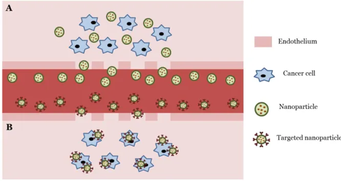
Figure 1 - Schematic representation of passive (A) and active (B) targeting of nanoparticles
to tumors. Figure adapted from 3. ... 28
Figure 2 - Representation of the hydrophilic and hydrophobic portions of a phospholipid. 29 Figure 3 - Schematic representation of monolayer structures formed by phospholipids: lipid bilayer (A), micelle (B). ... 30
Figure 4 - Schematic representation of two different types of liposomes: unilamellar vesicle (A) and multilamellar vesicle (B). ( ) represents a water molecule. ... 31
Figure 5 - Phosphatidic acid structure. ... 32
Figure 6 – Phosphatidylcholine structure. ... 32
Figure 7 - Dimyristoyl phosphatidylcholine and dipalmitoyl phosphatidylcholine structures. ... 33
Figure 8 – Chemical structure of the molecule cholesterol... 34
Figure 9 - Representation of molecular moietis of phospholipids associated with the chemical instability of liposomes. ... 38
Figure 10 - Structure of lysophosphatidylcholine; R = Fatty acid acyl chain. ... 38
Figure 11 - Possible causes of instability of liposomes. ... 39
Figure 12 - Mannitol (A) and sorbitol (B) structures... 41
Figure 13 - Apparatus used to prepare proliposomes by film deposition on carrier (from 49). The reproduction of this figure was authorized. ... 43
Figure 14 - Pressure-temperature equilibrium diagram. At the triple point, solid, liquid and vapor are in dynamic equilibrium. Liquid-vapor phase limit ends at the critical point. The normal freeze point is the temperature at which the liquid freezes at a pressure of 1 atm and the normal boiling point represents the temperature at which the liquid vapor pressure is 1 atm. Adapted from 52. ... 44
Figure 15 – Schematic representation of the stages in the spray drying technique (from 63). The reproduction of this figure was authorized. ... 45
Figure 16 - Xanthone scaffold (numbered according IUPAC). ... 48
Figure 17 - Scaffolds containing a γ-pyrone moiety: flavonoids (A) and chromones (B). ... 48
Figure 18 - LEM2 structure. ... 51
12
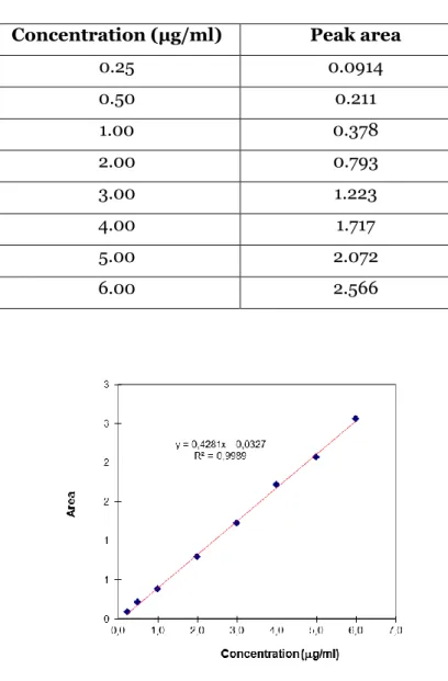
Figure 20 –Calibration curve to extrapolate LEM2 concentration values using HPLC
method. ... 65
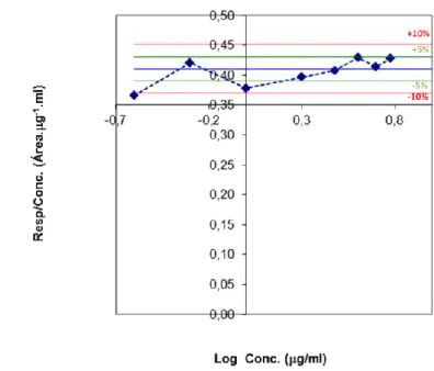
Figure 21 – Graphic of the standard deviation of concentration values of LEM2 standard
solutions. ... 66
Figure 22 - Graphic of response / concentration vs concentration logarithm. ... 66 Figure 23 – LEM2 standard solution chromatogram. ...67 Figure 24 – LEM2 standard solution chromatogram and UV spectrum at 242 nm for the
specific detection of the compound. ...67
Figure 25 – Box and whiskers plot of effective diameter of liposomes formed by hydration of
proliposomes produced by freeze drying, with different carrier : lipid weight ratio. Each box represents three individual batches. Statistical significance: *
p < 0.009, # 0.003. ... 69
Figure 26 – Box and whiskers plot of zeta potential of liposomes formed by hydration of
proliposomes produced by freeze drying, with different carrier : lipid weight ratio. Each box represents three individual batches. ... 70
Figure 27 –SEM images of the surface of mannitol (A-B) and proliposome powders without
drug (C-D) and with LEM2 (E-F) produce from the film deposition on carrier method. ... 72
Figure 28 - SEM images of the surface of mannitol (A-B) and proliposome powders without
drug (C-D) and with LEM2 (E-F) produce from the freeze drying method. ... 73
Figure 29 - SEM images of the surface of mannitol (A-B) and proliposome powders without
drug (C-D) and with LEM2 (E-F) produce from the spray drying method. ...74
Figure 30 - DSC thermogram of egg phosphatidylcholine (A), cholesterol (B), mannitol (C)
and LEM2 (D). ...76
Figure 31 – DSC thermograms from -20 ºC to 240 ºC at 10 ºC/min of proliposomes with no
drug (A), 0.8% LEM2 (B), 2% LEM2 (C) and 4% LEM2 (D) obtained with film deposition on carrier method. ... 77
Figure 32 - DSC thermograms from -20 ºC to 240 ºC at 10 ºC/min of proliposomes with no
drug (A), 0,8% LEM2 (B), 2% LEM2 (C) and 4% LEM2 (D) obtained with freeze drying method. ... 78
Figure 33 - DSC thermogram of spray dried mannitol from -20 ºC to 240 ºC at 10 ºC/min.79 Figure 34 - DSC thermograms from -20 ºC to 240 ºC at 10 ºC/min of proliposomes with no
drug (A), 2% LEM2 (B) obtained with spray drying method. ... 80
Figure 35 - CryoSEM images of liposomes formed by hydration of proliposomes produced by
the film deposition on carrier method, without drug (A-B) and with 2% of LEM2 (C-D) at x 25000 magnification. ... 82
13
Figure 36 - CryoSEM images of liposomes formed by hydration of proliposomes produced
by the freeze drying method, without drug (A-B) and with 2% of LEM2 (C-D) at x 25000 magnification. ... 82
Figure 37 - CryoSEM images of liposomes formed by hydration of proliposomes produced by
the spray drying method, without drug (A-B) and with 2% of LEM2 (C-D) at x 25000 magnification. ... 83
Figure 38 – Box and whiskers plot of effective diameter of liposomes formed by hydration of
proliposomes produced by film deposition on carrier, with no drug and with different percentages of LEM2. Each box represents three individual batches. ... 84
Figure 39 – Box and whiskers plot of effective diameter of liposomes formed by hydration of
proliposomes produced by freeze drying, with no drug and with different percentages of
LEM2. Each box represents three individual batches. ... 85 Figure 40 – Box and whiskers plot of effective diameter of liposomes formed by hydration of
proliposomes produced by spray drying, with no drug and with different percentages of
LEM2. Each box represents three individual batches. ... 85 Figure 41 – Box and whiskers plot of zeta potential of liposomes formed by hydration of
proliposomes produced by film deposition on carrier, with no drug and with different percentages of LEM2. Each box represents three individual batches. ... 87
Figure 42 – Box and whiskers plot of zeta potential of liposomes formed by hydration of
proliposomes produced by freeze drying, with no drug and with different percentages of
LEM2. Each box represents three individual batches. ... 87 Figure 43 – Box and whiskers plot of zeta potential of liposomes formed by hydration of
proliposomes produced by spray drying, with no drug and with different percentages of
LEM2. Each box represents three individual batches. ... 88 Figure 44 – Box and whiskers plot of entrapment efficiency of liposomes formed by
hydration of proliposomes produced by film deposition on carrier, with different percentages of LEM2. Each box represents three individual batches. ... 89
Figure 45 – Box and whiskers plot of entrapment efficiency of liposomes formed by
hydration of proliposomes produced by freeze drying, with different percentages of LEM2. Each box represents three individual batches. Statistical significance: *
p = 0.02. ... 90
Figure 46 – Box and whiskers plot of entrapment efficiency of liposomes formed by
hydration of proliposomes produced by spray drying, with 2% of LEM2. Each box represents three individual batches. ... 90
14
Figure 47 - DSC thermograms from -20 ºC to 240 ºC at 10 ºC/min of proliposomes with 2%
of LEM2 at the day of production (A) and at day 30 (B) obtained with film deposition on carrier method. ... 91
Figure 48 - DSC thermograms from -20 ºC to 240 ºC at 10 ºC/min of proliposomes with 2%
of LEM2 at the day of production (A) and at day 30 (B) obtained with freeze drying method. ... 92
Figure 49 - DSC thermograms from -20 ºC to 240 ºC at 10 ºC/min of proliposomes with 2%
of LEM2 at the day of production (A) and at day 30 (B) obtained with spray drying method. ... 93
Figure 50 – Box and whiskers plot of effective diameter of liposomes formed by hydration of
proliposomes produced by film deposition on carrier, with 2% of LEM2, at the day of production and at day 30. Each box represents three individual batches. ... 94
Figure 51 – Box and whiskers plot of effective diameter of liposomes formed by hydration of
proliposomes produced by freeze drying, with 2% of LEM2, at the day of production and at day 30. Each box represents three individual batches. Statistical significance: *
p = 0.005. .... 95
Figure 52 – Box and whiskers plot of effective diameter of liposomes formed by hydration of
proliposomes produced by spray drying, with 2% of LEM2, at the day of production and at day 30. Each box represents three individual batches. ... 95
Figure 53 – Box and whiskers plot of zeta potential of liposomes formed by hydration of
proliposomes produced by film deposition on carrier, with 2% of LEM2, at the day of production and at day 30. Each box represents three individual batches. Statistical significance: *
p = 0.018. ... 96
Figure 54 – Box and whiskers plot of zeta potential of liposomes formed by hydration of
proliposomes produced by freeze drying, with 2% of LEM2, at the day of production and at day 30. Each box represents three individual batches. ...97
Figure 55 – Box and whiskers plot of zeta potential of liposomes formed by hydration of
proliposomes produced by spray drying, with 2% of LEM2, at the day of production and at day 30. Each box represents three individual batches. ...97
Figure 56 – Box and whiskers plot of entrapment efficiency of liposomes formed by
hydration of proliposomes produced by film deposition on carrier, with 2% of LEM2, at the day of production and at day 30. Each box represents three individual batches. ... 98
Figure 57 - Box and whiskers plot of entrapment efficiency of liposomes formed by
hydration of proliposomes produced by freeze drying, with 2% of LEM2, at the day of production and at day 30. Each box represents three individual batches. ... 99
15
Figure 58 – Box and whiskers plot of entrapment efficiency of liposomes formed by
hydration of proliposomes produced by spray drying, with 2% of LEM2, at the day of production and at day 30. Each box represents three individual batches. ... 99
16
INDEX OF TABLES
Table I- Marketed liposomal products for cancer treatment. ... 35
Table II - Advantages and drawbacks of liposomes as drug delivery systems. ... 37
Table III - Anticancer drugs encapsulated in proliposomes. ...47
Table IV - Xanthone derivatives encapsulated in micro and nanoparticles. ... 49
Table V - Concentration of LEM2 standard solutions and respective peak areas. ... 65
Table VI - Characterization of liposomes formed from the proliposomal formulation PC : CH (3:1) produced by film deposition on carrier. ... 68
Table VII – DSC data of thermograms of egg phosphatidylcholine, cholesterol, mannitol and LEM2. ...76
Table VIII –DSC data of thermograms of LEM2, mannitol and proliposomes with no drug, and with 0.8%, 2% and 4% of LEM2, obtained with film deposition on carrier method. ... 77
Table IX – DSC data of LEM2, mannitol and proliposomes with no drug, and with 0,8%, 2% and 4% of LEM2, obtained with freeze drying method... 78
Table X –DSC data of LEM2, spray dried mannitol and proliposomes with no drug and with 2% LEM2 obtained with spray drying method. ... 80
Table XI – DSC data of proliposomes with 2% of LEM2 at the day of production and at day 30, obtained with film deposition on carrier method. ... 92
Table XII – DSC data of proliposomes with 2% of LEM2 at the day of production and at day 30, obtained with freeze drying method. ... 92
Table XIII – DSC data of proliposomes with 2% of LEM2 at the day of production and at day 30 obtained with spray drying method. ... 93
Table XIV - Characterization of liposomes formed by hydration of proliposomes with different carrier : lipid weight ratio, by freeze drying. ... 131
Table XV - Characterization of liposomes formed by hydration of film deposition on carrier proliposomes at the day of production. ... 132
Table XVI - Characterization of liposomes formed by hydration of freeze dried proliposomes at the day of production. ... 132
Table XVII - Characterization of liposomes formed by hydration of spray dried proliposomes at the day of production. ... 133
17
Table XVIII - Characterization of liposomes formed by hydration of film deposition on
carrier proliposomes at the day 30. ... 134
Table XIX - Characterization of liposomes formed by hydration of freeze dried proliposomes
at the day 30. ... 134
Table XX - Characterization of liposomes formed by hydration of spray dried proliposomes
18
INDEX OF SCHEMES
Scheme 1 - General synthesis of LEM2. r.t. = room temperature; MW = microwave; NBS =
N-bromosuccinimide; BPO = benzoyl peroxide; [(BMIm)BF4] = 1-butyl-3-methylimidazolium
tetrafluoroborate. ... 59
Scheme 2 - Friedel-Crafts acylation of 3,4,5-trimethoxytoluene (1) with 2-methoxybenzoyl
chloride (2) to obtain the benzophenone intermediate 3. r.t. = room temperature. ... 60
Scheme 3 - Cyclization of the benzophenone intermediate 3 to xanthone intermediate 4.
MW = microwave. ... 61
Scheme 4 - Wohl-Ziegler reaction of dibrominated xanthone intermediate 5 from xanthone
intermediate 4. NBS = N-bromosuccinimide; BPO = benzoyl peroxide. a isolated yield. ... 62
Scheme 5 - Synthesis of carbaldehydic xanthone derivative LEM2 from xanthone
intermediate 5. [(BMIm)BF4] = 1-butyl-3-methylimidazolium tetrafluoroborate. a isolated yield. ... 63
19
ABSTRACT
Xanthone derivatives are frequently isolated from natural sources, having a wide range of pharmacological activities. Thus, these structures have attracted great interest and a large variety of synthetic xanthone derivatives have emerged. LEM2 is a synthetic xanthone derivative with tested antitumor effect in different cell lines. Xanthone derivatives frequently present poor aqueous solubility and nanosystems might present an attractive strategy to overcome this limitation.
Liposomes represent a versatile system for drug delivery, in the nanometer and micrometer scale. Liposomes have already demonstrated to be adequate systems for the use in cancer chemotherapy. However, liposomes present physical and chemical instability, limiting their shelf-life. In this context, proliposomes, dry phospholipid powders, emerge as an alternative to overcome the instability of liposomes.
In this dissertation, proliposomal formulations were developed to encapsulate the synthetic xanthonic compound LEM2. Three methods were used to produce proliposomes: film deposition on carrier, freeze drying and spray drying. After their production, proliposomes were hydrated to form liposomes.
The three methods produced proliposomes which, on hydration, formed liposomes in the nanometer scale, which were efficient in encapsulating LEM2. It was found that 30 days after the production of proliposomes by film deposition on carrier and freeze drying, the liposomes obtained by their hydration present altered properties, reflecting some instability. On the contrary, the spray dried proliposomes presented good stability after 30 days of their production, presenting a promising strategy to obtain liposomes with improved stability.
20
RESUMO
Derivados xantónicos são frequentemente isolados de fontes naturais, tendo uma variedade de atividades farmacológicas. Por isso, estas estruturas têm atraído um grande interesse e uma grande variedade de derivados xantónicos sintéticos têm surgido. LEM2 é um derivado xantónico de origem sintética com efeito antitumoral testado em diferentes linhas celulares. Os derivados xantónicos apresentam frequentemente fraca solubilidade em sistemas aquosos e o uso de nanossistemas poderá representar uma estratégica atrativa para ultrapassar esta limitação.
Os lipossomas representam um sistema versátil para a libertação de fármacos, na escala manométrica e micrométrica. Os lipossomas já demonstraram ser adequados para uso em quimioterapia no cancro. No entanto, os lipossomas apresentam instabilidade física e química, limitando o seu tempo de vida útil. Neste contexto, os prolipossomas, pós de fosfolípidos secos, surgem como uma alternativa para ultrapassar a instabilidade dos lipossomas.
Nesta dissertação, foram desenvolvidas formulações de prolipossomas para encapsular o derivado xantónico sintético LEM2. Foram usados três métodos para produzor prolipossomas: film deposition on carrier, liofilização e spray drying. Após a sua produção, os prolipossomas foram hidratados e a para formar lipossomas.
Os três métodos produziram prolipossomas que, quando hidratados, conseguiram formar prolipossomas na escala nanométrica, que demonstraram ser eficientes na encapsulação do LEM2. Foi descoberto que 30 dias após a produção dos prolipossomas por film deposition on carrier and freeze drying, os lipossomas gerados pela sua hidratação apresentam propriedades alteradas, refletindo alguma instabilidade. Pelo contrário, os prolipossomas produzidos por spray drying apresentaram boa estabilidade ao fim de 30 dias, representando uma estratégia promissora para obter lipossomas com melhor estabilidade.
21
ABBREVIATIONS
ΔH – Enthaplpy variation BPO - Benzoyl peroxide
CEQUIMED – Centro de Química Medicinal da Universidade do Porto CH - Cholesterol
DMPC – Dimyristoyl phosphatidylcholine DPPC – Dipalmitoyl phosphatodylcholine DSC – Differential scanning calorimetry EPC - Egg phosphatidylcholine
EPR – Enhanced permeation and retention FD – Freeze drying
FDC – Film deposition on carrier
HPLC – High Performance Liquid Chromatography
IUPAC - International Union of Pure and Applied Chemistry LUV – Large unilamellar vesicles
MLV – Multilamellar vesicles MW – Microwave
NBS - N-bromosuccinimide PC – Phosphatidylcholine
22
RES – Reticuloendothelial system Rf – Retention factor
SD – Spray drying
SPC – Soya phosphatidylcholine SUV – Small unilamellar vesicles TLC – Thin layer chromatography Tm – Phase transition temperature
23
OUTLINE OF THE DISSERTATION
The present dissertation consists of seven chapters. This dissertation involves three main areas of research: synthesis of formylated xanthone LEM2, development of an HPLC method to quantify LEM2 and development of proliposomal formulations to encapsulate LEM2.
CHAPTER 1 – INTRODUCTION
The introductory chapter of the present dissertation is divided in four sections. In the first part, a briefly overview about cancer nanotechnology will be presented and in the second will be focused on liposomes as drug delivery systems suitable for application in cancer nanotechnology, with their advantages and drawbacks being highlighted. In the third part, proliposomes will be presented as a promising strategy to overcome the drawbacks presented by liposomes. And in the fourth part, a brief introduction to xanthone derivatives will be given and their use in nanossystems will be justified.
CHAPTER 2 – AIMS
Herein, the main objectives of the present dissertation are described.
CHAPTER 3 - RESULTS AND DISCUSSION
Results are subdivided in five sections. In the first part, the different reaction steps for the synthesis of carbaldehydic xanthone LEM2 will be described. The second part will present the development of a HPLC method for the quantification of LEM2. The third part will show the analysis of surface morphology and thermal behavior of proliposomes. The fourth part will show the characterization of the liposomes obtained by hydration of proliposomes. The fifth part will present stability results of proliposomes.
24
CHAPTER 4 – CONCLUSIONS
This chapter includes the general conclusions of the present dissertation.
CHAPTER 5 - MATERIAL AND METHODS
In this chapter, the experimental procedures for the synthesis of LEM2 will be detailed. The HPLC conditions for the development of an HPLC method to quantify LEM2 will be detailed. The procedures for the proliposome production and their analysis will be described in detail. The hydration of proliposomes and the characterization of the obtained liposomes will also be described. The conditions used to access the stability of proliposomes will be specified and the statistical tests and software will be identified.
CHAPTER 6 – REFERENCES
The references will be presented at the end of this dissertation. The references followed the American Chemical Society style guide. The main bibliographic research motors were ISI Web of Knowledge, from Thomson Reuters, Scopus, and Google.
CHAPTER 7 – APPENDICES
This section will include the resume of data for the characterization of liposomes obtained by hydration of proliposomes that were used to construct the box and whiskers plots used to present the results.
25
C
HAPTER
1
27
CHAPTER 1 – INTRODUCTION
1.1. Cancer nanotechnology
Cancer is one of the leading causes of mortality worldwide, with approximately 8.2 million related deaths in 2012 1. Cancer begins as localized disease but spreads to different
sites within the body, which difficults treatment. The most common treatments are chemotherapy, radiation and surgery. Chemotherapy in general presents a number of drawbacks, such as nonspecific distribution of antitumor drugs, inadequate drug concentrations reaching the tumor site, cytotoxicity, difficult monitoring of therapeutic responses and development of multiple drug resistances 2. Considering this, there is an
emerging necessity to develop alternatives to improve cancer treatment.
Ideally, the therapeutic agent should reach the tumor sites in the desired concentration to destroy the cancerous cells, while minimizing damage to normal cells 2.
Nanotechnology, which commonly refers structures that are up to several nanometers in size, emerges as one of the most promising fields in cancer therapy 3. Cancer nanotechnology is an
upcoming field concerning interdisciplinary research, involving biology, chemistry, engineering and medicine, and its applicability in cancer detection, diagnosis and treatment deserves considerable attention 2, 4.
Nanosystems applied to cancer treatment present unique properties: (i) they can themselves have therapeutic properties; (ii) they might carry a large amount of therapeutic agent; (iii) their surface can be modified with targeting ligands, increasing the affinity and specificity for target cells and tissues; (iv) they can accommodate multiple drug molecules for combinatorial cancer therapy and (v) can overcome drug resistance mechanisms 2.
Nanotechnology can use passive and active targeting strategies to specifically deliver drugs into cancer cells, therefore enhancing the anticancer effect, and simultaneously minimizing toxicity in normal cells. Passive targeting exploits the characteristics of tumor growth (Figure 1A), while active targeting is based on molecular recognition processes (Figure 1B) 2-3.
Passive targeting takes advantage of the size of nanoparticles and the anatomical and functional differences between normal and tumor vasculature to confine the drug delivery (Figure 1A). Commonly, the vasculature of tumors is highly heterogeneous, having areas of
28 vascular necrosis and densely vascularized areas supplying oxygen and nutrients which enhance tumor growth. Angiogenic blood vessels present a high proportion of proliferating endothelial cells with aberrant underlying basement membrane compared to normal blood cells. In addition, tumor tissues have a leaky and defective architecture, with increased fenestrations between adjacent endothelial cells and the microvessels with enhanced permeability. The tumor lymphatic system is also abnormal, leading to fluid retention in tumors and high interstitial pressure with an outward convective interstitial fluid, which results in metastasis. The combination of the leaky microvasculature and the poor lymphatic drainage results in the enhanced permeation and retention (EPR) effect. This induces the passive targeting of nanocarriers through their accumulation in the tumor at a higher concentration than in the plasma and other tissues, enhancing tumor cytotoxicity (Figure
1A) 2, 4.
Active targeting involves the functionalization of surface of nanoparticles with ligands to deliver the drug to the pathological site or to cross biological barriers based on molecular recognition processes (Figure 1B). The receptor for the ligand should be expressed exclusively on tumor cells for the recognition to occur in tumor microenvironment. Usually, the internalization occurs via receptor-mediated endocytosis 2, 4.
Figure 1 - Schematic representation of passive (A) and active (B) targeting of nanoparticles to tumors. Figure
29
1.2. Liposomes
Liposomes, first reported by Bangham et al. in 1965 5, are microscopic spherical
vesicles, in which an aqueous environment is entirely enclosed by a concentric bilayer of phospholipids 5-8. These vesicular systems are lyotropic liquid crystals with size varying from
20 nanometers to 20 micrometers 9-10. Liposomes have been extensively studied for their
application in drug delivery, drug targeting, controlled release and increased solubility of drugs 7, 10-11.
Liposomes are similar to biological membranes, since they are mainly composed of phospholipids, which are amphiphilic molecules with a hydrophilic head and a hydrophobic tail (Figure 2) 10.
Figure 2 - Representation of the hydrophilic and hydrophobic portions of a phospholipid.
Phospholipids are naturally prone to self-assembly, which leads to their spontaneous aggregation in aqueous environments. In the presence of water, phospholipids align themselves in a thermodynamically stable manner in planar bilayer sheets, minimizing the unfavorable interactions between the bulk aqueous phase and the long fatty acid chains. The heads of the phospholipids form a surface facing the water, while the hydrocarbon tails are repelled by water and face each other, creating a lipid bilayer (Figure 3A). Therefore, in a cell, two layers of heads are formed facing the outside and the inside of the cell, attracted to both aqueous environments. The hydrocarbon tails of both layers face each other, thus the resultant structure forms a bilayer (Figure 3A). When membrane phospholipids are disrupted, they can reassemble themselves into tiny spheres, smaller than a normal cell,
HYDROPHILIC HEAD
HYDROPHOBIC TAIL
30 either as monolayers or bilayers. The monolayer structures are called micelles (Figure 3B) and the bilayer structures are liposomes (Figure 4A) 8. The organization of amphiphiles in
the form of bilayer sheets occurs due to the high entropy of the system, caused by the interaction forces between the water and the hydrophobic hydrocarbon chains. These interactions are eliminated when the sheets fold themselves into sealed vesicles 8, 10, 12. Thus,
liposomes (Figure 4) form spontaneously when the phospholipids are exposed to an aqueous environment, since this is the more energetically stable form 13.
Figure 3 - Schematic representation of monolayer structures formed by phospholipids: lipid bilayer (A), micelle
(B).
Phospholipids confer an amphiphilic nature to liposomes, thus they have a hydrophilic inner core surrounded by a hydrophobic membrane. This vesicular organization allows the entrapment of both hydrophilic and hydrophobic drugs. Water soluble drugs may be solubilized in the internal aqueous compartment of the liposome which difficults and therefore slows down its passage through lipid bilayers. A hydrophobic drug can be dissolved in the hydrophobic part of the liposome or bind to the membrane. Usually, lipophilic drugs exhibit higher encapsulation efficiencies than hydrophilic drugs 8-9. Lipophilic drugs are more
likely to remain encapsulated during storage due to their partition coefficients. They associate with lipid bilayers, thus avoiding leaking out to the exterior water phase 9.
Liposomes can be classified according to the number of bilayers entrapping the internal aqueous volume into unilamellar or multilamellar vesicles. If liposomes have just one bilayer they are classified as unilamellar vesicles (Figure 4A), and their properties are similar to those of flat surfaces. Depending on their size, unilamellar vesicles can be separated into small unilamellar vesicles (SUV) with a diameter ranging from 20 to 100 nm, or large unilamellar vesicles (LUV), with a diameter from 100 nm to 1 µm. SUV exhibit large curvature, while LUV presents a low curvature. If there is more than one bilayer, liposomes
31 are considered to be multilamellar vesicles (MLV) (Figure 4B). MLV represent a heterogeneous group with respect to size and morphology of the liposomes. Usually, MLVs present a size range from 100 nm to 20µm. Each concentric layer of liposomes has a thickness of about 4 nm 9-10, 14. Typically, unilamellar vesicles are suitable for entrapment of hydrophilic
drugs in the internal-aqueous space, while MLVs are appropriate to entrap lipophilic drugs 10.
Figure 4 - Schematic representation of two different types of liposomes: unilamellar vesicle (A) and multilamellar
vesicle (B). ( ) represents a water molecule.
Liposomes, as drug delivery systems, need to show appropriate chemical and biological stability. Colloidal stable structures on equilibrium, such as liposomes, are less sensitive to external changes than equilibrium structures, such as micelles. Thereby, liposomes are suitable for pharmaceutical applications. Biological stability is related to the retention of the drug in its target and to the control of the clearance rate of liposomes from the blood system and from certain parts of the body. The clearance rate depends on the dose, size and surface charge of liposomes.
32
1.2.1. Composition of liposomes
Glycerophospholipids are commonly used phospholipids in liposomal formulations. Glycerophospholipids are composed of a glycerol molecule (C3H8O3) covalently attached to
two fatty acid chains (with variable levels of saturation) by ester linkages and to a highly polar or charged group by a phosphodiester linkage in the third carbon. Glycerol acts as a backbone, by its attachment to the fatty acid chains and to the phosphate group. The hydrophobic tail of phospholipids is composed by the two fatty acid chains and the hydrophilic head is made of glycerol, phosphate and a polar group 10, 15. Glycerophospholipids are derivatives of
phosphatidic acid (Figure 5), in which phosphate group bears a negative charge at neutral pH 15.
Figure 5 - Phosphatidic acid structure.
The polar head group of glycerophospholipids might be neutral, negatively charged or positively charged 15. The charge of phospholipids determine the overall surface charge of
liposomes 9. The most common phospholipid is phosphatidylcholine (PC), which has choline
as polar head group and presents a neutral net charge (Figure 6) 10, 15-16.
Figure 6 – Phosphatidylcholine structure.
33 Phospholipids might be natural or synthec. Phosphatidylcholine from natural sources includes soya phosphatidylcholine (SPC) and egg phosphatidylcholine (EPC). Dimyristoyl phosphatidylcholine (DMPC) and dipalmitoyl phosphatidylcholine (DPPC) are examples of synthetic phosphatidylcholine (Figure 7) 10.
Figure 7 - Dimyristoyl phosphatidylcholine and dipalmitoyl phosphatidylcholine structures.
The structure and flexibility of lipid bilayers are influenced by the temperature. Below physiological temperatures, the bilayer lipids are in a semisolid gel phase. In this state, the polar head groups are uniformly displayed at the surface and the acyl chains are packed, and with no motion. Above physiological temperatures, the hydrocarbon chains are in constant motion, producing a fluid state, also known as liquid-disordered state, in which the interior of the bilayer is more fluid than solid. At physiological temperatures, lipids are in the liquid-ordered state, where the acyl chains of lipids suffer less thermal motion, but there is a lateral movement in the bilayer plane. The phase transition temperature (Tm) is the temperature at
which occurs the transition between the semisolid gel phase and the liquid-disordered state, where the phospholipid bilayer becomes more leaky and flexible. Each type pf phospholipid has a specific Tm. The hydration of phospholipids above the Tm, allows them to assemble into
liposomes 10, 17.
Cholesterol, a steroid molecule (Figure 8), might be incorporated in liposome bilayers to modify the membrane fluidity, reduce the permeability of water soluble molecules through the membrane, and improve its stability 10. It functions as a fluidity buffer. Below Tm,
34 cholesterol makes the membrane more disordered and permeable, while above Tm, it causes
membrane organization, stabilizing it 14. Since the formation of liposomes is achieved by
hydration of phospholipids above their Tm, cholesterol enhances the rigidity of liposomes,
possibly by filling the gaps between the phospholipid molecules in the bilayer structures 10, 18.
Besides, the presence of cholesterol in the bilayer membranes improves its stability in the presence of biological fluids, such as blood/plasma. In the absence of cholesterol, liposomes tend to react with blood proteins, being destabilized. Cholesterol appears to reduce this interaction. However, the presence of cholesterol does not entirely prevent the loss of liposomal phospholipids 19.
Cholesterol molecules arrange themselves among the phospholipid molecules with the hydroxy group facing towards the water phase and the tricyclic ring trapped between the first few carbons of the fatty acyl chains, into the hydrocarbon core of the bilayer 19.
Figure 8 – Chemical structure of the molecule cholesterol.
1.2.2. Liposomes in cancer nanotechnology
In clinical applications, liposomes have proven to take advantage of the EPR effect to passively accumulate in regions of enhanced vasculature permeability, when their average diameter is <200 nm. Thus, the drug-mediated delivery by liposomes explores the overexpression of fenestrations in the cancer vasculature to increase drug concentration in tumor sites. This results in reduced side effects and toxicity of the encapsulated drugs as opposed to free drugs, as well as an increased therapeutic index 4, 11.
35 Table I- Marketed liposomal products for cancer treatment.
Product Drug Indications Reference(s)
Doxil/Caelyx (Johnson & Johnson) Myocet (Cephalon) Doxorubicin Kaposi’s sarcoma; ovarian cancer; breast cancer; multiple myeloma 20-23 DaunoXome (Galen) Daunorubicin Kaposi’s sarcoma 24 Marqibo (Talon) Vincristine Acute lymphoblastic leukemia 25-26
36 Liposomes might also prolong the residence time of drugs in circulation for an effective drug delivery. Cells of the reticuloendothelial system (RES) act as immunological barriers to effective targeting of nanoparticles. The surface of liposomes might be modified with the polymer polyetlylene glycol to avoid the uptake by the RES and increase the circulatory half-time. Therefore, the targeting of liposomes within the tumor is enhanced 4, 9.
Several liposomal products encapsulating drugs have been introduced in the market and many other drugs are in clinical trials. Table I shows the approved liposomal products for cancer treatment in the market. The parenteral route of administration is predominant for the clinically approved products, in particular intravenous administration 11.
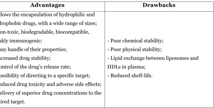
1.2.3. Advantages and drawbacks of liposomes
Liposomes have attracted considerable interest as drug delivery systems since they are versatile drug carriers, suitable for the encapsulation of both hydrophilic and hydrophobic substances. Also, these vesicular structures allow the encapsulation of both small molecules, with the size of an ion, and large molecules of several hundred thousand Daltons 9, 27.
Liposomes present a number of advantages over conventional dosage forms (Table II). They have shown to be relatively non‐toxic, biodegradable, biocompatible, weakly immunogenic (produce no antigenic or pyrogenic reactions). These properties, like size, charge and surface can be readily handled by the addition of new ingredients to the lipid mixture before the preparation of liposomes or by using different preparation methods. Liposome vesicles act as drug reservoirs, thus protecting drugs from the external environment, such as enzymes and inhibitors. These agents could lead to its inactivation of drugs encapsulated in liposomes. Encapsulation of drugs can increase their stability, avoiding rapid degradation 8-9. Also,
liposome formulations have the ability to control the drug release rate in the presence of biological fluids, retaining a relatively constant and, effective drug concentration in the circulation. Therefore, they can prevent undesirable side effects and reduce drug toxicity 9, 28.
The possibility of targeting liposomes to a particular type of cell or organ leads to the increase of its efficacy and therapeutic index, mainly due to the alteration of biodistribution. Manipulating liposomes for a selective uptake is another way of reducing drug toxicity and injurious side effects because of the minimized drug distribution of the drug. Besides, liposomal drug delivery systems enable the delivery of higher drug concentrations to the
37 desired target. Moreover, drug encapsulation results in enhanced pharmacokinetic properties, such as reduced elimination or prolonged residence time of the drug in systemic circulation 8-9.
Despite all the advantages of using liposomes as drug delivery systems, this strategy presents a few drawbacks mainly related to their large scale manufacture (Table II). There is a necessity to obtain large quantities of the product with reproducible properties and to demonstrate suitable stability during storage and before administration. However, liposomes exhibit poor chemical and physical stability, which restricts its storage for a long period 10, 27, 29-30.
Table II - Advantages and drawbacks of liposomes as drug delivery systems.
Advantages
Drawbacks
- Allows the encapsulation of hydrophilic and hydrophobic drugs, with a wide range of sizes; - Non-toxic, biodegradable, biocompatible, weakly immunogenic;
- Easy handle of their properties; - Increased drug stability;
- Control of the drug’s release rate;
- Possibility of directing to a specific target; - Reduced drug toxicity and adverse side effects; - Delivery of superior drug concentrations to the desired target.
- Poor chemical stability; - Poor physical stability;
- Lipid exchange between liposomes and HDLs in plasma;
- Reduced shelf-life.
Physical instability of aqueous dispersion of liposomes occurs due to vesicle aggregation and fusion, which leads to an alteration in vesicle size and loss of retained material. Problems associated with physical stability of liposomes are important to consider when analyzing the appearance, size and size distribution of liposomes 10, 31.
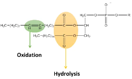
38 Figure 9 - Representation of molecular moietis of phospholipids associated with the chemical instability of
liposomes.
The chemical instability of these systems is caused by the formation of ice crystals in the liposome, which conduces to the destabilization of bilayers resulting in drug leakage. Chemical instability is associated with tendency of phospholipids in liposomal formulations to suffer hydrolysis and oxidation (Figure 9). Hydrolysis may occur in ester bonds linking the glycerol backbone to the fatty acids, leading to the disconnection of the hydrophobic chains. In the case of phosphatidylcholine, the hydrolysis might cause the formation of lysophosphatidylcholine (Figure 10), increasing the permeability of liposomes.
Figure 10 - Structure of lysophosphatidylcholine; R = Fatty acid acyl chain.
Therefore, it is important to keep the levels of lyso-phospholipids to a minimum during preparation and storage of liposomes. Oxidation might occur in the presence of unsaturated acyl chains, which could change the permeability of liposomes and their shelf life.
39 Oxidation of phospholipids might be minimized by protecting them from light or by the addition of antioxidants to the liposomes 10, 19, 31. Oxidation and hydrolysis of lipids may lead
to the formation of short-chain lipids and then less hydrophobic derivatives appear in the bilayers, resulting in compromised quality of liposomes. Besides, the described stability problems cause quicker liposome breakdown and altered drug release profile 32.
In addition, in plasma, liposomes are destabilized due to the lipid exchange between liposome and HDLs, leading to aggregation and leakage of the entrapped material 31.
The refered drawbacks of liposomal formulations (Figure 11) limit their clinical application. Indeed, there is a necessity of developing strategies to improve the characteristics of liposomes and, consequently, expanding their applications.
40
1.2. Proliposomes
Although liposomes have been widely used for drug delivery, the drawbacks cited above limit their application for medicinal purposes. To be commercialized, these systems need to be stable in the storage time and conditions, and persist intact and active until they reach the biological target. The factors affecting the stability of liposomes will affect their shelf life and their performance in vivo 27.
In an attempt to overcome the instability inherent to liposomes, Payne et al. introduced, in 1986, a dry phospholipid formulation as an alternative to conventional aqueous liposomes, named proliposome 29-30. This approach is based on the ability of membrane lipids
to form vesicles when they contact with water, enabling the conversion of the proliposome preparation into a liposomal dispersion by addition of an aqueous phase 27, 33.
Originally, proliposomes were defined by Payne et al. as dry, free-flowing granular products composed of drug and lipids which form an isotonic multilamellar liposomal suspension, when dispersed in water 30. However, the concept of proliposomes was expanded,
later in 1991, to include liquid phospholipid formulations that can generate liposomes upon addition of aqueous phase 34. These liquid formulations are concentrated ethanolic solutions
of phospholipids. Thus, proliposomes can be generally defined as powdered or liquid lipid formulations that can form liposomes upon addition of aqueous phase and shaking. Usually, powdered formulations are better suited to entrap lypophilic drugs because the greater part of drug locates into the liposomal lipid phase. Liquid lipid formulations are suitable for the entrapment both hydrophobic and hydrophilic drugs 10. The application of proliposomes is
extended to several administration routes 35-39.
In this work, attention will be given only to powdered lipid proliposomal formulations. Powdered proliposomes are composed of a carrier which is a water soluble porous powder, usually the polyol sugars mannitol or sorbitol (Figure 12), where phospholipids and the drug dissolved in organic solvent might be loaded 28.
41 Figure 12 - Mannitol (A) and sorbitol (B) structures.
Sterilized proliposomes can be stored in a dry state and then dissolved in aqueous solution to form a liposomal suspension when necessary 28. The conversion of proliposomes
into liposomes may take place in vivo by the effect of physiological fluids, or in vitro before the administration, by addition of a convenient hydrating fluid above the Tm of the lipid,
followed by shaking 10, 27.
Drying an organic solution of phospholipids and a carrier/drug results in the formation of particles involving a crystalline carrier/drug at the core encapsulated by a phospholipid shell 40. When an aqueous phase is added to proliposomes, the process of
dissolution/disintegration might occur by a progressive hydration of the lipid surface of proliposome. Then, liposomes “bud off” from the central core of the proliposome until hydration of the lipid and carrier dissolution is complete 29.
The fact that proliposomes are accessible in a dry powder form makes them easy to distribute, transfer, measure and store and consequently, a useful and economic delivery system 10, 27. Besides, proliposomes demonstrated controlled drug release, improved stability
and increased solubility relatively to conventional liposomes 31. Preparing liposomes by
conversion from proliposomes allows the encapsulation of a wide variety of drugs with different solubility in water and organic solvents and presents high encapsulation efficiencies when compared to other methods based on passive entrapment 33.
Several studies of drug incorporation in proliposomes revealed that these systems have the ability to convert a drug from its crystalline form to its amorphous state 35, 41-42. It is
established that drugs in amorphous state have higher solubility. In this state, thermodynamically unstable, since no energy is required to break up the crystal lattice while the dissolution process occurs, the drug release is enhanced when in that physical state 43-44.
42
1.3.1. Manufacturing processes
From the variety of manufacturing processes of powdered proliposomes, attention will be given to film deposition on carrier, freeze drying and spray drying.
1.3.1.1. Film deposition on carrier
This is the original method used by Payne et.al to produce proliposomes, which requires a modified rotary evaporator (Figure 13) 30. It involves a film of drugs and
phospholipids, which is deposited onto a porous, water soluble carrier material 10.
The selection of the carrier material is of great importance for formulation of powdered proliposomes by film deposition on carrier. It should be selected based on its solubility, porosity and ability to accommodate phospholipids on its surface. The particle size of the carrier influences the size and polydispersity of the generated liposomes 18.
The process involves two main steps. First, the selected carrier is placed in a round-bottom flask, attached to a rotary evaporator and dried under reduced pressure 45-46. In the
second step, a solution of drug and phospholipids in a volatile organic solvent is sprayed dropwise, from a separating funnel to a bed of carrier material, via feed tube (Figure 13) 47-48.
Thus, a thin film of phospholipid is coated onto the carrier surface and the organic solvent is evaporated to obtain a dry granular material 10, 27. The goal of modifying the rotary
evaporation unit is to ensure the efficient mixing of the formulation components and to monitor the temperature of the powder bed 30.
Preparation of proliposomes using film deposition on carrier method is one of the most cost-effective strategies to produce liposomes in large scale. Liposomes formed by this method have shown to be similar to conventional liposomes 31. However, the step of the
addition of the organic solution to the rotary evaporator and its evaporation makes this method slow and difficult to control 27.
43 Figure 13 - Apparatus used to prepare proliposomes by film deposition on carrier (from 49). The reproduction of
this figure was authorized.
1.3.1.2. Freeze drying
Freeze drying, also called lyophilization, implicates heat and mass transfer to and from the product that is being prepared. To use this technique, a solution of drug, lipids and a carrier material is placed in a freeze dryer, where the procedure occurs. This comprises three stages: the freezing stage, consisting in cooling the solution until it is frozen; a primary drying stage, in which the frozen solvent is removed by sublimation by action of the vacuum; and a second drying stage to remove the solvent that did not freeze 44, 50-51.
For the success of sublimation step, it is fundamental the avoidance of ice fusion. This may be achieved as long as one operates below the triple point of the pressure-temperature equilibrium of water, which occurs near the temperature 0 oC and at the absolute pressure of
4,59 mm Hg (Figure 14). At the triple point, water coexists simultaneously in solid, liquid and gaseous states. Below the triple point, it only coexists in the solid and the gaseous states (Figure 14), allowing the direct evaporation of the frozen particles, bypassing the liquid state of the water 44, 50-51.
44 Freeze drying allows the preservation of chemical properties of substances that might be affected by heat for a long period, since the procedure occurs at low temperatures. Also, using freeze drying, the drug is subjected to a minimal thermal stress during the solid particles formation, minimizing the risk of phase separation as soon as the solution is vitrified. Besides, lyophilized products have a sponge structure, allowing their rapid dissolution 44, 50. However, freeze drying is time-consuming and expensive 10.
Figure 14 - Pressure-temperature equilibrium diagram. At the triple point, solid, liquid and vapor are in dynamic
equilibrium. Liquid-vapor phase limit ends at the critical point. The normal freeze point is the temperature at which the liquid freezes at a pressure of 1 atm and the normal boiling point represents the temperature at which the liquid vapor pressure is 1 atm. Adapted from 52.
Freeze drying has already been employed for the drying of liquid liposomal formulations with a cryoprotectant, usually a carbohydrate molecule, in order to increase their stability. Minimizing the levels of residual water can improve the shelf-life of lyophilized liposomes and prevent the increase of vesicle size upon rehydration 53-54. Besides, when the
method of the film deposition on carrier is employed to produce proliposomal powders, some residual organic solvents may still remain in the formulation and freeze drying might be used to complete the drying of the powder product 47, 55-56. Also, Fei et al., reported the use of freeze
45
1.3.1.3. Spray Drying
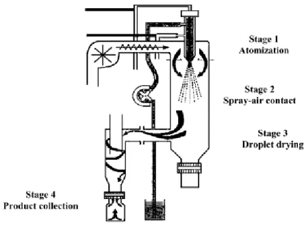
Spray drying has a great utility in pharmaceutical industry, mainly in the production of solid dispersions 43, 58, and recently, has also been explored to produce proliposomes for the
pulmonary delivery of drugs 38, 59-61. Spray drying is characterized by its ability to comprise in
one step the formation of particle and its drying, which allows a better control of the particle formation 18.
Spray drying converts liquid feed of the drug, which can be a solution, emulsion or suspension, into a dry powder 18. Spray drying involves the addition of the carrier material,
commonly a sugar, to an organic solution comprising the drug and lipids, and its subsequent spraying into a stream of heated air flow to remove the solvent 10, 58-59, 62. This process involves
four stages (Figure 15):
1) Atomization of the product into a spray nozzle; 2) Spray-air contact;
3) Drying of the spray droplets; 4) Collection of the solid product.
Figure 15 – Schematic representation of the stages in the spray drying technique (from 63). The reproduction of this figure was authorized.
46 The solutions subjected to spray drying are pumped and atomized into the drying chamber with a spray nozzle, and dried in a concurrent heated air flow that is later collected in a reservoir 43, 63. The large surface area of the droplets and the high temperature of the
drying air leads to the rapid solvent evaporation and formation of the solid dispersion in seconds, which might be fast enough to prevent phase separation and to allow thermolabile molecules to be converted into fine powder 10, 58.
Spray drying parameters exert a great influence in the powder characteristics, such as particle size, size distribution, shape, morphology and density, which might be optimized since spray drying has the ability to manipulate and control a variety of parameters, including the solvent composition, solute concentration, feed rates of solution and gas, temperature and droplet size 10, 18.
Spray drying usually produces drugs in the amorphous state, but occasionally the drug may be partially crystallized during processing 62.
Spray drying allows the production of a fine, dust free powder as well as agglomerated to particular specifications, within a narrow range of particles sizes, which makes it especially advantageous when it is necessary to prepare particles of uniform size and shape. Also it is cost effective, which makes it easy to scale up and suitable for both laboratory and industrial scale production of proliposomes 27, 43, 64.
Spray drying has also been used for the drying of liposomal formulations. Spray-dried liposomes of the drugs verapamil and metronidazole with mannitol as the carrier preserved the size distribution of liposomes and entrapment efficiency of the drug after a year of storage
65.
1.3.2. Proliposomes and anticancer drugs
It is known that the intravenous route is the preferred route for the administration of liposomes as drug delivery systems. The studies of proliposomal formulation to encapsulate anticancer drugs have also been applied to this route of administration. The anticancer drugs studied for encapsulation in proliposomal formulations are presented in Table III. The studied proliposomes exhibited good stability for at least 12 months 66. The encapsulation of
47 pharmacokinetic and tissue distribution profiles and increase the therapeutic efficiency when compared to an intravenous administration of the free drug 37, 67.
Table III - Anticancer drugs encapsulated in proliposomes.
Drug Indications Reference(s)
Adriamycin Solid tumors; malignant lymphomas; acute leukemia 68-69 Carboplatin
Ovarian cancer; small or non-small cell lung cancer; head and neck cancer; lung cancer
67
Docetaxel
Non-small cell lung cancer
48
1.3. Xanthones
Chemically, xanthones are a class of oxygenated heterocyclic compounds with a dibenzo--pyrone scaffold (Figure 16) 70-72.
Figure 16 - Xanthone scaffold (numbered according IUPAC).
Although xanthone (9H-xanthen-9-one) does not occur in nature, xanthone derivatives are frequently isolated as secondary metabolites from plants and microorganisms
71, 73. These oxygenated heterocycles are structurally related to other natural compounds with
the γ-pyrone scaffold: flavonoids (Figure 17A) and chromones (Figure 17B) 71.
Figure 17 - Scaffolds containing a γ-pyrone moiety: flavonoids (A) and chromones (B).
Xanthones derivatives from natural sources have attracted great interest due to associated large variety of pharmacological activities 71. However, naturally-occurring
xanthones present a relatively restricted structural diversity since the biosynthetic pathways limit the type and position of the substituents in the xanthone scaffold. Naturally occurring