C A S O C L Í N I C O
R A S H
,
F E V E R A N D P R O T E I N U R I AA F T E R A M O X I C I L L I N I N A S L E P A T I E N T
M. Couto,
*C. Duarte,
**A. Geraldes,
***Colleen Medeiros,
****L. Inês,
*****A. Malcata
******quently exposed to drugs, which, “per se” repre-sents a risk factor for DA8and thus normal control
would be inappropriate. Pope et al5studied DA in
SLE patients using self-report data from age and sex matched controls with inflammatory arthritis. It was found that, with the exception of skin rash for sulfonamides other DA were not more frequent in SLE. The other possibilities for the reported diffe-rences would be diffediffe-rences in the ethnicity of con-trol groups and the means of ascertaining allergic manifestations,5with medical record review1
repor-ting lower frequencies than self-reported.5Finally,
making the attribution of any allergic manifestation must deal with the fact that drug2,3may
exacerba-te or even induce SLE.9For instance, sulfonamides
may cause flares in SLE patients.9In addition some
manifestations, such as fever, rash and oral ulcers, might be attributed to DA but in fact may be a fea-ture of SLE exacerbation.
Case report
A 30-year-old Portuguese female law student pre-sented to our emergency room with a 3-day history of a progressive maculopapular rash that started over her legs, odynophagia, minimally productive cough and non-bloody diarrhoea 5-6 times a day. Three days before her admission, she had consul-ted her dentist for dental pain and was given the combination of amoxicillin 875 mg and clavulanic acid 125 mg bid. She had a history of a rash to pe-nicillin. After taking the second tablet, she develo-ped chills, malaise, myalgia and a rash over lower extremities.
The patient had a 12-year history of SLE mani-fested by malar rash, photosensitivity, oral ulcers and alopecia, leukopenia, polyarthritis and WHO class IV diffuse membranoproliferative nephritis. When she was seen 2 months before, her SLE disea-se activity measured by SLAM-R was 3 (Table I) with stable doses of prednisone 20 mg qd and azathio-prine 50 mg bid. She was also on captopril 50 mg
*Rheumatology Fellow at the Department of Rheumatology, Coimbra University Hospital and Assistant Professor of Physiology, Coimbra University School of Medicine, Coimbra, Portugal
**Rheumatology Fellow at the Department of Rheumatology, Coimbra University Hospital, Coimbra, Portugal
***Immuno-Allergology Fellow at the Department of Immunology and Allergy, Coimbra University Hospital, Coimbra, Portugal **** Clinical Medicine Pharmacist, Boston Medical Centre, Boston, Massachusetts USA
*****Consultant Rheumatologist at Department of Rheumatology, Coimbra University Hospital and Assistant Professor of Rheumatology at Health Sciences School of Medicine, Beira Interior University, Covilhã Portugal ******Head of the Department of Rheumatology, Coimbra University Hospital and Assistant Professor of Rheumatology at Coimbra University School of Medicine, Coimbra, Portugal
Abstract
We report a case of severe type IV hypersensitivity reaction to amoxicillin, which occurred in a person with a 12-year history of SLE. The present case il-lustrates the wide differential diagnosis in a SLE patient who presents with an allergic drug reac-tion. The attribution of the presenting symptoms to the underlying SLE and/or to the drugs used to tre-at SLE and coexisting conditions is a major chal-lenge.
Keywords: Systemic Lupus Erythematosus; Drug
Hypersensitivity; Amoxicillin; Simvastatin; Rhab-domyolysis.
Introduction
Allergic drug reactions may be increased in Syste-mic Lupus Erythematosus (SLE) patients. Some studies show that drug allergies (DA) are similar in SLE and controls,1,2others suggest that SLE patients
have an increased risk of DA.3-7One explanation for
discrepancies between these studies is the choice of controls and whether they are normal indivi-duals or disease controls. SLE patients are more
fre-M. C O U T O E C O L.
bid, simvastatin 20 mg qd, furosemide 20 mg qd and folic acid 5 mg qd.
In the emergency room, her physical examina-tion showed an alert but ill woman, bilateral con-junctival haemorrhages, generalized purpuric rash, and a temperature of 39.5º C. Her pulse was 111/min, her blood pressure was 103/70 mm Hg; the rest of her physical examination was normal.
Laboratory tests showed high values of serum creatinine (4.6 mg/L) and C-reactive protein (29.8 mg/dL), hypoalbuminemia (2.9 g/dL), eleva-ted SGOT (311 U/L), SGPT (86 U/L) and CPK (24220 U/L). Her lymphocytes (1.1x10³/µl) and
pla-telets (57x103/µL) were low (Table II). There was no
eosinophilia. Her urine dipstick showed 3+ protei-nuria and hematuria. Her arterial blood gases showed pH=7.39 [7.35-7.45]; paO2=118 mm Hg
[85-108]; pCO2=21.2 mm Hg [35-45] and HCO3=
12.7 mmol/L [21-29]. The chest X-ray showed dif-fuse bilateral lung interstitial infiltrates.
The presumptive diagnoses at emergency room were sepsis, drug allergic reaction to the antibio-tic, rhabdomyolysis with secondary acute renal fai-lure and a SLE flare. She received intravenous fluids, paracetamol, bicarbonate, imipenem, a 250 mg iv bolus of methylprednisolone followed by oral prednisone 60 mg qd, intramuscular clemastin and supportive care.
After admission to the rheumatology ward, prednisone was tapered from 60 mg qd to 20 mg over 13 days and she was given hydroxyzine 25 mg qd, furosemide 20 mg bid and albumin 1 g tid. Her skin lesions were treated with topical 0.1% betame-thasone. By the third day she became afebrile, wi-thout constitutional symptoms, skin lesions beca-me vesicular and epidermolysis occurred. Antibio-therapy was stopped at the 8thhospital day.
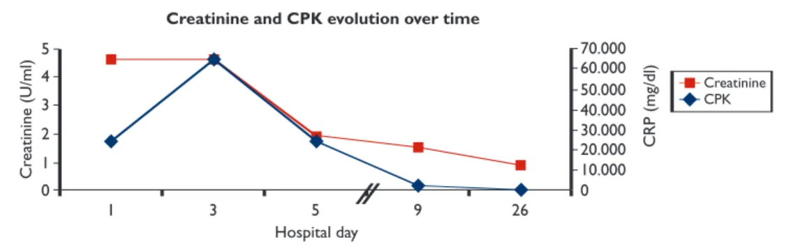
Labo-ratory values normalized as she recovered (Figures 1 and 2). Sputum was unobtainable and a subse-quent chest film showed clearing of the infiltrates present at admission.
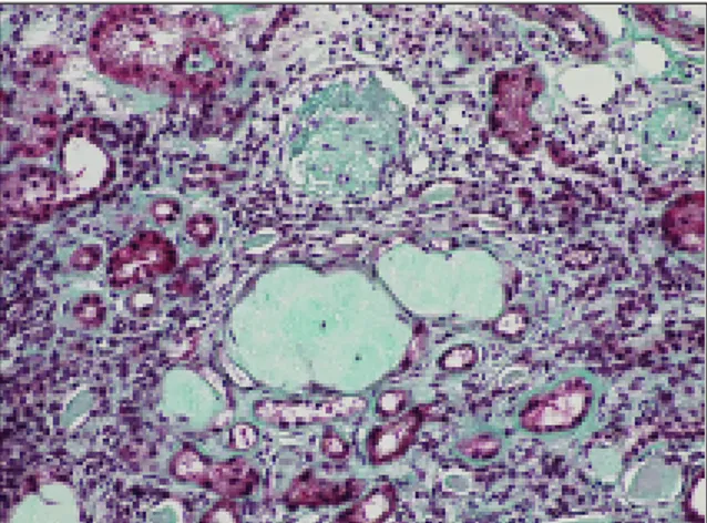
Blood, urine and stool cultures, serology for EBV, CMV, parvovirus B19 and coxsackie were negative. Anti-dsDNA antibody was 34.2 U/ml [<4.2 U/ml], C3 0.78 g/L [0.9-1.8] and C4 0.2 g/L [0.1-0.4]. She had a proteinuria of 2.88 g/day, which persisted after creatinine and CPK normalization. Except for proteinuria, urinalysis did not show abnormaliti-es (Table I); myoglobin, red cell casts and dysmor-phic red blood cells were absent. Her disease acti-vity measured by SLAM-R was 9. A repeat renal biopsy showed WHO class IV-C diffuse proliphera-tive glomerulonephritis, with diffuse and
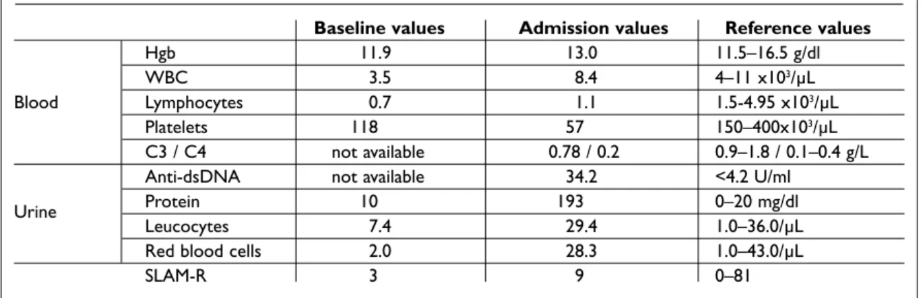
segmen-Table I. Comparison of laboratorial values at baseline and on admission
Baseline values Admission values Reference values
Hgb 11.9 13.0 11.5–16.5 g/dl
WBC 3.5 8.4 4–11 x103/µL
Blood Lymphocytes 0.7 1.1 1.5-4.95 x10³/µL
Platelets 118 57 150–400x103/µL
C3 / C4 not available 0.78 / 0.2 0.9–1.8 / 0.1–0.4 g/L
Anti-dsDNA not available 34.2 <4.2 U/ml
Urine Protein 10 193 0–20 mg/dl
Leucocytes 7.4 29.4 1.0–36.0/µL
Red blood cells 2.0 28.3 1.0–43.0/µL
SLAM-R 3 9 0–81
Hgb- haemoglobin;WBC- White blood cells; SLAM-R: SLE Activity Measure-Revised
Table II. Laboratorial values on admission
Reference Value values Creatinine 4.6 0.6-1 mg/dl CRP 29.8 0-0.82 mg/l Albumine 2.9 3.5-5 g/dl SGOT 311 5-34 U/L SGPT 86 5-34 U/L CK 24220 29-168 U/L Hgb 13.0 11.5-16.5 g/dl Leukocytes 8.4x10³ 4-11x103/µL Lymphocytes 1.1x10³ >1.5x10³/µL Platelets 57x103 150-400x103/µL
S L E A N D A L L E R G I C D R U G R E A C T I O N
tal sclerosis without active lesions (Figures 3 and 4). Intradermal skin tests confirmed type IV hyper-sensitivity to amoxicillin, ampicilin and dicloxa-cillin.
The patient was discharged at the 28thhospital
day treated with prednisolone 10 mg qid, furosemi-de 40 mg bid, enalapril 5 mg qid and azathioprine 50 mg bid. After discharge, the Nephrologist decided to change azathioprine for mycophenolate mofetil 1 g bid, stopped after one year due to lack of bene-fit. She continues her follow-up at the Rheumato-logy and NephroRheumato-logy Outpatient Clinics. She main-tains mild chronic renal insufficiency with a SLAM-R score of 3 (low disease activity) at last visit.
Discussion
In this patient several diagnoses had to be consi-dered. These were in order of likelihood: 1) amoxi-cillin hypersensitivity; 2) sepsis; 3) rhabdomyolysis from simvastatin precipitated by amoxicillin he-patotoxicity; 4) sepsis causing a lupus flare; 5) amo-xicillin- induced lupus flare or 6) some
combina-tion of the above.
Drug allergy (DA) to amoxicillin was the most probable precipitating event considering the pati-ent’s previous history, the temporal sequence of events and because allergic reactions to β-lactamic drugs are common. Our patient presented a type IV hypersensitivity reaction, which can occur from one hour to several days after re-exposure to the drug, more commonly 48-72 hours after.10This
re-action differs from an immediate hypersensitivity reaction, which generally appears within 12 minu-tes and no longer than one hour of an antigen chal-lenge with urticaria with or without angioedema or anaphilaxis.1There is evidence that the longer the
interval between drug intake and appearance of the reaction the less the probability of being IgE mediated. The patient had no family or personal history of atopy, which has been associated with an increased risk of DA.2The onset of maculopapular
lesions occurring after the re-exposure to antibio-tics is typical of Type IV hypersensitivity reactions.3
As the mechanism is not IgE-mediated, measuring IgE specific to amoxicillin would not help confirm the diagnosis. The use of skin testing, such as
in-0 1 1359 26 2 4 3 5 0 60.000 50.000 40.000 30.000 20.000 10.000 70.000 Cr ea tinine (U /ml) CR P (mg/ dl) Hospital day
Creatinine and CPK evolution over time
Creatinine CPK
Figure 1. Evolution of creatinine and CPK over time
0 200 13 59 26 400 600 800 1.000 0 25 30 20 15 10 5 35 SGO T a nd SG T P (U/ ml) CR P (mg /d l) Hospital day
CRP, SGOT and SGTP over time
SGOT SGTP CRP
M. C O U T O E C O L.
tradermal or patch testing is somewhat controver-sial and some contend that is particularly not help-ful in autoimmune diseases like SLE.3
Oral challenge test with amoxicillin is the diag-nostic gold standard test,4but in this patient it was
contraindicated because she had a severe event and testing was potentially dangerous or even fa-tal. In this case, the positive intradermal test to amoxicillin helped to confirm the diagnosis.
The other major differential was sepsis from her dental infection. High fever, hypotension and ele-vated CRP made sepsis diagnosis very likely and thus the decision to treat her immediately with an-tibiotic. Knowledge of a previous allergy to Benzyl penicillin could have prevented this hospitalizati-on; penicillin, amoxicillin and, to a lesser extent cephalosporins share extensive cross-reactivity.5
One might also question the use of imipenem du-ring her hospitalization as it is a β-lactamic and its cross-reactivity is not fully known.6,7
Rhabdo-myolysis is uncommon in amoxicillin DA, but is a known and rare adverse effect of simvastatin and thought to be dose-related. Hepatotoxicity has been described in amoxicillin and liver injury could interfere with the metabolism of simvastatin re-sulting in higher drug levels leading to muscle ne-crosis. Acute renal failure develops in 30-40% of patients with rhabdomyolysis. Suggested mecha-nisms include precipitation of myoglobin and uric acid crystals within renal tubules, decreased glo-merular perfusion, and the nephrotoxic effect of ferrihemate (formed upon dissociation of myoglo-bin in the acidic environment of the renal pa-renchyma).8
Infection2,7and drugs,9including antibiotics can
trigger a systemic lupus flare or the patient could have had a lupus flare not related to these. The pre-sence of chills, neutrophilia, leucocytosis are thought to be markers of infection rather than SLE11and our patient had these features. The
pati-ent had SLE class IV diffuse glomerulonephritis since ten years before. On this admission, presen-ce of fever, hypocomplementemia, lymphopenia, thrombocytopenia, elevated anti-dsDNA and al-most nephrotic-range proteinuria (compared to her baseline) favours a lupus flare. A non-renal lu-pus flare is likely to have occurred. The reversal of acute renal failure could relate to improvement of the drug reaction but a lupus nephritis flare could also have improved with the increase in steroid therapy. However, the argument for a lupus nephri-tis flare is weak, since the renal biopsy showed no evidence of active lesions. Furthermore, the trial with mycophenolate mofetil produced no impro-vement, as expected from the biopsy.
In conclusion, the most likely causal pathway for the most severe manifestations was a type IV hyper-sensitivity reaction due to amoxicillin causing he-patotoxicity leading to increased simvastatin pre-cipitating rhabdomyolysis and acute renal failure. Also, a non-renal lupus flare and possibly an infec-tion likely occurred in this complex clinical case.
Correspondence to: Maura Couto
Department of Rheumatology, Coimbra University Hospital, Coimbra, Portugal
E-mail: [email protected] Figure 3. Two hyaline glomeruli with global sclerosis;
chronic interstitial nephritis with fibrosis (PAS staining; magnification: 20x)
Figure 4. Two hyaline glomeruli and peritubular sclerosis
with epithelial atrophy (Masson tricromic staining; magnification: 20x)
S L E A N D A L L E R G I C D R U G R E A C T I O N
Acknowledgments
We are extremely grateful to Dr Matthew Liang for his insightful comments that much contributed for the case discussion.
References
1. Adkinson NF Jr, Yunginger JW, Busse WW, Bochner BS, Holgate ST, Simons FER. Drug Allergy. In Middle-ton`s Allergy, principles and practice. New York: Mos-by Co, 2003: 1679-1694.
2. Pope J. Frequency of adverse drug reactions in pa-tients with sytemic lupus erythematosus. J Rheum 2003; 30: 480-484.
3. Brockow K, Romano A, Blanca M, Ring J, Pichler W, Demoly P. General considerations for skin test proce-dures in the diagnosis of drug hypersensitivity. Al-lergy 2002; 57: 45-51.
4. Aberer W, Bircher A, Romano A et al. Drug provoca-tion testing in the diagnosis of drug hypersensitivity reactions: general considerations. Allergy 2003; 58: 854-863.
5. Zandman-Goddard G, Shoenfeld Y. Infections and SLE. Autoimmunity 2005; 38: 473-485.
6. Gomez MB, Torres MJ, Mayorga C, Demoly P, DeWeck A. Immediate allergic reactions to betalac-tams: facts and controversies. Curr Opin Allergy Clin Immunol 2004; 4:261-266.
7. Kishiyama JL, Adelman DC. The cross-reactivity and immunology of beta-lactam antibiotics. Drug Saf 1994; 10:318-327.
8. Craig S. Rhabdomyolysis. eMedicine: Specialties: Emergency Medicine. Updated in 2008.
9. Rubin RL. Drug-induced lupus. In Wallace, D J.; Hahn, BH. Dubois' Lupus Erythematosus. Baltimore: Lippincott Williams & Wilkins, 2007: 874-875. 10. Pichler WJ. Drug Hypersensitivity reactions:
Classifi-cation and relationship to T-Cell activation. In Drug Hypersensitivity. Basel: Karger, 2007: 168-189. 11. Stahl NI, Klippel JH, Decker JL. Fever in systemic
lu-pus erythematosus. Am J Med 1979; 67:935-940.