RESUMO.- [Parâmetros clínicos, hematológicos, de
pro-teína plasmática total e fibrinogênio em pinguins-de --magalhães (Spheniscus magellanicus)pré e pós-reabi -litação.] Pinguins-de-magalhães (Spheniscus magellanicus) geralmente chegam debilitados às praias do Brasil durante o inverno. A hematologia fornece informações valiosas so-bre estado clínico e imunidade dos animais. Os objetivos do presente estudo foram determinar o perfil hematológico, de proteína plasmática total (PPT) e fibrinogênio de pinguins --de-magalhães jovens e adultos no PROAMAR e no CETAS--SC, relacionando esses resultados com o estado de saúde e possibilidade de sobrevivência dos animais. Foram avalia
-Clinical, hematological, total plasma protein and fibrinogen
parameters of magellanic penguins (
Spheniscus
magellanicus
) pre- and post-rehabilitation
1Angela M. Coraiola2*, Cristiane K.M. Kolesnikovas3, Ricardo Krul4, Paulo R. Mangini5
and Rosangela Locatelli-Dittrich2
ABSTRACT.- Coraiola A.M., Kolesnikovas C.K.M., Krul R., Mangini P.R. & Locatelli-Dittrich R. 2014. Clinical, hematological, total plasma protein and fibrinogen parameters of magellanic penguins (Spheniscus magellanicus) pre- and post-rehabilitation. Pes-quisa Veterinária Brasileira 34(Supl.1):43-48. Laboratório de Patologia Clínica Veterinária, Departamento de Medicina Veterinária, Universidade Federal do Paraná, Rua dos Funcio -nários 1540, Curitiba, PR 80035-050, Brazil. E-mail: [email protected]
Magellanic penguins (Spheniscus magellanicus) usually arrive in poor body conditions at Brazilian beaches during the winter. Hematology provides valuable information about clinical and immunity status of the animals. The aims of this study were to determine the hematologic, total plasma protein (TPP) and fibrinogen profiles of young and adult ma -gellanic penguins in PROAMAR and CETAS-SC, relating these results with the state of health and survival possibility of the animals. In Paraná 14 animals were evaluated in pre and ei -ght in post-rehabilitation and 29 animals were evaluated in Santa Catarina after rehabilita -tion. Before rehabilitation, all animals showed weakness. In hematological exams of these animals, we found that anemia was present in 83% of the penguins that died and 50% of those which survived. The heterophils/lymphocytes (H/L) ratio was 3.87±0.57 in animals that died, significantly higher than the average of 2.20±0.30 for animals that survived. The -se two parameters are u-seful to as-sess the survival possibility of animals to rehabilitation. The body condition score was positively correlated with hematocrit and TPP, and negative -ly correlated with H/L ratio. After rehabilitation, the values were similar to other animals of the family Spheniscidae, with averages ranging from 1.64 to 1.90x10⁶ erythrocytes/µL;
43.38 to 48.80% of hematocrit; 12.45 to 13.52g/dL of hemoglobin; 8,684 to 14,011 leu -kocytes/µL; 4,767 to 8,041 heterophils/µL; 3,215 to 4,951 lymphocytes/µL; 95 to 655 eo -sinophils/µl; 179.8 to 277.9 monocytes/µL; 141 to 184.9 basophils/µL; and 1.26 to 1.74 of H/L ratio. These parameters can therefore be used as reference values and release para -meters for young and adult Magellanic penguins in captivity on the rehabilitation centers. INDEX TERMS: Spheniscus magellanicus, penguin, seabirds, hemograma, anemia, H/L ratio.
1 Recebido em 28 de setembro de 2014.
Aceito para publicação em 15 de dezembro de 2014.
2 Laboratório de Patologia Clínica Veterinária, Departamento de Medici -na Veterinária, Universidade Federal do Paraná (UFPR), Rua dos Funcio -nários 1540, Curitiba, PR 80035-050, Brasil. [email protected] (*Autor para correspondência), [email protected].
3 Associação R3 Animal, Rod. João Gualberto Soares s/n, Florianópolis, SC 88061-500, Brasil. [email protected]
4 Laboratório de Aves Marinhas, Centro de Estudos do Mar, Universidade Federal do Paraná (UFPR), Cx. Postal 61, Pontal do Paraná, PR 83255-976, Brasil. [email protected]
dos no Paraná 14 animais na pré e oito na pós-reabilitação e 29 animais em Santa Catarina após a reabilitação. Antes da reabilitação, todos os animais apresentavam debilidade. Nos exames hematológicos desses animais, observou-se que a anemia estava presente em 83% dos pinguins que foram a óbito e em 50% dos que sobreviveram. A relação heterófi -los/linfócitos (H/L) foi de 3,87±0,57 nos animais que foram a óbito, significativamente maior que a média de 2,20±0,30 dos animais que sobreviveram. Esses dois parâmetros são úteis na avaliação da possibilidade de sobrevivência dos animais à reabilitação. O escore corporal apresentou corre-lação positiva com hematócrito e PPT, e correcorre-lação negativa com relação H/L. Após a reabilitação os valores foram seme -lhantes aos de outros animais da família Spheniscidae, com médias variando de 1,64 a 1,90 x10⁶ eritrócitos/µL; 43,38 a
48,80% de hematócrito; 12,45 a 13,52g/dL de hemoglobi -na; 8.684 a 14.011 leucócitos/µL; 4.767 a 8.041 heterófilos/ µL; 3.215 a 4.951 linfócitos/µL; 95 a 655 eosinófilos/µL; 179,8 a 277,9 monócitos/µL; 141 a 184,9 basófilos/µL; e 1,26 a 1,74 de relação H/L. Esses parâmetros, portanto, po -dem ser utilizados como valores de referência e parâmetros para soltura para pinguins-de-magalhães jovens e adultos em cativeiro nos centros de reabilitação.
TERMOS DE INDEXAÇÃO: Spheniscus magellanicus, pinguim,aves
marinhas, hemograma, anemia, relação H/L.
INTRODUCTION
Magellanic penguins (Spheniscus magellanicus Forster, 1781) breed during the summer on the Patagonian main -land and the Falk-land Is-lands, and in winter they migrate north, reaching southern Brazil and Uruguay (Gandini et al. 1998, Pütz et al. 2007). The animals that arrive on beaches in Brazil are generally weak due to the long journey, diffi -culty in finding food, oil contamination and diseases (Silva --Filho & Ruoppolo 2006, Rodrigues et al. 2010).
Hematology is useful for assessing and diagnosing di -seases in birds (Coles 1986, Campbell 2007). Nevertheless, few studies have been conducted to determine hematologi -cal parameters of penguins in Central and South America. There are reference haematological values for Rock-hopper Penguins (Eudyptes crestatus), gentoo penguins ( Pygosce-lis papua) and the magellanic penguins on the Patagonian coast in the wild (Hawkey et al. 1989), Humboldt penguins (Spheniscus humboldti) in captivity and free-living in the coastal regions of Chile (Wallace et al. 1995, Villouta et al. 1997), free-living blue penguins (Eudyptula minor) (Ser-gent et al., 2004) and free-living galapagos penguins ( Sphe-niscus mendiculus) (Travis et al. 2006). No study assessed the animals during rehabilitation or established hematolo-gic parameters to determine release.
In a study conducted by Rodrigues et al. (2010) with magellanic penguins arriving along the coast of Rio Grande do Sul, contaminated by oil or not, the authors found that hematocrit and body weight of young animals are helpful indicators for the assessment of the health status and sur-vival chances during rehabilitation.
Currently, there are standardized protocols for treat -ment and rehabilitation of magellanic penguins in Brazil;
however, the clinical and hematological values used for de -termining release criteria of these animals are from resear-ch on rehabilitation of other species (Ruoppolo et al. 2004). This study was aimed at determining hematologic, total plasma protein (TPP) and fibrinogen profiles of young and adult magellanic penguins, pre and post rehabilitation, and relating the parameters to animals’ health and survival.
MATERIALS AND METHODS
The study was conducted at Pontal do Parana, in the state of Pa
-rana, with 14 magellanic penguins received in PROAMAR (Project for Studies and Recovery of Birds, Mammals and Reptiles), from the Center of Sea Research, Federal University of Paraná (UFPR) in the years 2010 and 2011; and at Florianopolis, in the State of Santa Catarina, with 29 magellanic penguins received in CETAS (Screening Center of Wild Animals), from the Brazilian Institute of Environment and Renewable Natural Resources (IBAMA) in 2011. The project was approved by the Ethics Committee on Animal Use - CEUA/UFPR under number 046/2010 and by (ICMBio) SISBIO
# 25013-2.
The rehabilitation period in both sites was from one to two months. In PROAMAR the animals were received in the period from June to October, and blood collections were carried out in
the beginning of the rehabilitation process and in the end of the
process from August to October. In CETAS-SC, the penguins were received from June to September, and the blood collections were carried out from September to November, in the end of the reha -bilitation process.
Animal handling during rehabilitation was different in the two locations. In PROAMAR, in the first days the animals were fed approximately 100g of fish per day, and this diet was increased gradually to about 500g of fish per day. The Penguins were fed discarded trash fish from shrimp fisheries or frozen sardines. Nu
-tritional supplements were provided with thiamine (vitamin B1) (75mg five times per week), and vitamins A (0.25ml twice a week) and E (0.5ml once a week). The most debilitated animals were fed liquid diets with the same supplementation.
In CETAS-SC, each animal was fed 500g of frozen sardines per day, and they were also provided nutritional supplement with thiamine (75mg doses three times per week). When debilitated, the animals were fed up to 10 sardines per day, plus oral rehydra
-tion. When they showed to be extremely weak, they received Pote
-nay Gold B12® (1ml) and two doses of Iron Injectable Ferrodex®,
with one week interval.
Deworming was performed at both sites after stabilizing the
animals. Drontal Plus® tablets were administered twice (1/4 of
660mg tablet) at an interval of 15 days.
Sanitary handling of the animals was performed similarly in the two sites, with daily cleaning of the rooms and swimming pool with water and sodium hypochlorite.
The release criteria after rehabilitation were different. In Pro
-amar, the general condition of the animals was assessed, including body weight measurements and inspection of the feathers (fea
-ther loss or damage), posture and good performance in the pool. In CETAS-SC, a complete clinical examination (with emphasis on auscultation) was performed using IFAW (International Found for Animal Welfare) criteria, which consider the general condition of the animals, waterproofing properties of feathers, hematocrit not less than 38%, retention of leukocytes in capillaries maximum of 2% and total protein level greater than or equal to 3g/dl (Silva
--Filho & Ruoppolo 2006).
-ter animal’s arrival) and in the end of the rehabilitation period.
The animals received in Santa Catarina were assessed only in the end of the rehabilitation period using physical examinations, body weight measurements and blood sample collections. The monitoring protocols were different due to the recovery criteria
used by the rehabilitation centers.
Physical examination was performed in physical restrained animals, assessing buccal and eye mucosa, inspecting their bodies
for injuries or oil stains and palpating the pectoral muscles to
de-termine the body condition allocating a body score, ranging from 1 (extremely thin, when the keel is very prominent and observed without palpation), 2 (thin, when the keel is not very prominent but can be felt) or 3 (normal, when the keel is not prominent or
easily felt).
Blood samples were collected after an 8 hour fast (PROAMAR)
and 24 hour fast (CETAS-SC) by puncturing the median metatarsal
vein. The fasting protocols were different due to different feeding methods used in the sites. Blood samples of approximately 3ml were collected from each animal into tubes containing the anti
-coagulant heparin. Blood smears were made immediately after sample collection on samples without anticoagulant.
The samples were transported under refrigeration to the La
-boratory of Veterinary Clinical Pathology, UFPR, for analysis. The hemogram included the erythrogram (erythrocytes count, hematocrit and hemoglobin concentration and RBC indices), the leucocyte count (Total leukocyte and differential leukocyte count) and thrombocytes count. Furthermore, the plasma concentra
-tions of TPP were determined and the plasma fibrinogen levels were estimated.
Erythrocytes and leukocytes counts were performed using
a hemocytometer (Improved Neubauer chamber®) with blood
samples diluted 1:20 in cresyl blue. The thrombocyte count
es-timate was performed by slide estimation method, determining the average number of thrombocytes per field. The hematocrit (Hct) was measured immediately using micro-haematocrit me -thod and blood centrifugation in capillary tubes at 9000 rpm for
5 minutes (Coles 1986). After completion of the hematocrit, plas
-ma was used to determine TPP concentrations and esti-mate the concentration of Fibrinogen in plasma by refractometry (Millar et al. 1971). Hemoglobin concentration was measured using cyano
-methaemoglobin method with centrifugation before reading the absorbance by spectrophotometer (Coles 1986).
The MCV (mean corpuscular volume) and MCHC (mean cor
-puscular hemoglobin concentration) were measured according to
Wintrobe (1933).
Blood extensions were stained with Wright stain and the sli
-des were examined with an optical microscope. The erythrocytes were examined for their color, size, shape, immaturity and for the
presence of inclusion bodies or parasites. A hundred erythrocytes
per slide were examined for determining erythrocytic nuclear ab -normalities (ENA) (micronuclei and budding) to calculate the
per-centage of ENA/100 erythrocytes. Differential leukocyte counts were performed on 100 leukocytes (heterophils, basophils, eosi
-nophils, lymphocytes and monocytes) and they were assessed for the presence of toxic changes, immature or degenerated cells and
the presence of inclusion bodies or parasites. Obtained cell counts
were used for calculation of the relative proportion of heterophils to lymphocytes. (H/L ratio).
The parameters were analyzed statistically with the software
BioEstat 5.0 using the Student’s t-test and linear regression analysis.
RESULTS
Fourteen penguins were monitored, 13 young and one adult, at PROAMAR. Physical examination before rehabi
-litation revealed that all animals had green diarrhea. Six animals that were severely debilitated died during the first week. The remaining eight animals were slightly to mode -rately debilitated and recovered within two months. Two penguins had skin lesions suggestive of trauma and four showed signs of oil contamination.
Twenty-nine magellanic penguins were monitored at CETAS in the State of Santa Catarina, in 2011, 19 of them were adults and 10 were young and all of them were cli -nically healthy. The rehabilitation period of these animals was from one to two months.
The results of haematological examinations, TPP, Fibri -nogen, body condition score and body weight of the animals, pre and post-rehabilitation, in PROAMAR and CETAS-SC are shown in Table 1. The blood smear-estimated thrombocyte count was not performed in animals from CETAS-SC, since all blood smears showed platelets aggregation.
In pre-rehabilitation, comparing the parameters of the animals that survived and those who came to death, there was a significant difference by Student’s t test (p <0.05) in the mean relative values of heterophils and lymphocytes and H/L ratio (Table 2).
Comparing pre and post-rehabilitation data only from the animals that survived in Parana, there were significant differences (p <0.05) between means of body weight, abso -lute and relative values of eosinophils and TPP (Table 3).
DISCUSSION
No significant difference (p>0.05) was identified in animal’s body weight pre-rehabilitation between animals who survived and those who died, which differs from the study conducted by Rodrigues et AL (2010), who found that the magellanic penguins received at the rehabilitation center in Rio Grande do Sul had significantly lower body weight on arrival, and that the animals that died during rehabilitation had lower body weight on arrival than those who survived.
After rehabilitation, the body weight of the animals in Parana was significantly lower than those in Santa Catari -na. This difference may have been due to different feeding methods used in the sites. The animals in Parana were fed discarded trash fish from shrimp fisheries. On the other hand, the animals in Santa Catarina were fed only sardines.
The main fish species discarded at Pontal do Parana are: Cathorops spixii, Selene setapinnis, Cynoscion spp.., Larimus breviceps, Paralonchurus brasiliensis and Stellifer spp. (Cat-tani et al. 2011). The sardines have higher levels of lipids, proteins and calories than Cynoscion spp. and P. brasilien-sis (Andrade et al. 2009, Martins & Oetterer 2010, Minozzo 2010). This fact may have been responsible for the higher body weight observed in animals after rehabilitation in Santa Catarina.
In the erythrogram of penguins assessed after reha-bilitation, there was significant variation (p<0.05) in the parameters according to age (young or adult) and site (or Parana Santa Catarina). The samples were collected at the two sites almost at the same time, so probably there was no variation in parameters influenced by the season. However, the feeding methods were different in the sites, which may have been the cause of the differences observed.
The values of erythrocytes count, hemoglobin count and hematocrit of these animals were similar to those obtained from other Family Spheniscidae penguins, captive and free living (Hawkey et al. 1989, Villouta et al. 1997, Sergent et al. 2004), including magellanic penguins (Hawkey et al. 1989).
Despite not having anemia, the penguins showed higher MCV values and the MCHC values were lower than those found in the family Spheniscidae’s literature (Hawkey et al. 1989, Villouta et al. 1997, Sergent et al. 2004), including free-living magellanic penguins, which showed MCV values of 215±34 fL and MCHC values of 33,1±2,8% (Hawkey et al. 1989).
The TPP of the animals in Parana was similar to healthy Humboldt penguins in captivity (Villouta et al. 1997) and healthy free-living blue penguins (Sergent et al. 2004). The animals in Santa Catarina showed TPP values significantly higher (p<0.05), possibly due to the sardine diet.
The levels of erythrocytes count, hemoglobin count and hematocrit in the penguins before rehabilitation in Parana indicate the presence of anemia, as they were significantly lower (p<0.05) than in the penguins assessed after rehabi -litation in Santa Catarina.
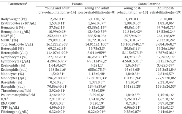
Table 1. Means and standard deviations of haematological parameters, total plasma protein, fibrinogen and body weight of young and adult magellanic penguins (Spheniscus magellanicus)
received at PROAMAR and CETAS-SC, pre- and post-rehabilitation
Parameters* Parana Santa Catarina
Young and adult Young and adult Young post- Adult post-pre-rehabilitation(n=14) post-rehabilitation(n=8) rehabilitation(n=10) rehabilitation(n=19) Body weight (kg) 2,26±0,1a 2,81±0,15b 3,39±0,1c 3,5±0,08c Erythrocytes (x106/µL) 1,53±0,11a 1,64±0,07ab 1,90±0,06b 1,83±0,06b Hematocrit (%) 37,5±2,15a 43,38±1,15ab 48,8±1,04b 47,74±0,71b Hemoglobin (g/dL) 10,99±0,93a 12,45±0,52ab 12,84±0,42ab 13,52±0,24b MCV1 (fL) 252,4±14,45a 266,5±8,95a 257,9±6,9a 264,1±6,69a MCHC2 (%) 29,09±1,54a 28,72±0,97a 26,3±0,57a 28,32±0,24a Total leukocyte (/µL) 16.122±2.368a 14.011±1.100ab 10.100±948,1ab 8.684±868,7b Heterophil (%) 69,21±2,84a 56,75±3,3b 50,8±3,29b 54,26±1,96b Heterophils (/µL) 11.407±1.902a 8.041±959ab 5.115±573,2b 4.767±516,1b Lymphocytes (%) 27,14±2,31a 36,25±3,51ab 44,9±3,52b 37,26±2,14b Lymphocytes (/µL) 4.204±637,7a 4.951±496,2a 4.568±531,3a 3.215±365,2a Eosinophils (%) 1,64±0,62ab 4,5±1,1a 1,0±0,49b 3,42±0,69ab Eosinophils (/µL) 243,5±116a 655±175,7b 95±48,65a 265,3±51,84a Monocytes (%) 1,5±0,51a 1,12±0,48a 1,8±0,84a 2,84±0,57a Monocytes (/µL) 196,2±88,28a 179,8±87,33a 181±90,94a 277,9±78,06a Basophils (%) 0,5±0,23a 1,37±0,5ab 1,5±0,4ab 2,21±0,44b Basophils (/µL) 70,86±46,81a 184,9±59,6a 141±38,28a 159,5±26,53a Thrombocytes/field 3,92±0,41a 4,75±0,59a - Policromatophils/field 3,46±0,59a 3,59±0,6a 1,8±0,33b 1,45±0,16b H/L3 2,91±0,37a 1,74±0,26b 1,26±0,17b 1,62±0,16b ENA/1004 0,93±0,3a 0,5±0,19a 0,7±0,3a 0,89±0,28a TPP5 (g/dL) 4,99±0,29a 6,15±0,28b 6,96±0,21b 6,81±0,12b Fibrinogen (g/dL) 0,32±0,04a 0,22±0,04ab 0,28±0,07ab 0,14±0,04b * Values are expressed as mean ± standard deviation. abc Different letters in each row indicate statistically significant diffe-rences (p<0,05). 1 MCV = Mean corpuscular volume, 2 MCHC = Mean corpuscular hemoglobin concentration, 3 H/L = hetero -phil/lymphocyte ratio, 4 ENA/100 = erythrocytic nuclear abnormalities in 100 erythrocytes, 5 TPP = Total plasma protein.
Table 2. Means and standard deviations of haematological parameters, total plasma protein, fibrinogen and body weight pre-rehabilitation of magellanic penguins (Spheniscus
magellanicus) received at PROAMAR
Parameters* Death (n=6) Survival (n=8) Body weight (kg) 2,13±0,14a 2,35±0,13a Erythrocytes (x106/µL) 1,52±0,2a 1,54±0,13a Hematocrit (%) 33,5±3,27a 40,5±2,5a Hemoglobin (g/dL) 9,93±1,48a 11,79±1,19a MCV1 (fL) 228,1±17,7a 270,7±19,99a MCHC2 (%) 29,18±2,08a 29,02±2,32a Total leukocyte (/µL) 19.988±3.486a 13.223±2.986a Heterophil (%) 75,83±3,81a 64,25±3,19b Heterophils (/µL) 15.114±2.815a 8.627±2.223a Lymphocytes (%) 21,17±2,06a 31,63±2,91b Lymphocytes (/µL) 4.335±860,8a 4.107±960a Eosinophils (%) 1,67±1,48a 1,62±0,26a Eosinophils (/µL) 310,2±279,4a 193,5±32,33a Monocytes (%) 1,17±0,98a 1,75±0,56a Monocytes (/µL) 207,2±186,2a 188±82,01a Basophils (%) 0,17±0,17a 0,75±0,37a Basophils (/µL) 22,17±22,17a 107,4±80,09a Thrombocytes/field 3,85±0,79a 3,97±0,47a Policromatophils/field 3,43±0,97a 3,49±0,8a
H/L3 3,87±0,57a 2,2±0,3b
ENA/1004 1,5±0,56a 0,5±0,27a TPP5 (g/dL) 4,73±0,6a 5,17±0,26a Fibrinogen (g/dL) 0,27±0,04a 0,36±0,06a * Values are expressed as mean ± standard deviation. ab Different
In the pre-rehabilitation, the combination of anemia and hypoproteinemia was more frequent in those that died (83%) than in those that survived (50%). Likewise, Rodri -gues et al. (2010), who monitored magellanic penguins du -ring rehabilitation in Rio Grande do Sul, found significantly lower hematocrit in the animals that died than in those who survived.
Erythrocytic nuclear abnormalities (ENA) were taken as biomarkers to assess exposure to petroleum hydrocar -bons and their derivatives (Netto et al. 2000). Yet, in the present study there was no correlation between the pre -sence of ENA and oil contamination.
After rehabilitation, the total leukocyte count in animals in Parana was significantly higher (p<0.05) than the young and adult animals in Santa Catarina. Villouta et al. (1997) found that the total leukocyte count varied according to the time of captivity in Humboldt penguins, and there was no evidence of disease in the animals. This study revealed that newly captured penguins had a mean total leukocyte count of 11,700±5,600 leukocytes/µL. After three weeks of cap -tivity this value increased to 15,900±4600 leukocytes/µL and after seven weeks of captivity it was 9,800±2,400 leu -kocytes/µL. The variation in the number of total leukocyte observed in the animals of this study can therefore be con-sidered normal.
In post-rehabilitation the animals showed a higher number of heterophils that lymphocytes, which was also
observed in healthy Humboldt penguins free-living (Walla -ce et al. 1995) and in captivity (Villouta et al., 1997), free --living blue penguins (Sergent et al. 2004) and free--living galapagos penguins (Travis et al. 2006).
Before rehabilitation, leukocytosis was more frequent in penguins that died (66%) than in those that survived (25%). All the animals that died showed relative and absolute hete -rophilia and relative lymphopenia. According to Hawkey et al. (1989), the absolute number of lekocyte is relatively stable in healthy adult birds, while the heterophils count is much more susceptible to variations under the influence of stress or microbial infection. The penguins with leukocytosis that survived showed heterophilia and absolute lymphocytosis.
The H/L ratio in animals post-rehabilitation was signi -ficantly lower (p<0.05) than in pre-rehabilitation. In the -se animals, the mean H/L ratio of penguins that died was 3.87±0.57, significantly different (p<0.05) from the mean 2.20±0.30 of those that survived.
Leukocyte profiles can be directly related to stress and the levels of circulating corticosteroids (Davis et al. 2008). Corticosteroid-induced leukocytosis reveals mature hete -rophilia (slight to moderate) and lymphopenia (Campbell 2007). Therefore, the heterophils/lymphocytes (H/L) ratio has been proposed as a sensitive measure of chronic stress in birds, although it also changes in case of diseases (Davis et al. 2008).
Hawkey et al. (1985) found that the H/L ratio in he -althy gentoo penguins was 2.3, whereas in animals with pododermatitis it was 4.4, demonstrating the correlation between this parameter and the disease severity. These data demonstrate that interpreting changes in leukocyte profiles can be problematic, especially if infection status is not known. The two factors are closely related: the stress is known for increasing vulnerability to disease and diseases for increasing stress (Davis et al. 2008).
Eosinophils numbers varied significantly (p<0.05) in post-rehabilitation among young (95±48.65/µL) and adult animals (265.3±51.84/µL) received in Santa Catarina and (655±175.72/µL) in Parana. This aspect has been observed by other authors in family Spheniscidae penguins. Villouta et al. (1997) found wide variation in this type of cells in he -althy Humboldt penguins, and animals in captivity for three days had an average of 400±600 eosinophils/µL, while ani -mals in captivity for 15 weeks had 1,100±900 eosinophils/ µL. Similarly, comparing pre and post-rehabilitation of ani -mals that survived in Parana, there were significant differen -ces (p<0.05) in relative and absolute values of eosinophils.
In mammals, eosinophils are responsible for immediate hypersensitivity reactions and for fighting parasitic agents. In birds, however, there is little correlation between eosi -nophilia and parasitic infections, and these cells seem not to contribute to acute hypersensitivity reactions. In this case, eosinophilia is often associated with inflammatory response (Mitchell & Johns 2008). In animals from this present study there was no apparent correlation between inflammatory response and eosinophilia.
The monocyte counts found in animals after rehabili-tation were slightly smaller, and the basophil values were similar to those previously reported for the family
Sphe-Table 3. Means and standard deviations of haematological parameters, total plasma protein, fibrinogen and body weight
pre and post-rehabilitation of magellanic penguins
(Spheniscus magellanicus) that survived at PROAMAR
Parameter* Pre-rehabilitation Post-rehabilitation
(n=8) (n=8)
Body weight (kg) 2,35±0,13a 2,81±0,15b Erythrocytes (x106/µL) 1,54±0,13a 1,64±0,07a Hematocrit (%) 40,5±2,5a 43,38±1,15a Hemoglobin (g/dL) 11,79±1,19a 12,45±0,52a MCV1 (fL) 270,7±19,99a 266,5±8,95a MCHC2 (%) 29,02±2,32a 28,72±0,97a Total leukocyte (/µL) 13.223±2.986a 14.011±1.100a Heterophil (%) 64,25±3,19a 56,75±3,3a Heterophils (/µL) 8.627±2.223a 8.041±959a Lymphocytes (%) 31,63±2,91a 36,25±3,51a Lymphocytes (/µL) 4.107±960a 4.951±496,2a Eosinophils (%) 1,62±0,26a 4,5±1,1b Eosinophils (/µL) 193,5±32,33a 655±175,7b Monocytes (%) 1,75±0,56a 1,12±0,48a Monocytes (/µL) 188±82,01a 179,8±87,33a Basophils (%) 0,75±0,37a 1,37±0,5a Basophils (/µL) 107,4±80,09a 184,9±59,6a Thrombocytes/field 3,97±0,47a 4,75±0,59 Policromatophils/field 3,49±0,80a 3,59±0,6a
H/L3 2,2±0,3a 1,74±0,26a
ENA/1004 0,5±0,27a 0,5±0,19a TPP5 (g/dL) 5,17±0,26a 6,15±0,28b Fibrinogen (g/dL) 0,36±0,06a 0,22±0,04a * Values are expressed as mean ± standard deviation. ab Different let
niscidae penguins (Hawkey et al. 1989, Wallace et al. 1995, Villouta et al. 1997, Sergent et al. 2004, Travis et al. 2006).
The function of Fibrinogen in birds, as in mammals, is activating the blood coagulation system and promoting systemic inflammatory response with nonspecific criteria (Harr, 2010). However, no correlations between increased levels of Fibrinogen and inflammatory response were ob -served in the animals assessed.
CONCLUSIONS
The body condition score was positively correlated with hematocrit and TPP, and negatively correlated with the H/L ratio.
The presence of anemia in magellanic penguins in their pre-rehabilitation indicates higher risk of death. Penguins with high H/L ratios (3.87±0.57) before rehabilitation are possibly at greater risk of death than those with lower H/L ratios (2.20±0.30). Feeding the penguins a diet of supple -mented sardines during rehabilitation seems to be more effective for full recovery and subsequent release.
The data suggest that the hematologic criteria for ani-mals release should be hematocrit above 45%, TPP greater than or equal to 6.0g/dL, total leukocyte count less than 10,000/µL and (H/L) ratio less than 1.5.
REFERENCES
Andrade G.Q., Bispo E.S. & Druzian J.I. 2009. Avaliação da qualidade nu -tricional em espécies de pescado mais produzidas no Estado da Bahia. Ciênc. Tecnol. Aliment. 29(4):721-726.
Campbell T.W. 2007. Hematologia de aves, p.215-247. In: Thrall M.A. (Ed.), Hematologia e Bioquímica Clínica Veterinária. Roca, São Paulo. Cattani A.P., Santos L.O., Spach H.L., Budel B.R. & Gondim-Guanais J.H.D.
2011. Avaliação da ictiofauna da fauna acompanhante da pesca do ca-marão sete-barbas do município de Pontal do Paraná, litoral do Paraná, Brasil. Bolm Inst. Pesca, São Paulo, 37(2):247-260.
Coles E.H. 1986. Veterinary Clinical Pathology. 4th ed. W.B. Saunders Com-pany, Philadelphia. 486p.
Davis A.K., Maney D.L. & Maerz J.C. 2008. The use of leukocyte profiles to measure stress in vertebrates: a review for ecologists. Funct. Ecol. 22:760-772.
Gandini P., Frere E. & Boersma P.D. 1998. Status and conservation of the magellanic penguin Spheniscus magellanicus in Patagonia, Argentina.
Bird Conserv. Int. 6:307-316.
Harr K.E. 2010. Overview of avian hemostasis, p.703-708. In: Weiss D.J. & Wardrop J. (Eds), Schalm’s Veterinary Hematology. 6th ed. Blackwell Publishing, Ames.
Hawkey C.M., Horsley D.T. & Keymer I.F. 1989. Haematology of wild penguins (sphenisciformes) in the Falkland Islands. Avian Pathol. 18:495-502.
Hawkey C.M., Henderson G.M. & Hart M.G. 1985. Haematological findings in captive gentoo penguins (Pygoscelis papua) with bumblefoot. Avian Pathol. 14:251-256.
Martins W.S. & Oetterer M. 2010. Correlação entre o valor nutricional e o preço de oito espécies de pescado comercializadas no estado de São Paulo. Bolm Inst. Pesca, São Paulo, 36(4):277-282.
Millar H.T., Simpson J.G. & Stalker A.L. 1971. An evaluation of the heat pre -cipitation method of fibrinogen estimation. J. Clin. Pathol. 24:827-830. Minozzo M.G. 2010. Patê de pescado: Alternativa para incremento da
pro-dução nas indústrias pesqueiras. Tese de Doutorado em Tecnologia de Alimentos, Setor de Tecnologia, Universidade Federal do Paraná, Curi -tiba, PR. 206p.
Mitchell E.B. & Johns J. 2008. Avian hematology and related disorders. Vet. Clin. North Am., Exotic Anim. Pract. 11:501-522.
Netto A.D.P., Moreira J.C., Dias A.E.X.O., Arbilla G., Ferreira L.F.V., Oliveira A.S. & Barek J. 2000. Avaliação da contaminação humana por hidro-carbonetos policíclicos aromáticos (HPAs) e seus derivados nitrados (NHPAs): uma revisão metodológica. Quim. Nova 23(6):765-773. Pütz K., Schiavini A., Rey A.R. & Lüthi B.H. 2007. Winter migration of
magellanic penguins (Spheniscus magellanicus) from the southernmost distributional range. Mar. Biol. 152:1227-1235.
Rodrigues S.C., Adornes A.C., Santos-Filho E.A., Silva-Filho R.P. & Colares E.P. 2010. Surviving probability indicators of landing juvenile magel-lanic penguins arriving along the southern Brazilian coast. Braz. Archs Biol. Technol.53(2):419-424.
Ruoppolo V., Adornes A.C., Nascimento A.C. & Silva-Filho R.P. 2004. Reabi -litação de pinguins afetados por petróleo. Revta Clín. Vet. 51(9):78-83. Sergent N., Rogers T. & Cunningham M. 2004. Influence of biological and
ecological factors on hematological values in wild little penguins, Eu-dyptula minor. Comp. Biochem. Physiol. A, Mol. Integr. Physiol. 138:333-339.
Silva-Filho R.P. & Ruoppolo V. 2006. Sphenisciformes (pinguim), p.309-323. In: Cubas Z.S., Silva J.C.R. & Catão-Dias J.L. (Eds), Tratado de Ani -mais Selvagens: medicina veterinária. Roca, São Paulo.
Sopezki M.S., Silva B.Z., Silveira D.T., Leite A.M., Silva-Filho R.P. & Bobro -wski V.L. 2007. Estudo da relação heterófilo/linfócito como marcador de estresse em pinguim-de-magalhães (Spheniscus magellanicus). Anais 16º Congresso de Iniciação Científica da UFPel, Pelotas, RS. (Re -sumo)
Travis E.K., Vargas F.H., Merkel J., Gottdenker N., Miller R.E. & Parker P.G. 2006. Hematology, serum chemistry, and serology of galapagos pen -guins (Spheniscus mendiculus) in the Galápagos Islands, Ecuador. J.
Wildl. Dis. 42(3):625-632.
Villouta G., Hargreaves R. & Rtveros V. 1997. Haematological and clinical biochemistry findings in captive humboldt penguins (Spheniscus hum-boldti). Avian Pathol. 26(4):851-858.
Wallace R.S., Teare J.A., Diebold E., Michaels M. & Willis M.J. 1995. Hema -tology and plasma chemistry values in free-ranging humboldt penguins (Spheniscus humboldti) in Chile. Zoo Biol. 14:511-316.