RESUMO.- [Intoxicação acidental e experimental por Closantel em ovinos no Uruguai.] Descreve-se um surto de intoxicação por Closantel em ovinos no Uruguai. O surto ocorreu em um lote de 1300 cordeiros que foram dosados
com uma solução de Closantel 10%, por via oral. Do total, 148 apresentaram sinais clínicos de intoxicação e 14 mor-reram. Os sinais clínicos incluíam midríase, nistagmo, refle -xo pupilar negativo, cegueira bilateral, pressão da cabeça contra objetos e desvio lateral da cabeça. Lesões macroscó-picas não foram observadas. Histologicamente havia vacuo-lização citoplasmática das células ganglionares e nas célu-las das camadas nuclear interna e externa. Na retina havia, também, diferentes graus de atrofia. Vacuolização e dege -neração axonal foram observados no nervo óptico, com áre-as multifocais de fibrose e infiltrado de linfócitos e céluláre-as Gitter. Quatro ovinos receberam experimentalmente duas, quatro e 10 vezes a dose terapêutica de Closantel (0,1 g/kg de peso vivo). Apenas os animais que receberam 10 vezes a dose recomendada apresentaram sinais clínicos. O exame histológico nos ovinos experimentais mostrou resultados
Accidental and experimental Closantel intoxication in
Uruguayan sheep
1Rodolfo Rivero2*, Carolina Matto2, Mauro Pereira Soares3 and María de Lourdes Adrien4
ABSTRACT.- RiveroR., Matto C., Soares M.P. & Adrien M.L. 2015. Accidental and experi- mental Closantel intoxication in Uruguayan sheep. Pesquisa Veterinária Brasileira 35(7):599-604. Laboratorio Regional Noroeste “Dr. Miguel C. Rubino”, Dirección de Labo-ratorios Veterinarios of Ministerio de Ganaderia, Agricultura y Pesca, Ruta 3, Km 369, Cx. Postal 57.037, CP 60.000, Paysandú, Uruguay.E-mail: [email protected]
An outbreak of Closantel intoxication in sheep in Uruguay is described. The outbreak occurred in a group of 1300 weaning lambs treated orally with a 10% solution of Closan -tel. One hundred forty eight lambs showed clinical signs of intoxication and 14 died. The clinical signs included mydriasis, nystagmus, and negative pupillary reflex, bilateral blind -ness, bump into objects, and lateral movement of the head. No macroscopic lesions were observed. The histological lesions of the retina were cytoplasmic vacuolization in ganglion cells and in cells of the inner and outer nuclear layers with different degrees of atrophy. Vacuolization and axonal degeneration were observed in the optic nerve, with multifocal areas of fibrosis and infiltration by lymphocytes and Gitter cells. To reproduce the intoxica -tion, four sheep were given two, four and 10 times the therapeutic dose of Closantel (0.1g/ kg of BW). Only the animals receiving 10 times the recommended dose showed clinical signs. The histological examination of the lesions in experimental sheep showed similar results to those described in the accidental outbreak, except for the absence of optic nerve fibrosis and inflammation, characterizing an acute phase. Axonal myelin sheaths loss, fibro -blasts and collagen fibers were observed in the ultrastructural study of the optic nerve of accidental intoxicated animals. The optic nerve of experimentally intoxicated animals had vacuoles that separated the myelin sheaths of axons. To prevent outbreaks it is suggested to weigh the animals before Closantel administration to avoid errors in dose calculation. INDEX TERMS: Closantel, antiparasitic overdose, poisoning, blindness, sheep.
1 Received on December 3, 2014.
Accepted for publication on June 1, 2015.
2 Laboratorio Regional Noroeste “Dr. Miguel C. Rubino”, Dirección de Laboratorios Veterinarios, Ministerio de Ganaderia, Agricultura y Pesca, Ruta 3, Km 369, Cx. Postal 57.037, CP 60.000, Paysandú, Uruguay. *Corres -ponding author: [email protected]
3 Laboratório Regional de Diagnóstico, Faculdade de Veterinária (FV), Universidade Federal de Pelotas (UFPel), Campus Universitário s/n, Pelo-tas, RS 96015-560, Brazil.
semelhantes aos do surto, com exceção da ausência de fi -brose e infiltrado inflamatório do nervo óptico, caracteri -zando um quadro agudo. Foram observadas a perda da bai-nha de mielina dos axônios e a presença de fibroblastos e fibras colágenas no estudo ultra-estrutural do nervo óptico de animais intoxicados espontaneamente. No nervo óptico de animais intoxicados experimentalmente havia vacúolos que separavam as bainhas de mielina dos axônios. Para evi-tar surtos, sugere-se pesar os animais antes da administra-ção de Closantel para evitar erros no cálculo da dose.
TERMOS DE INDEXAÇÃO: Closantel, sobredosagem, intoxicação,
cegueira, ovinos.
INTRODUCTION
Closantel is an antiparasitic drug belonging to the salicyla -nilide class used to control Haemonchus contortus, mature and immature forms of Fasciola hepatica and larvae of Oes-trus ovis (Gill et al. 1999). This drug is toxic to many spe -cies, including dogs (McEntee et al. 1995), sheep and goats (Gill et al. 1999, Barlow et al. 2002, Van der Lugt & Venter 2007, Ecco et al. 2008). A combination of Closantel and al-bendazole was also reported to be toxic for sheep and goats (Obwolo et al. 1989).
Accidental poisoning occurs when the drug is admi-nistered in amounts that are 2-6 times the recommended dose (Borges et al. 1999, Barlow et al. 2002). In all of these cases, the exact dose that induced intoxication was only estimated.
The clinical signs described in intoxicated sheep inclu -de lack of coordination, weakness, recumbency, mydriasis, blindness, walking in circles, depression, dark green diar-rhea and death (Obwolo et al. 1989, Borges et al. 1999, Gill et al. 1999, Barlow et al. 2002).
The most important macroscopic findings are restricted to the ocular globe and central nervous system. When they are observable, they include brain inflammation and con -gestion (Borges et al. 1999) and narrowing of the intraca-nalicular and rostral segments of the optic nerve (Gill et al. 1999, Barlow et al. 2002). The microscopic findings include bilateral degenerative changes in the retina, with detach-ment of the pigdetach-ment epithelium, loss of the photoreceptor cells and depletion of both outer and inner nuclear layers. Severe bilateral vacuolar degeneration of the intraorbital optic nerve, infiltration with Gitter cells and some perivas -cular cuffing may be observed (Barlow et al. 2002). Myelin edema in the optic nerve and optic tracts, with scattered foci of myelinic edema within the brainstem and cerebellar peduncles also was described (Gill et al. 1999).
The aim of this study was to describe the epidemiology, clinical signs, gross and histologic findings, and ultrastruc -tural features of an outbreak of accidental Closantel poi-soning in sheep and the experimental reproduction of the intoxication.
MATERIALS AND METHODS
Description of the outbreak. The Northwestern Regional La
-boratory of DILAVE “Miguel C. Rubino” was consulted in reference
to a case of collective neurological clinical signs in sheep.
Epide-miological information was obtained through interviews with the veterinarian responsible for the case and visits to the farm. Five affected sheep were necropsied between 2 and 12 months after the onset of clinical signs. All organs, including the central
ner-vous system, ocular bulb and optic nerve were fixed in 10% buffe
-red formalin, embedded in paraffin, sectioned at 5µm and stained with haematoxylin and eosin.
Experimental reproduction. Five Corriedale crossbreed
male sheep were used. These sheep were 6-year-old, castrated, with body weights (BW) between 37.5 and 44.5 kg. The treat
-ments performed are presented in Table 1. Daily clinical exami -nation was performed.
Between 5 and 10 minutes after the death of sheep 3 and 4,
a necropsy was performed and material was collected, including the central nervous system, ocular bulb and optic nerve for histo
-logical and ultrastructural study.
Ultrastructural study. Two sheep of the accidental outbreak that presented chronic clinical signs of blindness eight months af-ter the administration of Closantel were euthanized and
necrop-sied. Small fragments of the central nervous system, ocular bulb
and optic nerve were collected between 5 to 10 minutes after
eu-thanasia and placed in a solution of 2% glutaraldehyde and 2% paraformaldehyde buffered with 0.4 M cacodylate (pH 7.4). The tissues were subsequently fixed in osmium tetroxide 1% buffe
-red in 0.4 M sodium cacodylate (pH 7.4). Lastly, they were placed in Epon 812. The sections of tissue were stained with methylene blue. Ultrathin sections were stained with lead citrate and uranyl acetate and were examined by electron microscope Zeiss EM 10 at 60 kV.
The same procedure was performed for the collection of ma
-terial for ultrastructural study in the case of the experimentally
intoxicated sheep 3 and 4.
RESULTS
Description of the outbreak
The outbreak occurred on a farm in Tacuarembó, Uru -guay (31°43´5.22”S- 55°59´6.93”0) in 2011. From a total of 1300 Corriedale sheep, 148 lambs (morbidity 14%) pre -sented neurological clinical signs twenty days after oral administration of 10% Closantel (January 2011). Fourteen lambs died from accidents secondary to the neurological signs such as blindness and lack of coordination. Clinical signs included mydriasis, nystagmus, negative papillary reflex to light, bilateral blindness, lateral movement of the head and bump into objects (Fig.1AB). The ataxia was in -tensified when the animals were moved.
No gross lesions were found in the necropsies perfor-med on the five lambs. Microscopic lesions were main -ly restricted to the retina, characterized by cytoplasmic vacuolization of ganglion cells and cells of the inner and
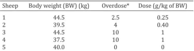
Table 1. Experimental reproduction: Identification of sheep, body weight and Closantel dose
Sheep Body weight (BW) (kg) Overdose* Dose (g/kg of BW)
1 44.5 2.5 0.25
2 39.5 4 0.40
3 44.5 10 1
4 37.5 10 1
5 40.0 0 0
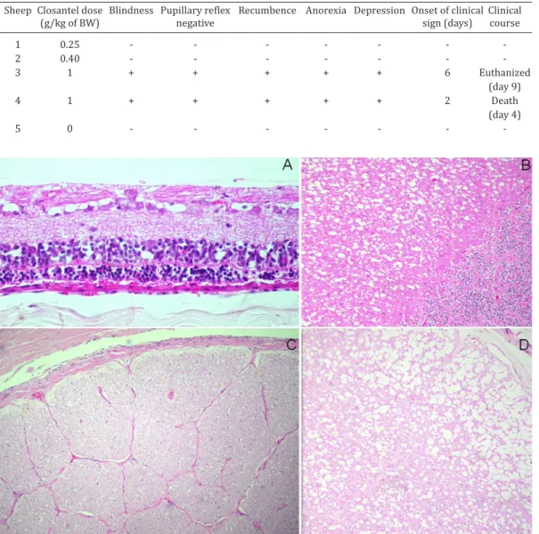
Fig.1. (A) Sheep accidentally intoxicated by Closantel bumping into a wire fence due to bilateral blindness. (B) Sheep with
marked dilatation of the pupil (mydriasis).
Fig.2. Sheep. Retina. Accidental Closantel outbreak. (A) Cyto -plasmic vacuolization of gan-glion cells and cells of the inner
and outer nuclear layer. HE,
obj. 40x. (B) Melanin pigment migration towards the inner re-gion of the retina. HE, obj. 25x. (C) Retina: Negative control animal. HE, obj. 10x. * Referen-ces: Pigmented epithelium (1),
Outer nuclear layer (2), Outer
plexiform layer (3), Inner nu
-clear layer (4), Inner plexiform layer (5), Ganglion cells (6), Layer of optic nerve fibers (7), HV = vitreous, E = sclera.
Fig.3. Sheep. Optical nerve. Accidental Closantel outbreak. (A) Cross section of the retrobulbar portion of the optic nerve. Fibroblast proliferation, neovascularization and Gitter cells (arrow). HE, obj.40x. (B) Infiltrate of lymphocytes and Gitter cells (arrow). HE, obj.40x.
outer nuclear layer. The outer plexiform layer also sho -wed vacuolization of the axons of the inner nuclear layer neurons (Fig.2A), associated with areas of pigmented epi-thelial hyperplasia and melanin pigment migration to the inner layers (Fig.2B). In the central nervous system, there was vacuolization (status spongiosus) of the white matter of the brain.
The optic nerve showed axonal degeneration (axonal vacuolization) with multifocal areas of fibrosis in the intra
-orbital portion and a lymphocytic monocytic (Gitter cells) infiltrate (Fig.3A and 3B).
Experimental reproduction
Bilateral edema of the intracanalicular portion of the optic nerve was macroscopically observed only in sheep 4. The histological lesions were similar in the sheep 3 and 4. There was vacuolization of ganglion cells and the cells of the inner and outer plexiform layers of the retina (Fig. 4A). In the central nervous system, there was vacuolization of the white matter of the cerebellum and brainstem. Vacuoli -zation and degeneration of axons was observed in the optic nerve (Fig.4B and 4D).
Ultrastructural study
In the optic nerve of accidental intoxicated sheep, most axons had lost their myelin sheath (demyelination). Fibro -blasts and collagen fibers were also observed replacing axons (Fig.5).
The lesion at the optic chiasm was similar to that obser -ved in the optic nerve (Fig.6), except for the lack of fibro -blasts and collagen fibers.
In the experimental reproduction, the spongy areas in
Fig.4. Sheep. Experimental reproduction of Closantel intoxication. (A) Sheep 3. Retina: cytoplasmic vacuolization of ganglion cells and
cells of the inner and outer nuclear layer. HE, 200x. (B) Sheep 4. Cerebellum: vacuolization of the white matter. HE, obj. 40x. (C).
She-ep 5. Optic nerve: negative control animal. HE, obj.20x. (D) Sheep 4. Optic nerve: vacuolization and axonal degeneration. HE, obj.40x. Table 2. Experimental reproduction: Closantel dose, clinical signs and evolution
Sheep Closantel dose Blindness Pupillary reflex Recumbence Anorexia Depression Onset of clinical Clinical
(g/kg of BW) negative sign (days) course
1 0.25 - - -
2 0.40 - - -
3 1 + + + + + 6 Euthanized
(day 9)
4 1 + + + + + 2 Death
(day 4)
5 0 - - -
-Fig.5. Sheep. Optical nerve. Accidental Closantel outbreak. (A*)
Axons without myelin sheath, (A) axon with myelin sheath.
The presence of fibroblast (F) and collagen fibers (C). Trans
Fig.7. Sheep. Experimental reproduction of Closantel intoxication.
Optic nerve. Axon with myelin layers separated by a large vacuole. Separation of the layers of myelin (detailed image). Transmission electron microscopy.
Fig.6. Sheep. Optic chiasm. Accidental Closantel outbreak. Swollen
axons without myelin sheath (A*) and a single axon with mye
-lin sheath (A). Transmission electron microscopy.
histological lesions were atrophy of the retina, pigment epithelial hyperplasia and cell pigment migration. Same le -sions have been reported by Van der Lugt & Venter (2007) in sheep and goats 56-70 days after of administration of Closantel. Degeneration of the optic nerve, with prolife-ration of fibrous tissue, was also observed. Van der Lugt & Venter (2007) also described vacuolar degeneration of myelin and eventually irreversible fibrosis of the optic ner -ve with contraction.
The spongy appearance of the white matter of the brain and spinal cord was also reported in spontaneous cases by Furlan et al. (2009). The spongy status induced by Closan -tel in the central nervous system is due to the formation of vacuoles between the layers of myelin. This lesion is also observed as a result of intoxication by substances such as ammonia (Cho & Leipold 1977) and copper (Morgan 1973), by ingestion of plants such as Stypandra imbricate (Huxta-ble et al. 1980, Main et al. 1981) and Helichrysum argyros-phaerum (Basson et al. 1975, Van der Lugt et al. 1996) or
by ingestion of fungi such as Stenocarpella maydis (Keller -man et al. 1991, Prozesky et al. 1994). The physiopathology of the separation of myelin layers is not known (Van der Lugt & Venter 2007). However, some authors report that oligodendrocyte death may play a role in the separation of myelin after administration of aniline in mice (Okazaki et al. 2001).
The retinopathy observed in Closantel poisoning, as well as in intoxication by Stypandra imbricata and Heli-chrysum argyrosphaerum, in sheep and goats occurs due to acute degenerative damage to the layers of the retina.The -se injuries would be a direct effect of the active substances and not a secondary effect of the optic nerve neuropathy (Gill et al. 1999).
Three possible causes have been suggested for the neuropathy induced by Closantel (Van der Lugt & Venter 2007), Stypandra imbricate (Huxtable et al. 1980) and Heli-chrysum argyrosphaerum (Van der Lugt et al. 1996). These
would be the direct effect on axons, edematous nerve com-pression within the optical channel, or a combination of both causes (Huxtable et al. 1980).
According to the literature, blindness is the primary cli -nical sign presented in animals intoxicated by Closantel as observed in the outbreak presented here, followed in some cases by ataxia and the death of some animals (Gill et al. 1999, Barlow et al. 2002, Van der Lugt & Venter 2007, Ecco et al. 2008, Furlan et al. 2009). In this accidental case, the-re wethe-re no deaths in the affected sheep probably because of the acute intoxication, consistent with findings by other authors (Borges et al. 1999, Gill et al. 1999, Furlan et al. 2009); this could be related to the dose utilized. The deaths that occurred were secondary accidents due to the blind -ness and the lack of coordination of the animals.
It was not possible to identify the toxic dose that caused cases of accidental intoxication. The recommended Closan -tel dose for sheep is 0.1g/kg of BW (Radostits et al. 2007). In some reports of intoxication, it has been estimated that the overdose sufficient to produce clinical signs must have been 3 to 6 times more the therapeutic dose (Borges et al. 1999) and, in other reports, 2 to 5 times more the thera-the optic nerve observed in thera-the histological study corres
-ponded to vacuoles that separated the myelin sheaths of axons (Fig.7). The vacuoles (single or multiple) were diffe -rent sizes and empty. Axonal swelling was also observed.
DISCUSSION
The lesions observed in both accidental and experimental intoxication are similar to those reported in Closantel into-xication (Gill et al. 1999, Barlow et al. 2002, Van der Lugt & Venter 2007). The lesions were mainly restricted to the retina, optic nerve and white matter of the central nervous system. The clinical signs manifested by these sheep were related to the specific lesions found.
peutic dose (Barlow et al. 2002). It is presumed that the dose error in the outbreak could have occurred because adult sheep (average 40 kg of BW) were dosed together with lambs (average 15 kg of BW) and the adult dose was administered in both cases. The fact that adult animals were not affected reinforces this idea. In other reports of Closantel-related poisonings, young animals, especially those younger than one year, are the main affected category (Van der Lugt et al. 1996, Borges et al. 1999, Barlow et al. 2002, Furlan et al. 2009).
Differential diagnoses of blindness in sheep should be undertaken in relation to other drugs such as rafoxanide (Prozesky & Pienaar 1977). Other diseases, such polioence -phalomalacia as a result of vitamin B1 deficiency and lead poisoning, also induce blindness, causing a characteristic laminar cortical necrosis of the gray matter. Other diseases that can also cause neurological clinical signs should also be differentiated, such as Listeria monocytogenes infection,
Coenurus cerebralis, Visna virus infection, rabies, intoxica -tion by Halimium brasiliense, toxemia of pregnancy in ewes
and intoxication by Ramaria flavo-brunnescens (Riet-Cor-rea & Méndez 2007, van der Lugt et al. 1996).All of these diseases have a particular epidemiology as well as gross and microscopic lesions that distinguish them from Closan-tel intoxication, with the exception of rafoxanide that could be confused. It is critical to send the ocular bulb, optic ner-ve and central nervous system to the laboratory for proper diagnosis of this type of intoxication.
In this outbreak, the epidemiology, clinical signs, gross and microscopic findings confirm the diagnosis of intoxi -cation by Closantel. It is recommended that, prior to the administration of this anthelmintic drug, maximum and minimum weight must be determined, especially in young animals. They should be dosed in small group sizes based on body weight to avoid dosing errors.
REFERENCES
Barlow A.M., Sharpe J.A.E. & Kincaid E.A. 2002. Blindness in lambs due to inadvertent closantel overdose. Vet. Rec. 151:25-26.
Basson P.A., Kellerman T.S., Albl P., Von Maltitz L.J., Miller E.S. & Welman W.G. 1975. Blindness and encephalopathy caused by Helichrysum
argy-rosphaerum DC. (Compositae) in sheep and cattle. Onderstepoort J. Vet.
Res. 42(4):135-147.
Borges A.S., Mendes C.N., Andrade A.L., Machado G.F. & Peiro J.R. 1999. Optic neutropathy in sheep associated with overdosage of closantel. Vet. Human Toxicol. 41(6):378-380.
Cho D.Y. & Leipold H.W. 1977. Experimental spongy degeneration in calves. Acta Neuropathol (Berl.). 39:115-127.
Ecco R., Barros C.S.L. & Graça D.L. 2008. Alterações oftálmicas associadas à intoxicação experimental por closantel em caprinos [Ophtalmic chan-ges associated with the experimental poisoning by closantel in caprine]. Arq. Bras. Med. Vet. Zootec. 60(1):42-50.
Furlan F.H., Lucioli J., Borelli V., Fonteque J.H., Stolf L., Traverso S.D. & Gava A. 2009. Intoxicação por closantel em ovinos e caprinos no Estado de Santa Catarina [Poisoning by closantel in sheep and goats in the State of Santa Catarina, Brazil]. Pesq. Vet. Bras. 29(1):89-93.
Gill P.A., Cook R.W., Boulton J.G., Kelly W.R., Vanselow B. & Reddacliff L.A. 1999. Optic neuropathy and retinopathy in closantel toxicosis of sheep and goats. Aust. Vet. J. 77(4):259-261.
Huxtable C.R., Dorling P.R. & Slatter D.H. 1980. Myelin oedema, optic neu -ropathy and retinopathy in experimental Stypandra imbricata toxicosis. Neuropathol. Appl. Neurobiol. 6(3):221-232.
Kellerman T.S., Prozesky L., Schultz R.A., Rabie C.J., van Ark H., Maartens B.P. & Lübben A. 1991. Perinatal mortality in lambs of ewes exposed to cultures of Diplodia maydis (= Stenocarpella maydis) during gestation. Onderstepoort J. Vet. Res. 58:297-308.
Main D.C., Slatter D.H., Huxtable C.R., Constable I.C. & Dorling P.R. 1981.
Stypandra imbricata (“blindgrass”) toxicosis in goats and sheep clinical
and pathologic findings in 4 field cases. Aust. Vet. J. 57(3):132-135. McEntee K., Grauwels M., Clerex C. & Henroteaux M. 1995. Closantel intoxi
-cation in a dog. Vet. Human Toxicol. 37:234-236.
Morgan K.T. 1973. Chronic copper toxicity of sheep: an ultrastructural study of spongiform leucoencephalopathy. Res. Vet. Sci. 15:88-95. Obwolo M.J., Odiawo G.O. & Ogaa J.S. 1989. Toxicity of a closantel-alben
-dazole mixture in a flock of sheep and goats. Aust. Vet. J. 66(7):229-230. Okazaki Y., Yamashita K., Sudo M., Tsuchitani M., Narama I., Yamaguchi R. & Tateyama S. 2001. The progression and recovery of neurotoxicity in -duced by a single oral dose of aniline in rats. J. Toxicol. Pathol. 14:19-28. Prozesky L., Kellerman T.S., Swart D.P., Maartens B.P. & Schultz R.A. 1994. Perinatal mortality in lambs of ewes exposed to cultures of Diplodia
maydis (= Stenocarpella maydis) during gestation. A study of the central
nervous system lesions. Onderstepoort. J. Vet. Res. 61(3):247-253. Prozesky L. & Pienaar J.G. 1977. Amaurosis in sheep resulting from treat
-ment with rafoxanide. Onderstepoort. J. Vet. Res. 44(4):257-260. Radostits O.M., Gay C.C., Hinchcliff P.D. & Constable P.D. 2007. Veterinary
Medicine. 10thed. Saunders Elsever, Spain. 2156p.
Riet-Correa F. & Méndez M.C. 2007. Plantas e micotoxinas que afetam a pele e outros órgãos [Plant and mycotoxins that affect the skin and other organs], p.206-209. In: Riet-Correa F., Schild A.L., Lemos R. & Borges J.R.J. (Eds), Doenças de ruminantes e equídeos [Diseases of ruminants and equines]. 3ªed. Editorial Palotti, Brasil.
Van der Lugt J.J., Olivier J. & Jordain P. 1996. Status spongiosis, optic neu-ropathy, and retinal degeneration in Helichrysum argyrosphaerum poi-soning in sheep and a goat. Vet. Pathol. 33:495-502.