Effects of
Bothrops moojeni
venom on
Leishmania amazonensis
promastigote forms
Castilhos P (1, 2), Pereira CG (1), Silva ALN (1), Napolitano DR (2), Oliveira F (2), Souza MA (1)
(1) Laboratory of Molecular Biology, Institute of Biomedical Sciences, Federal University of Uberlândia, Uberlândia, Minas Gerais State, Brazil; (2) Laboratory of Biophysics, Federal University of Uberlândia, Uberlândia, Minas Gerais State, Brazil.
Abstract: In the present work, the effect of Bothrops moojeni venom fractions on Leishmania promastigotes was evaluated. The snake venom was fractionated into five protein fractions (E1 to E5), by ion exclusion chromatography, that were used to treat Leishmania amazonensis and Leishmania braziliensis promastigote forms whereas the viability and nitric oxide production were evaluated. It was observed that E5 venom fraction strongly inhibited Leishmania amazonensis nitric oxide production, while in Leishmania braziliensis the nitric oxide production was enhanced in all doses. Bothrops moojeni crude venom reduced the viability of both parasites in a dose-dependent manner and a peptide of 64 kDa was apparently degraded. Bothrops moojeni E5 venom fraction only reduced the viability and nitric oxide production of Leishmania amazonensis and no protein degradation was observed. Thus, these results suggest that Bothrops moojeni E5 venom fraction may offer components with a promising antileishmanial therapeutic application.
Key words: Leishmania spp., Bothrops moojeni, snake venom, nitric oxide.
O
R
IG
IN
A
L
P
A
P
E
R
INTRODUCTION
Human parasitic infections – including malaria, sleeping sickness, Chagas’ disease, leishmaniasis, filariasis and schistosomiasis – have a devastating impact on global health and economic development (1). Leishmaniasis is a parasitosis caused by several species of the protozoa Leishmania and it is currently endemic in 88 countries (2, 3). The parasites are phagocytized by macrophages and converted into amastigotes, which multiply within and survive in parasitophorous vacuoles of these cells and infect surrounding macrophages (4). Moreover, the disease progression depends on which species of Leishmania is involved, as well as on the genetics and immune status of the host (5).
Parasites have different strategies to escape the host defense system and also to take advantage of host biochemical factors. In trypanosomatids,
a nitric oxide (NO) pathway mediates protection against apoptotic death (6). Although macrophages trigger the host defense mechanism to neutralize the parasite, evidence shows that NO pathway, from L (L.) amazonensis, participates on parasite-host interaction, whereas a correlation between NO production and the amount of metacyclic forms in the culture of infective forms was found (7). The involvement of Leishmania spp. and NO pathway in host-macrophage interaction showed the importance of both NO pathways in the parasite-host interplay.
All these drugs are limited to some extent by their toxicity, lack of efficacy in endemic areas, and difficulty of administration because of their long term treatment and high cost (9). The effect on parasite viability was evaluated with several drugs in vitro and in vivo. For example, treatment with imidocarb reduced parasite burden in experimental L. amazonensis infections in mice (10). Also, it has been demonstrated that Brazilian propolis reduced L. amazonensis infection in macrophages (4). Currently, the chemotherapy remains the mainstay for the leishmaniasis control, since effective vaccines have yet been developed (11).
In order to develop alternative tools to subdue infectious diseases, several substances have been tested for treating the parasite, such as snake venoms, aiming at the development of future drugs that disrupt the viability of protozoan. Snake venoms comprise a complex mixture of proteins that present various physiologic effects and provoke systemic alterations such as systemic bleeding, coagulopathy, hypovolemia, hemodynamic instability and shock, and acute renal failure (12, 13). Bothrops moojeni is frequently found in central and south-eastern Brazil, throughout the Cerrado morphoclimatic domain, and its venom contains a wide range of proteases, which may be characterized as coagulant, anticoagulant or fibrinolytic factors that inhibit cytokines and modulate the immune system of the host (14, 15). Bothrops jararaca venom leads to cell death of epimastigote forms of Trypanosoma cruzi induced by stress, and Bothrops moojeni crude venom was demonstrated to kill Leishmania spp. in vitro (16). A myotoxic phospholipase A2 homologue from B. moojeni venom, called MjTX-II, was characterized and displayed antiparasitic effects against Schistosoma mansoni and Leishmania spp. Thus, MjTX-II is a promising candidate for future therapeutic applications (17). In the present study, we evaluated the effect of B. moojeni venom and its fractions on viability, protein profile and on nitric oxide production of Leishmania promastigotes in vitro.
MATERIALS AND METHODS
Parasites
Leishmania (Leishmania) amazonensis (IFLA/
Control Center of the municipal authority of São Paulo, São Paulo state, Brazil and Leishmania (Viannia) braziliensis (MHOM/BR/75/M2903 strain) was obtained from the Infectious Diseases Center of the Federal University of Espírito Santo, Vitória, Brazil. Promastigote forms were grown in a brain heart infusion (BHI) medium (Oxoid, England) supplemented with 10% of fetal bovine serum (FBS) and 1 µg/mL gentamicin (Cultilab, Brazil) at 25°C. Once the culture reached the stationary phase, the parasites were harvested and centrifuged at 3,000 x g at 4°C for 15 minutes and then washed four times by centrifugation in a sterile phosphate buffered saline solution (PBS) pH 7.2. The concentration of parasites was adjusted to 2 x 10⁵/mL in BHI medium.
Fractionation of Bothrops moojeni Venom by Ion Exchange Chromatography
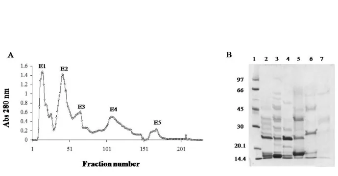
Desiccated Bothrops moojeni venom was purchased from Bio-Agents Serpentarium (Batatais, Brazil) and stored at –20°C until use. B. moojeni crude venom (200 mg) was dissolved in 0.05 M ammonium bicarbonate buffer (AMBIC), pH 7.8, and clarified by centrifugation at 10,000 x g for ten minutes. The supernatant was chromatographed on a DEAE Sephacel column (Sigma Chemical Co., USA) (1.7 X 15 cm), previously equilibrated with 0.05 M AMBIC (pH 7.8) and eluted with a concentration gradient (0.05 M to 0.3 M) of the same buffer. Fractions of 3.0 mL/tube were collected (flow rate of 20 mL/ hour) and their absorbances were read at 280 nm. The samples were pooled, lyophilized and stored at –20°C until the moment of use. Protein concentration was determined by Itzhaki and Gill (18) method using bovine serum albumin as standard.
Gel Electrophoresis
trypsin inhibitor (20.1 kDa) and α-lacto albumin (14.4 kDa). The slab gels were stained with Coomassie blue R-250. The relative molecular mass of the proteins was estimated by Kodak 1D image analysis software (USA).
Leishmania Promastigotes Treated with
Bothrops moojeni Crude Venom and Fractions
Leishmania promastigotes were cultured in BHI medium, in 96-well plates (2 x 10⁵ parasites/ well) and incubated at 25°C for 72 hours with different doses of Bothrops moojeni crude venom (25, 12.5, 3.125, 1.56 µg) or venom fractions (2.5, 1.25, 0.625, 0.3125 µg) or amphotericin B (25, 12.5, 6.25, 3.125, 1.56 µg) in triplicate or were not treated (BHI medium only; negative control). Parasite viability was analyzed by 3-(4.5-dimethylthiazol-2-yl)-2.5-diphenyltetrazolium bromide assay (MTT). After centrifugation of the plates the supernatant was collected and NO production was determined. These samples of treated parasites were also used to analyze the effect of Bothrops moojeni venom on protein profile of Leishmania. After centrifugation at 2,000 x g for
ten minutes, the pellet was treated with sodium dodecyl sulphate (SDS) and 2-mercaptoethanol and heated at 100°C for three minutes and run in 14 % polyacrylamide gel.
Leishmania Promastigote Viability Tests
Parasite viability was estimated by MTT assay (20). Briefly, L. amazonensis and L. braziliensis cultures in 96-well plates (treated or not with B. moojeni venom, E1, E2, E3, E4, E5 fractions or amphotericin B for 72 hours) were treated with 10 μL of MTT (Sigma-Aldrich, USA) at 5 mg/ mL and 90 μL BHI and incubated at 25°C for four hours. The reaction was stopped with 100 µL of isopropyl alcohol with 2.5 mM chloridric acid and incubated for ten minutes and the absorbance was read at a plate reader (Molecular Devices, USA) at 570 nm. All the assays were done in triplicate. The viability rate was calculated by the following equation:
% viability = ABS test x 100 ABS negative control
Nitric Oxide Measurement
NO production in supernatants of Leishmania culture was estimated by accumulation of NO2– (21). Briefly, 50 μL of Griess reagent [1 % sulfanilamide in 2.5 % H3PO4 and 0.1 % naphthylethylenediaminedihydrochloride (NEED) in 2.5 % H3PO4 (v/v)] was added to 50 μL of each sample in a 96-well plate and incubated at 25°C for 20 minutes. Blank reference and standard curve were determined. The absorbance was measured at 540 nm using a plate reader (Molecular Devices, USA). Nitrite content (μM) was quantified by extrapolation from sodium nitrite standard curve in each experiment. All the assays were performed in triplicate.
Statistical Analysis
Comparisons with control were performed by analysis of variance (one-way ANOVA). Significant differences were analyzed by the Tukey’s Multiple Comparison test (Statistics Package for Social Sciences, version 10.0). Differences were considered to be significant when p < 0.05.
RESULTS
Profile of B. moojeni Crude Venom and Protein Fractions
B. moojeni venom was fractionated into five protein fractions by ion exchange chromatography in a DEAE Sephacel column and the resulting fractions were denominated E1, E2, E3, E4 and E5 (Figure 1 – A). After electrophoresis of these proteins in polyacrylamide gel at the same concentration (Figure 1 – B), E1 was shown to contain peptides markedly stained, with molecular weight of 14, 15 and 24 kDa (Figure 1 – B). E2 fraction presented strongly stained bands corresponding to 15 e 24 kDa. Three major bands were also observed for E3 fraction, with molecular weights of 36, 25, 15 kDa. E4 showed bands modestly stained with 15, 27 and 46 kDa. The protein pattern of E5 was observed as bands with lower intensity but clearly stained with molecular weights of 15 and 35 kDa (Figure 1 – B).
Effect of B. moojeni Venom and Protein Fraction on Leishmania
B. moojeni crude venom and its fractions were
moojeni crude venom and E5 fraction presented inhibitory activities on parasite viability in a dose-dependent manner (Figure 2). L. amazonensis was demonstrated to be sensitive to crude venom (≥ 0.78 µg) and E5 fraction (≥ 0.156 µg) (Figure 2 – A and B). L. braziliensis was affected only by higher doses of crude venom (25 and 12.5 µg) and lower concentration of E5 fraction (0.156 µg), and showed enhancement in viability at the highest doses of E5 fraction (Figure 2 – A and B). Besides, both Leishmania strains were sensitive to amphotericin B in all analyzed doses (Figure 2 – C). Statistical analysis among amphotericin B, crude venom and E5 fraction showed to be significant (p < 0.001). On the other hand, comparison between venom and E5 fraction showed no difference.
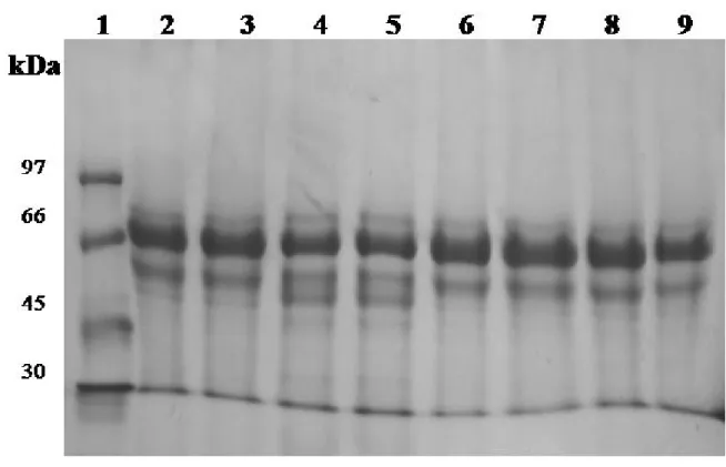
In addition, the protein pattern of Leishmania treated with crude venom, E5 fraction and amphotericin-B was analyzed on SDS-PAGE. An interesting protein profile of L. amazonensis treated with crude venom was observed when compared to non-treated parasites. More specifically, a peptide with 64 kDa was apparently degraded in two other peptides of 56 and 51 kDa (Figure 3 – A). However, when L. amazonensis and L. braziliensis were treated with E5 fraction or amphotericin B, no protein degradation was observed (Figure 3 – A and B).
Nitric Oxide Production by Leishmania
after Treatment with B. moojeni Venom and Protein Fractions
NO levels were determined in supernatants from cultures of L. amazonensis and L. braziliensis treated with B. moojeni crude venom, E5 fraction or amphotericin B in different doses. By analysis of L. amazonensis supernatants, it was observed that crude venom (Figure 4 – A) and E5 fraction, mainly, (Figure 4 – B) strongly inhibited NO production in different doses when compared with venom and amphotericin B (p < 0.001). On the other hand, when supernatants from L. braziliensis were analyzed (Figure 4), crude venom showed irregular inhibition of NO production (Figure 4 – A), while it was observed enhancement in NO production by parasites treated with E5 fractions (Figure 4 – B) at any concentration. Treatment of L. braziliensis with amphotericin-B led to enhance of NO production in a dose-Figure 4. Effect of treatment on NO production by
L. amazonensis (PH8) and L. braziliensis (M2903). NO levels were determined in supernatants from cultures of Leishmania treated with (A) B. moojeni crude venom, (B) B. moojeni venom E5 fraction and (C) amphotericin B, in different doses, or without treatment (medium). Columns shown nitrite (NO2) levels (means ± standard deviations from three independent experiments) expressed in µM.
DISCUSSION
B. moojeni Venom Fractioning and Its Effect on Parasite Viability and Protein Pattern Expression
Biochemical studies in Leishmania spp. have focused on the extracellular promastigote form, instead of the intracellular amastigote form, since promastigotes are easier to culture in vitro. In our study, the assessment of the effects of B. moojeni venom on Leishmania promastigotes was carried out after DEAE-Sephacel chromatography. Venom fractionation by ion exclusion chromatography resulted in five fractions. A strongly stained band of 15 kDa observed in E1 fraction could probably be myotoxin (22, 23). E2 fraction presented a band corresponding to 24 kDa, and it could be metalloproteinase and the proteins present in this fraction showed substantial proteolytic activity towards azocasein and fibrinogen (data not shown). Three major bands were observed for E3 fraction, and the 36 kDa peptide appears to belong to the proteases family with an anti-thrombosis effect (24). When analyzing the protein pattern of E5 fraction, bands with lower intensity but clearly stained with molecular weight of 15 and 35 kDa were observed. However, these fractions that were detected could be corresponding to L-amino acid oxydase (LAAO), a 70 kDa protein, responsible for the yellow stain of the crude venom and also known for its anti-parasite effect (25).
In this study, the effect of B. moojeni venom on Leishmania viability was tested using MTT assay for cell viability after incubation with crude venom and its fractions obtained by chromatography. We demonstrated that crude venom reduces viability of both species, although L (L.) amazonensis was demonstrated to be sensitive to crude venom (≥ 0.78 µg) and L (V.) braziliensis was demonstrated to be sensitive only in the presence of higher doses of crude venom (25 and 12.5 µg). Among the fractions, only E5 affects L. amazonensis viability (≥ 0.156 µg). These results indicate that the crude venom and its fractions act differently on each species of parasite. It is possible to explain the aforementioned differences by considering the contribution of the parasite genotype to parasite virulence and pathogenesis (26).
B. moojeni venom also influenced the protein pattern of Leishmania spp. As a variable protein profile for Leishmania spp. treated or not with B. moojeni venom and its fractions was observed
by SDS-PAGE. In fact, Leishmania proteins identified by various methods in various species have a wide range of characteristics (27). A band of 64 kDa presented a different profile after treatment with crude venom. These results suggest that B. moojeni venom changed the proteins from Leishmania either by inducing the production of new proteins or leading to degradation of proteins commonly produced. It was also observed that the crude venom changed the protein pattern of components from BHI medium supplemented with 10% FSB (data not shown). Based on these observations, we believe that the venom probably modified the medium components and then affected parasite growth due to environment changes. Moreover, it could lead to a parasite physiologic alteration with impact on its adaptation (28).
Crude Venom and Its Fractions Inhibited NO Production in Cultures of Leishmania amazonensis
Amphotericin B acts on ergosterol, a steroid present in the membrane of Leishmania, by increasing the permeability of the cell membrane. As consequence, it promotes an ion flux into the parasite, thus leading to parasite death (29). B. moojeni venom appears to act in a different way. Gonçalves et al. (30) demonstrated mitochondrial alterations by electronic microscopy after treatment with Bothrops jararaca venom and a possible explanation for the observed mitochondrial swelling may be that venom inhibited the parasite respiratory chain, thus lowering ATP levels, leading to mitochondrial swelling and finally to parasite death.
This report demonstrated in vitro effects of B. moojeni crude venom and its protein fractions on L. amazonensis and L. braziliensis promastigote forms. Our data corroborate results previously obtained by Tempone et al. (25), who showed that L-amino acid oxidase from crude venom of B. moojeni presented a in vitro killing effect against Leishmania spp. Our data showed that in vitro treatment of L. amazonensis promastigotes with B. moojeni E5 venom fraction and crude venom inhibited NO production and modified the protein expression profile. In addition, we observed reduced promastigote viability after treatment. Taken together, these data suggest that B. moojeni E5 venom fraction could have components with a promising leishmanicidal activity.
COPYRIGHT
© CEVAP 2011
SUBMISSION STATUS
Received: November 5, 2010.
Accepted: February 10, 2011.
Abstract published online: February 10, 2011.
Full paper published online: May 31, 2011.
CONFLICTS OF INTEREST
There is no conflict.
FINANCIAL SOURCE
The National Council for Scientific and Technological Development (CNPq) and The State of Minas Gerais Research Foundation (Fapemig) provided the financial grants.
CORRESPONDENCE TO
MARIA A. SOUZA, Instituto de Ciências Biomédicas, Universidade Federal de Uberlândia, Av. Pará, 1720, Campus Umuarama, bloco 6T07, Uberlândia, MG, 38.400-902, Brasil. Phone: +55 34 3218 2549. Fax: +55 34 3218 2333. Email: [email protected].
REFERENCES
1. Peters N, Sacks D. Immune privilege in sites of chronic infection: Leishmania and regulatory T cells. Immunol Rev. 2006;213(1):159-79.
2. Murray HW, Berman JD, Davies CR, Saravia NG. Advances in leishmaniasis. Lancet. 2005;366(9496):1561-77.
3. World Health Organization (WHO) [homepage]. Geneva: WHO; 2011 [cited 2010 Nov 5]. Leishmaniasis. Available from: www.who.int/ leishmaniasis/en/.
4. Ayres DC, Marcucci MC, Giorgio S. Effects of Brazilian propolis on Leishmania amazonensis. Mem Inst Oswaldo Cruz. 2007;102(2):215-20. 5. McConville MJ, Handman E. The molecular
basis of Leishmania pathogenesis. Int J Parasitol. 2007;37(10):1047-51.
6. Piacenza L, Peluffo G, Radi R. L-Arginine-dependent suppression of apoptosis in
Trypanosoma cruzi: contribution of the nitric
oxide and polyamine pathways. Proc Nat Acad Sci. 2001;98(13):7301-6.
7. Genestra M, Guedes-Silva D, Souza WS, Cysne-Finkeltein L, Soares-Bezerra RJ, Monteiro FP, et al. Nitrite oxide synthase (NOS) characterization
in Leishmania amazonensis axenic amastigotes.
Arch Med Res. 2006;37(3):328-33.
8. Amato VS, Tuon FF, Bacha HA, Amato Neto V, Nicodemo AC. Mucosal leishmaniasis. Current scenario and prospects for treatment. Acta Trop. 2008;105(1):1-9.
9. Davies CR, Kaye P, Croft SL, Sundar S. Leishmaniasis: new approaches to disease control. Brit Med J. 2003;326(7385):377-82.
10. Rodrigues FH, Afonso-Cardoso SR, Gomes MAB, Belleti ME, Rocha A, Guimarães AHB, et al. Effects of imidocarb and levamisole on the experimental infection of BALB/c mice by
Leishmania (Leishmania) amazonensis. Vet Par. 2006;139(1-3):37-46.
11. Palatnik-de-Sousa CB. Vaccines for leishmaniasis in the fore coming 25 years. Vaccine. 2008;26(14):1709-24.
12. Estevão-Costa MI, Diniz CR, Magalhães
metalloproteinases mutalysin i and ii on several components of the hemostatic and fibrinolytic
systems. Thromb Res. 2000;99(4):363-73.
13. Gutiérrez JM, Escalante T, Rucavado A. Experimental pathophysiology of systemic alterations induced by Bothrops asper snake venom. Toxicon. 2009;54(7):976-87.
14. Marsh NA, Williams V. Practical applications of snake venom toxins in haemostasis. Toxicon. 2005;45(8):1171-81.
15. Costa LA, Fornari MC, Berardi VE, Miles HA, Diez RA. In vivo effect of snake phospholipase A2 (crotoxin + cardiotoxin) on serum IL-1a, TNF-a and IL-1ra level in humans. Immunol Lett. 2001;75(2):137-41.
16. Deolindo P, Teixeira-Ferreira AS, Melo EJT, Arnholdt ACV, Souza W, Alves EW, et al.
Programmed cell death in Tripanosoma cruzi
induced by Bothrops jararaca venom. Mem Inst
Oswaldo Cruz. 2005;100(1):33-8.
17. Stábeli RG, Amui SF, Sant’ana SD, Pires MG,
Nomizo A, Monteiro MC, et al. Bothrops
moojeni myotoxin-II, a Lys49-phospholipase A2
homologue: An example of function versatility of snake venom proteins. Comp Biochem Physiol
Part C Toxicol Pharmacol.2006;142(3-4):371-81.
18. Itzhaki RF, Gill DM. A micro-biuret method for estimating proteins. Anal Biochem. 1964;9(4):401-10.
19. Laemmli UK. Cleavage of structural proteins during the assembly of the head of bacteriophage T4. Nature. 1970;227(5259):680-5.
20. Mosmann T. Rapid colorimetric assay for cellular growth and survival: application to proliferation and cytotoxicity assays. J Immunol. 1983;65(1-3):55-63.
21. Green LC, Wagner DA, Glogowki J, Skipper PL, Wishnor JS, Tannenbaum SR. Analysis of nitrate, nitrite, and [15N] nitrate in biological fluids. Anal Biochem. 1982;126(1):131-8.
22. Soares AM, Andrião-Escarso SH, Ângulo Y, Lomonte B, Gutiérrez JM, Marangoni S, et al. Structural and functional characterization of a myotoxin I from Bothrops moojeni (caissaca) snake venom. Arch Biochem Biophys. 2000; 373(1):7-15.
23. Soares AM, Rodrigues VM, Homsi-Brandeburgo MI, Toyama MH, Lombardi FR, Arni RK, et al. A rapid procedure for the isolation of the
Lys-49 myotoxin II from Bothrops moojeni
(caissaca) venom: biochemical characterization, crystallization, myotoxic and edematogenic activity. Toxicon. 1998;36(3):503-14.
24. Bernardes CP, Santos-Filho NA, Costa TR, Gomes MS, Torres FS, Costa J, et al. Isolation and structural characterization of a new fibrin(ogen) olytic metalloproteinase from Bothrops moojeni
snake venom. Toxicon. 2008;51(4):574-84. 25. Tempone AG, Andrade HF Jr, Spencer PJ,
Lourenço CO, Rogero JR, Nascimento N.
Bothrops moojeni venom kills Leishmania spp.
with hydrogen peroxide generated by its L-amino acid oxidase. Biochem Biophys Res Commun. 2001;280(3):620-24.
26. Smith DF, Peacock C, Cruz AK. Comparative genomics from genotype to disease phenotype in the leishmaniasis. Inter J Parasitol. 2007;37(11):1173-86.
27. Kubar J, Fragaki K. Leishmania proteins derived from recombinant DNA: current status and next steps. Trends Parasitol. 2006;22(3):111-6.
28. Clayton CE. Life without transcriptional control? From fly to man and back again. EMBO J. 2002;21(8):1881-8.
29. Ellis D. Amphotericin B: spectrum and resistance. J Antimicrob Chem. 2002;49(Suppl. 1):7-10. 30. Gonçalves AR, Soares MJ, Souza W, DaMatta RA,
Alves EW. Ultrastructural alterations and growth inhibition of Trypanosoma cruzi and Leishmania
major induced by Bothrops jararaca venom.