The role of complement in the early
phase of
Leishmania
(
Leishmania
)
amazonensis
infection in BALB/c mice
1Laboratório de Patologia de Doenças Infecciosas, LIM-50,
Departamento de Patologia, Faculdade de Medicina, Universidade de São Paulo, São Paulo, SP, Brasil
2Microbiology and Tumorbiology Center, Karolinska Institute, Stockholm, Sweden 3Departamento de Patologia, Faculdade de Medicina Veterinária e Zootecnia,
Universidade de São Paulo, São Paulo, SP, Brasil M.D. Laurenti1,
A. Örn2,
I.L. Sinhorini3 and
C.E.P. Corbett1
Abstract
Complement-depleted and -non-depleted BALB/c mice were inocu-lated with Leishmania (Leishmania) amazonensis promastigotes into the hind footpad to study the role of the complement system in cutaneous leishmaniasis. Total serum complement activity was meas-ured by hemolytic assay and C3 fragment deposit at the inoculation site was determined by direct immunofluorescence in the early period of infection, i.e., at 3, 24, 48 h and 7 days post-infection. The inflammatory reaction and the parasite burden were evaluated in the skin lesion at 7 and 30 days post-infection. Total serum complement activity decreased in the early phase of infection, from 3 to 24 h, in non-depleted mice compared to non-infected and non-depleted mice. C3 fragment deposit at the site of parasite inoculation was present throughout the period of infection in non-depleted mice. In contrast, no C3 fragment deposit was observed at the inoculation site in complement-depleted mice. Complement-depleted mice showed a significant decrease in the inflammatory response and a significant increase in the number of parasites (70.0 ± 5.3 vs 5.3 ± 1.5) at 7 days of infection (P < 0.05). A higher number of parasites were also present at 30 days of infection at the inoculation site of complement-depleted mice (78.5 ± 24.9 vs 6.3 ± 5.7). These experiments indicate that complement has an important role at the beginning of experimental cutaneous leishmaniasis caused by L. (L.) amazonensis by controlling the number of parasites in the lesion.
Correspondence
M.D. Laurenti
Departamento de Patologia Faculdade de Medicina, USP Av. Dr. Arnaldo, 455 1º andar, Sala 1209 01246-903 São Paulo, SP Brasil
Fax: +55-11-3081-7799 E-mail: [email protected]
Presented at the I Symposium on Advances in Medical Research, Institute of Medical Investigation Laboratories, HC-FMUSP, São Paulo, SP, Brazil, March 21-22, 2003.
Research supported by LIM-50, HC-FMUSP, FINEP and CAPES (PDEE 0025/95-20).
Publication supported by FAPESP.
Received June 12, 2003 Accepted October 14, 2003
Key words
•Cutaneous leishmaniasis •Leishmania (Leishmania)
amazonensis
•Complement •Pathology
Introduction
Leishmaniasis develops when Leishma-nia survives the nonspecific defense mech-anism or innate immunity, such as phagocy-tosis by inflammatory cells and the activity of the complement system. Subsequently,
the control or the progress of the lesions depends on the cell immune response.
promastigotes activate complement via the classical pathway (1,2), but most reports show that Leishmania activation of comple-ment is antibody independent by activation of lectin or alternative complement pathway (3,4). Promastigotes of cutaneous strains of Leishmania are susceptible to lysis by nor-mal serum, regardless of the presence of a specific antibody, suggesting that comple-ment activation in vitro occurs through the alternative pathway (5). The susceptibility of the parasite to complement lysis also de-pends on the growth phase of the culture. Most of the promastigotes in the logarithmic phase of culture growth are lysed by the action of normal human serum, while pro-mastigotes in the stationary phase, depend-ing on the parasite species, may not be lysed (6). Serum resistance of infective promasti-gotes is not caused by their failure to bind and activate complement since both loga-rithmic and stationary forms have the ability to do it efficiently (7). Instead, serum resis-tance appears to be linked to the expression of surface antigens which differ between these two phases of parasite growth (8,9). Amastigotes of cutaneous strains are more susceptible to complement lysis than vis-ceral strains, suggesting the important role of complement factors in limiting the infec-tion in the skin (10).
On the other hand, opsonization of para-sites by fragments of complement, such as iC3b and C3b, may enhance phagocytosis by specific receptors on the macrophage cell surface, which favors parasite survival, since phagocytosis through CR1 and CR3 receptors is considered to be inefficient in triggering the oxidative burst (11). In sup-port of these data, an imsup-portant role of complement invisceral leishmaniasis estab-lishment in hamsters has been previously shown (12).
In order to study the role of the comple-ment system in cutaneous leishmaniasis, complement-depleted and -non-depleted BALB/c mice were inoculated
subcutane-ously with promastigotes of Leishmania (Leishmania) amazonensis. Total serum complement activity and the C3 deposit in the skin lesion were studied together with the features of the inflammatory reaction and the parasite burden in the skin lesion during the early period of infection.
Material and Methods
Animals
Male BALB/c mice aged 8 to 10 weeks from the General Colony of the São Paulo University Medical School were kept in our laboratory during the experiments.
Parasite
Promastigotes of L. (L.) amazonensis, HSJD-1 strain, were used. Promastigotes in the stationary phase from the 3rd culture passage in supplemented RPMI 1640 medi-um (10% fetal calf sermedi-um, 5 mM HEPES, 50 µg/ml gentamicin, and 100 U/ml penicillin) were harvested and washed three times in sterile saline (1500 g for 10 min). The para-site concentration was adjusted to 2 x 10⁸ promastigotes/ml and 50 µl was injected subcutaneously into the hind footpads of the mice.
Infection
and 48 h after parasite inoculation. A non-infected group was used as control. Sera and fragments from the inoculation site were taken at 3, 24 and 48 h and 7 days after inoculation to evaluate the complement ac-tivity and C3 fragment deposit, respectively. Fragments of skin were taken on the 7th and 30th day of infection to evaluate the inflam-matory reaction and the parasitism. Sera from non-infected BALB/c mice were collected as control for evaluation of complement he-molytic activity.
Complement hemolytic titration
The total complement activity was stud-ied in each serum individually by the usual hemolytic assay, using triethanolamine buf-fer solution (TBS) (13) and the values are reported as the mean for five animals per group. Blood was collected from the retro-orbital sinus by puncture, immediately im-mersed in ice, and centrifuged (200 g for 10 min at 5ºC), and the sera obtained were stored at -80ºC until use. The sera which presented high spontaneous hemolysis were discarded. One hundred microliters hemoly-sin at 1/50 dilution (anti-sheep red blood cell polyclonal antibody, produced and kindly provided by Dr. Tambourgi, Immunochemi-cal Laboratory of Instituto Butantan, São Paulo, SP, Brazil, which reacts at up to 50 times dilution as determined by hemaggluti-nation assay), 300 µl 5 x 10⁸ sheep red blood cell solution in TBS, 100 µl test sera and 300 µl TBS, all kept in tubes in an ice bath, were added. Tubes were shaken and then incu-bated in a water bath at 37ºC for 1 h. Next, 200 µl saline solution was added to the tubes in an ice bath and the tubes were centrifuged at 5ºC (200 g for 10 min). The absorbance of the supernatant solution was measured with a spectrophotometer at 541 nm (Micronal Protometer, São Paulo, SP, Brazil). Percent lysis was calculated by considering the posi-tive and negaposi-tive controls. One hundred mi-croliters of distilled water was added to the
positive control tube instead of 100 µl test serum, and 100 µl TBS was added to the negative control tube instead of 100 µl test serum.
Direct immunofluorescence assay
Complement C3 fragment deposits were evaluated by direct immunofluorescence methods using fluorescein-conjugated anti-mouse C3 goat IgG (Cappel, Aurora, OH, USA) at a concentration of 80 µg/ml, in frozen sections of tissue obtained from the inoculation site.
Histopathological studies
The lesion and the parasitism at the in-oculation site were evaluated on the 7th and 30th day of infection by light microscopy in paraffin-embedded section stained with he-matoxylin and eosin. Morphometric analy-sis was performed at three different levels of each section per animal using a graticule eyepiece in an area of 1 mm2 and an Olympus planapochromatic immersion objective lens (100X) (12).
Limiting dilution assay for parasite quantification
based on Poisson statistics using the ELIDA software.
Statistical analysis
The one-way ANOVA multiple range test was used with the SigmaStat Statistical Software version 1.0 to analyze the differ-ences in complement activity in the sera and the morphometric data of the lesion between experimental groups. Differences were con-sidered significant when P < 0.05.
Results
Serum complement evaluation
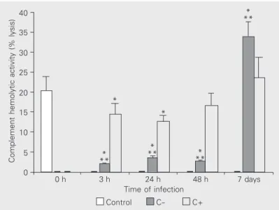
A decrease in complement hemolytic ac-tivity in the sera of mice infected with the parasite compared with non-infected con-trols was observed between 3 and 24 h (P < 0.05; Figure 1). At 48 h and at 7 days of infection, the complement activity in the sera was similar to control. Complement-depleted mice compared with non-Complement-depleted mice infected with Leishmania presented very low serum complement activity at 3, 24 and 48 h (P < 0.05). On the 7th day after infection, the complement hemolytic activ-ity was higher in depleted mice than in non-depleted and control mice.
Tissue C3 fragments deposit
The C3 fragment deposit at the parasite inoculation site was diffuse in the inflamma-tory foci. It was persistent and characterized by a moderate deposit between 3 to 24 h and mild at 48 h and 7 days post-infection in non-depleted mice. In complement-non-depleted mice, no C3 deposit was observed at 3, 24 and 48 h or at 7 days (Table 1).
Inoculation site histopathology
A significant decrease in the inflamma-tory response was observed in
complement-Table 1. C3 fragment deposits at the Leishmania (L.) amazonensis subcuta-neous inoculation site of complement-depleted (C-) and complement-non-depleted (C+) BALB/c mice at 3, 24, 48 h and 7 days post-infection.
Mouse strain Groups Time post-infection
3 h 24 h 48 h 7 days
BALB/c C- - - -
-C+ ++ ++ + +
The C3 fragment deposit was analyzed by direct immunofluorescence using FITC-conjugated goat anti-mouse C3. The method showed diffuse features in the inflammatory foci and intensity was scored as negative (-), mild (+), moderate (++) and intense (+++) on the basis of the controls. Data are from three separate experiments yielding similar results.
Figure 1. Complement hemolytic activity as percent lysis in the sera of comple-ment-depleted (C-) and complement-non-depleted (C+) BALB/c mice infected with Leishmania (L.) amazonensis promastigotes. Data are representative of three separate experiments yielding similar results. Data are reported as the mean ± SD for 5 animals per group. *P < 0.05 between control and infected + non-depleted mice and between control and infected + depleted mice. **P < 0.05 between infected + depleted and infected + non-depleted mice (one-way ANOVA).
Complement hemolytic activity (% lysis)
40
35
30
25
20
15
10
5
0
* **
*
** *
** *
*
0 h 3 h 24 h 48 h 7 days Time of infection
Control C- C+
* **
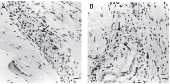
depleted mice on the 7th day of infection (P < 0.05; Figure 2A), characterized by fewer polymorphonuclear (PMN) cells and mono-nuclear cells compared with non-depleted mice. The number of parasites was signifi-cantly higher in depleted mice on the 7th day of infection (P < 0.05; Figure 2B). On the 30th day of infection, the inflammatory pro-cess was mostly characterized by mono-nuclear cells and was similar in both groups. However, more PMN cells were seen in complement-depleted mice. Morphometric analysis showed that parasitism was signifi-cantly increased in depleted animals (P < 0.05) (Figure 3A,B).
Parasite quantification
The number of viable parasites at the inoculation site was higher in complement-depleted than in non-complement-depleted mice on the 7th and 30th day post-infection (P < 0.05; Figure 4).
Discussion
The evolution of the infection in BALB/c mice infected with L. (L.) amazonensis showed a consumption of complement he-molytic activity in the sera at the beginning of the infection, between 3 and 24 h, with
Figure 2. Morphometric analy-sis of the Leishmania (L.) ama-zonensis subcutaneous inocu-lation site of de-pleted (C-) and complement-non-depleted (C+) BALB/c mice, showing the inflamma-tory reaction (A) and the para-site burden of the lesion (B). Data are representative of three separate experiments yielding similar results. Data are re-ported as the mean ± SD for 5 animals per group. MO = mono-nuclear cells, PMN = polymor-phonuclear cells. *P < 0.05 compared to non-depleted ani-mals (one-way ANOVA).
Number of cells/mm
2
70 60 50
40 30
20 10 0
*
*
*
7
Time of infection (days) 30
C- PMN C- MO C+ PMN C+ MO
A
Number of parasites/mm
2
120 100 80 60 40 20
*
*
0
7
Time of infection (days) 30
B
C- C+
B
B
B
B
B
recovery from 48 h onwards. The presence of the C3 component at the subcutaneous inoculation site persisted throughout the study period, but varied from moderate to discrete. The activation of the complement system during the acute phase of Leishmania infec-tion may contribute to resistance or to
sus-Figure 3. Histopathology of an
Leishmania (L.) amazonensis
subcutaneous inoculation site at 7 days post-infection (H&E, magnification 40X). A, Comple-ment-depleted mice showing high number of parasites sorounded by inflammatory cells (arrow). B, Non-depleted mice showing few parasites in the dermal inflammatory reac-tion (arrows). Bars = 50 µm.
ceptibility, since parasites inoculated into a vertebrate host lead to an acute inflamma-tory process with participation of humoral and cellular factors (12). Leishmania can activate the classical and alternative comple-ment pathways simultaneously, in vitro. C3 binding by L. amazonensis promastigotes via the classical and alternative pathways is very rapid, reaching a maximum within 2 to 3 and 3 to 3.5 min, respectively. The real-time kinetics of Leishmania promastigote lysis in normal human serum showed that lysis is an extremely rapid reaction and 10% of L. amazonensis promastigotes in the sta-tionary phase showed a degree of comple-ment resistance (16).
In vitro experiments have shown that promastigotes of the cutaneous strain of Leishmania are susceptible to lysis by nor-mal serum by activation of the complement alternative pathway (4,5). Amastigotes of cutaneous strains are more susceptible to complement lysis than visceral strains, sug-gesting a specific role of the complement factors limiting the infection in the skin (10). Therefore, complement activation in the early phase of L. (L.) amazonensis infection in BALB/c mice in vivo, with systemic con-sumption and local deposit at the inoculation site, could be a factor acting in parasite destruction on the skin.
Adsorbed normal human serum shows alternative pathway C3 deposition capacity,
89.5% for L. amazonensis and 66.3% for L. donovani, similar to that seen in normal human serum-EGTA, 86.5% for L. amazo-nensis and 47% for L. donovani. Promasti-gotes preincubated with normal human se-rum-EDTA or IgM-EDTA, conditions that prevent promastigote mannan-binding lectin (MBL) and C-reactive protein binding, and subsequently incubated with adsorbed nor-mal human serum, trigger classical pathway C3 deposition. In contrast, promastigotes preincubated with IgG-EDTA contribute only 18% of total C3 bound. These data indicate that in normal human serum, natural IgM anti-Leishmania antibodies are the main trig-gering factor for classical pathway activa-tion by promastigotes, and that serum MBL and C-reactive protein do not contribute sig-nificantly to this process (16). On the other hand, MBL levels correlated directly with the probability of developing visceral leish-maniasis and affected the functions of L. chagasi-infected monocytes since the mono-cytes infected with MBL-opsonized Leish-mania chagasi promastigotes secreted high levels of TNF-α and IL-6 (17), cytokines
which can inhibit the leishmanicidal capac-ity of macrophages infected with L. mexi-cana (18,19).
The development of the inflammatory reaction in the early phase of infection could also be considered to be a factor contributing to parasite destruction, since a large number of PMN cells migrate to the inoculation site during the first hours after infection (12). Complement is involved in the phagocytosis of Leishmania by PMN cells. In vitro experi-ments have shown that these cells are effi-cient in killing phagocytized parasites through a high oxidative burst mechanism (20).
In order to establish the role of comple-ment in L. (L.) amazonensis mouse infec-tion, mice were depleted of complement us-ing CVF. The efficiency of CVF was con-firmed by the significant decrease in comple-ment activity in the sera when comparing depleted and non-depleted groups in the early Number of parasites (x1000)/hind footpad
400
C- C+ 300
200
100
0
*
*
7 30
Time of infection (days) Figure 4. Number of viable
para-sites in the skin lesion caused by
Leishmania (L.) amazonensis at 7 and 30 days after infection in complement-depleted (C-) and complement-non-depleted (C+) BALB/c mice. Parasites were measured by limiting dilution. Data are representative of three separate experiments yielding similar results. Data are reported as the mean ± SD for 5 animals per group. The 95% confidence
phase of infection, up to 48 h. The high complement activity by the 7th day of infec-tion in complement-depleted mice compared to non-depleted mice may have occurred due to a high systemic stimulus after intravascu-lar complement activation with CVF, which acts as C3-covertase leading to systemic C3 consumption (21). The complement activity in non-infected, complement-depleted and non-depleted animals did not show signifi-cant differences on the 7th day after CVF injection. However, the complement activity in depleted mice was slightly higher than in animals which did not receive CVF (data not shown).
C3 fragments were not detected by direct immunofluorescence in the tissue from de-pleted mice during the first week after infec-tion, in spite of complement recovery in the sera by the 7th day of infection. Morphomet-ric analysis during the histopathologic ex-amination of the inoculation site showed a decrease in the inflammatory response and an increase in parasite burden in depleted mice late in the acute phase, 7 days post-infection. A limiting dilution assay confirmed these results, since more viable parasites were observed in the skin of depleted ani-mals. These data suggest that the presence of complement at the inoculation site during the early phase of infection could have a role in the control of parasite spreading in BALB/c mice infected with L. (L.) amazonensis, since in vitro experiments showed complement activation on the surface of L. amazonensis promastigotes leading to extracellular para-site lysis (22,23).
For some species of Leishmania, such as L. major and L. enrietti, the complement-dependent binding of promastigotes to mac-rophages represents the main mechanism of adhesion. In other species, such as L. amazo-nensis, the binding to macrophages occurs efficiently, even in the absence of serum,
and the addition of exogenous complement enhances binding only slightly (24). A sig-nificant percentage of these organisms sur-vive within the macrophages, a fact probably due to an inherent resistance of these organ-isms to macrophage-mediated killing mech-anisms.
In order to establish the role of comple-ment during the early phase of Leishmania infection in the evolution of disease, the histopathology and parasitism of the lesion were studied on the 30th day of infection. No differences were observed in the number of mononuclear cells between depleted and non-depleted mice, but a higher number of PMN cells was present in the lesions of comple-ment-depleted mice. The number of viable parasites was higher in the lesions of de-pleted mice. The higher amount of PMN cells in the lesion of complement-depleted mice may be due to a higher parasitism, since fragments of parasites may act as a chemotactic factor for these cells (25) and may represent a late attempt by the host to destroy the parasite.
Differences between visceral and cuta-neous parasite strains have been described with respect to both complement pathway activation (26) and effect of in vitro lysis by normal serum (5). In experimental visceral leishmaniasis, complement was reported to be an important factor for L. (L.) chagasi promastigotes to escape from the inocula-tion site to the viscera (12). On the other hand, the present experiments showed that complement has a role in the control of parasite spreading in the cutaneous leish-maniasis lesion caused by L. (L.) amazonen-sis infection in BALB/c mice.
Acknowledgments
References
1. Navin TR, Krug EC & Pearson RD (1989). Effect of immunoglobulin M from normal human serum on Leishmania donovani promasti-gote agglutination, complement-mediated killing, and phagocytosis by human monocytes. Infection and Immunity, 57: 1343-1346. 2. Dominguez M & Toraño A (1999). Immune adherence-mediated
opsonophagocytosis: the mechanism of Leishmania infection. Jour-nal of Experimental Medicine, 189: 25-35.
3. Green PJ, Feizi T, Stoll MS, Thiel S, Prescott A & McConville MJ (1994). Recognition of the major cell surface glycoconjugates of
Leishmania parasites by the human serum mannam-binding pro-tein. Molecular and Biochemical Parasitology, 66: 319-328. 4. Mosser DM & Edelson PJ (1984). Activation of the alternative
complement pathway by Leishmania promastigotes: parasite lysis and attachment to macrophages. Journal of Immunology, 132: 1501-1505.
5. Mosser DM, Burke SK, Contavas EE, Wedgwood JF & Edelson PJ (1986). Leishmania species: Mechanisms of complement activation by five strains of promastigotes. Experimental Parasitology, 62: 394-404.
6. Franke ED, McGrevy PB, Katz SP & Sacks DL (1985). Growth cycle dependent generation of complement resistant Leishmania pro-mastigotes. Journal of Immunology, 134: 2713-2718.
7. Puentes SM, Sacks DL, Silva RP & Joiner KA (1990). Complement binding by two developmental stages of Leishmania major promas-tigotes varying in expression of a surface lipophosphoglucan. Jour-nal of Experimental Medicine, 167: 887-902.
8. Silva RP, Hall BF, Joiner KA & Sacks DL (1989). CR1, the C3b receptor, mediates binding of infective Leishmania major metacy-clic promastigotes to human macrophages. Journal of Immunology, 143: 617-622.
9. Sacks DL, Broclin TN & Turco SJ (1990). Developmental modifica-tion of the lipophosphoglycan from Leishmania major promasti-gotes during metacyclogenesis. Molecular and Biochemical Parasi-tology, 42: 225-234.
10. Hoover DL, Berger M, Macy CA, Hockmeyer WT & Meltzer MS (1984). Killing of Leishmania tropica amastigotes by factors in nor-mal human serum. Journal of Immunology, 132: 893-897. 11. Wright SD & Siverstein SC (1983). Receptors for C3b and iC3b
promote phagocytosis but not the release of toxic oxygen from human phagocytes. Journal of Experimental Medicine, 158: 2016-2023.
12. Laurenti MD, Corbett CEP, Sotto MN, Sinhorini IL & Goto H (1996). The role of complement in the acute inflammatory process in the skin and in host-parasite interaction in hamsters inoculated with
Leishmania (Leishmania) chagasi. International Journal of Experi-mental Pathology, 77: 15-24.
13. Whaley K (1985). Measurement of complement. In: Whaley K
(Edi-tor), Methods in Complement for Clinical Immunologists. Butler & Tanner Ltd., Frome and London, UK, 77-159.
14. Titus RG, Marchand M, Boon T & Louis JA (1985). A limiting dilution assay for quantifying Leishmania major in tissue of infected mice.
Parasite Immunology, 7: 545-555.
15. Lima HC, Bleyenberg JA & Titus RG (1997). A simple method for quantifying Leishmania in tissues of infected animals. Parasitology Today, 13: 80-82.
16. Dominguez M, Moreno I, López-Trascasa M & Toraño A (2002). Complement interaction with trypanosomatid promastigotes in nor-mal human serum. Journal of Experimental Medicine, 195: 451-459. 17. de Miranda Santos IKF, Costa CHN, Krieger H et al. (2001). Mannan-binding lectin enhances susceptibility to visceral leishmaniasis. In-fection and Immunity, 69: 5212-5215.
18. Barral-Netto M, Badaro R, Barral A, Almeida RP, Santos SB, Badaro F, Pedral-Sampaio D, Carvalho EM, Falcoff E & Falcoff R (1991). Tumor necrosis factor (cachectin) in human visceral leishmaniasis.
Journal of Infectious Diseases, 163: 853-857.
19. Hatzigeorgiou DE, He S, Sobel J, Grabstein KH, Hafner A & Ho JL (1993). IL-6 down-regulates the cytokine-enhanced antileishmanial activity in human macrophages. Journal of Immunology, 151: 3682-3692.
20. Pearson RD & Steigbigel RT (1981). Phagocytosis and killing of the protozoan Leishmania donovani by human polymorphonuclear leu-cocytes. Journal of Immunology,127: 1438-1443.
21. Regal JF, Frase DG, Anderson DE & Solem LE (1993). Enhancement of antigen-induced bronchoconstriction after intravascular comple-ment activation with cobra venom factor - Reversal by granulocyte depletion. Journal of Immunology, 150: 3496-3505.
22. Barral Neto M, Roters SB, Sherlock I & Reed SG (1987). Destruction of Leishmania mexicana amazonensis promastigotes by normal human serum. American Journal of Tropical Medicine and Hygiene, 37: 53-56.
23. Nunes AC, Almeida Campos FR, Horta MF & Ramalho Pinto FJ (1997). Leishmania amazonensis promastigotes evade complement killing by interfering with the late steps of the cascade. Parasitol-ogy,115: 601-609.
24. Mosser DM & Rosenthal LA (1993). Leishmania-macrophage inter-actions: multiple receptors, multiple ligands and diverse cellular responses. Seminars in Cell Biology,4: 315-322.
25. Sorensen AL, Kharazmi A & Nielsen H (1989). Leishmania interac-tion with human monocytes and neutrophils: modulainterac-tion of the chemotatic response. Acta Pathologica, Microbiologica et Immuno-logica Scandinavica, 97: 754-760.