RESUMO.- [Detecção de vírus entéricos em pâncreas e baço de frangos com a síndrome de nanismo e raqui-tismo (RSS).] A doença entérica é um problema multifa-torial em galinhas que causa alterações gastrointestinais, conversão alimentar elevada e deficiência de crescimento.
Nos últimos anos, os vírus entéricos foram associados à doença entérica; casos reportados mostraram a infecção de um único vírus e também infecções concomitantes du-rante os surtos sugerindo a presença de múltiplos fatores etiológicos nas doenças entéricas. Este estudo mostra uma alta taxa de detecção dos vírus entéricos em amostras de pâncreas e baço de um surto de enterite e má-absorção em 16 lotes de frangos (n=80 frangos). O vírus de nefrite aviá -ria (ANV) foi o vírus mais detectado, estando presente em 75% dos lotes seguido pelo rotavírus aviário grupo A (ART--A) em 68,75% dos casos, e pelo astrovirus (CAstV) e par-vovírus aviários (ChPV), ambos em 43,75% das amostras. Os vírus estavam presentes no pâncreas dos lotes positivos em percentuais elevados: 100% para ART-A e CAstV; 91,7% para ANV, e em 57,14% para ChPV. Em contraste, somente 16,7% e 57,14%, em amostras de intestino, foram positi-vos para ANV e CAstV, respectivamente. Reovírus aviário
Detection of enteric viruses in pancreas and spleen of
broilers with runting-stunting syndrome (RSS)
1Luis Fabian N. Nuñez2, Silvana H. Santander Parra2, Claudete S. Astolfi-Ferreira2, Claudia Carranza3, David I.D. De La Torre2,4, Antonio C. Pedroso5
and Antonio J. Piantino Ferreira3*
ABSTRACT.- Nuñez L.F.N., Santander Parra S.H., Astolfi-Ferreira C.S., Carranza C., De La Tor -re D.I.D., Pedroso A.C. & Fer-reira A.J.P. 2016. Detection of enteric viruses in pancreas and spleen of broilers with runting-stunting syndrome (RSS). Pesquisa Veterinária Brasilei-ra 36(5):595-599. Departamento de Medicina Veterinária Preventiva e Saúde Animal, Facul-dade de Medicina Veterinária e Zootecnia, UniversiFacul-dade de São Paulo, Av. Prof. Dr. Orlando Marques de Paiva 87, São Paulo, SP 05508-270, Brazil. E-mail: [email protected]
Enteric disease is a multifactorial problem in chickens, which causes gastrointestinal alterations, elevated feed conversions and impairment. In the last years, several enteric viruses were implicated in enteric disease; case reports have shown their presence alone or in concomitant infections during outbreaks and have suggested that they might be de -termining factors in the aetiology of enteric disease. This study shows high detection rates of enteric viruses in the pancreas and spleen in samples from an outbreak of enteritis and malabsorption in 16 chicken flocks (n=80 broilers). Avian nephritis virus (ANV) was the most ubiquitous virus, present in 75% of the flocks followed by avian rotavirus group A (ART-A) with 68.75%, and by chicken astrovirus (CAstV) and chicken parvovirus (ChPV) in 43.75% of samples. Viruses were present in the pancreas of positive flocks at extremely high rates: 100% for ART-A, 91.7% for ANV, 100% for CAstV and 57.14% for ChPV. By contrast, only 16.7% and 57.14% of intestine samples were positive for ANV and CAstV, respectively. Avian reovirus (AReo) and avian adenovirus group 1 (FAdV-1) were not de -tected. These results suggest that high viral detection rates in pancreas samples may be a result of viremia during enteric disease, with subsequent damage of the exocrine pancreas, leading to runting-stunting syndrome (RSS).
INDEX TERMS: Broiler, enteric viruses, detection, pancreas, spleen, runting-stunting syndrome, RSS.
1 Received on July 27, 2015.
Accepted for publication on April 19, 2016.
2 Departamento de Patologia, Faculdade de Medicina Veterinária e Zoo-tecnia (FMVZ), Universidade de São Paulo (USP), São Paulo, SP 05508-270, Brazil.
3 Departamento de Medicina Veterinária Preventiva e Saúde Animal (VPS), FMVZ-USP, São Paulo, SP 05508-270, Brazil. *Corresponding au -thor: [email protected]
4 Faculdad de Medicina Veterinaria y Zootecnia, Universidad Central del Ecuador, Quito, Ecuador.
(AReo) e o adenovírus do grupo 1 (FAdV-1) não foram de-tectados. Estes resultados sugerem que os elevados percen -tuais de vírus detectados em amostras de pâncreas podem estar associados à viremia durante a doença entérica, com subsequente lesão no pâncreas exócrino das aves levando ao desenvolvimento da síndrome de nanismo e raquitismo. TERMOS DE INDEXAÇÃO: Frangos, vírus entéricos, detecção, pân -creas, baço, síndrome de nanismo e raquitismo, RSS.
INTRODUCTION
Enteric disease in poultry is a multifactorial problem that is not fully understood. This disorder (also known as malabsorption syndrome and as runting-stunting syn -drome or RSS) is a multifactorial disease of chickens which results in gastrointestinal alterations along with elevated feed conversion and decreased body weight (Rebel et al. 2006, Saif 2013, Nuñez et al. 2016a, Nuñez et al. 2016b). Bacterial, protozoal and viral agents are known to be in -volved in the development of outbreaks, which are also related to flock management and to the immunological status of flocks (Nuñez & Ferreira 2013, Moura-Alvarez et al. 2013, Moura-Alvarez et al. 2014). In the last years, en -teric viruses, such as chicken astrovirus (CAstV), chicken parvovirus (ChPV), avian rotavirus (ART), fowl adenovirus type I (FAdV I) and avian reovirus (AReo) have been in -creasingly implicated. This report is showing its presence alone or in concomitant infections during outbreaks and suggesting that they play an important role in the aetiology of enteric disease (Zsak et al. 2013). These recent reports have led to the development of experimental models to recreate the disease and to try to determine its etiopatho -genicity (Zsak et al. 2013, Decaesstecker et al. 1989, Nuñez et al. 2015a). However, due to the complex nature of this disease, the exact aetiology remains unknown.
The objective of this study was to detect viruses asso -ciated with RSS in Brazilian commercial chicken flocks.
MATERIALS AND METHODS
Description of the problem. In late 2012, 16 broiler flocks
from the state of Minas Gerais, Brazil, were reported to be excret -ing undigested feed and aqueous feces and to be exhibiting delayed growth and increased mortality, at variable rates. Necropsy re -vealed severe congestion of the duodenal walls compatible with duodenitis, and runting-stunting syndrome(RSS) was mooted.
Sampling. Five broilers aged 8 to 28 days randomly, chosen from each affected flock described above, were submitted to the Avian Diseases Laboratory at the FMVZ-USP, São Paulo/SP, Brazil, giving a total of 80 birds analyzed. A separate pool of each organ (gastrointestinal tract, spleen or pancreas) was obtained from the five sampled birds from each flock, so were collected 16 samples (pool) of intestinal tract, 16 samples (pool) of pancreas and 10 samples (pool) of spleen, giving a total of 42 samples, all samples were submitted to molecular analyses for the presence of enteric viruses.
Nucleic acid extraction for viral detection. Tissues from the intestine, pancreas or spleen of five (05) birds of each flock were pooled separately and homogenized; pooled samples from each tissue type and from each flock were homogenized separately and diluted in 1.5-mL microcentrifuge tubes containing 0.1 M sterile PBS, pH 7.4, at a 1:1 proportion. These diluted samples were then frozen at -20°C for 10 minutes and thawed at 56°C for 1 minute, and this process was repeated three times, including homogeniz -ation between freeze-thaw cycles and vortexing for 20 seconds. For DNA extraction, the samples were centrifuged at 12.000xg for 20 minutes at 4°C, and for RNA extraction, the samples were cent -rifuged for 30 minutes under the same conditions. The resulting supernatants were stored at -20°C until analysis. DNA and RNA were extracted from the supernatants using TRIzol® (Invitrogen,
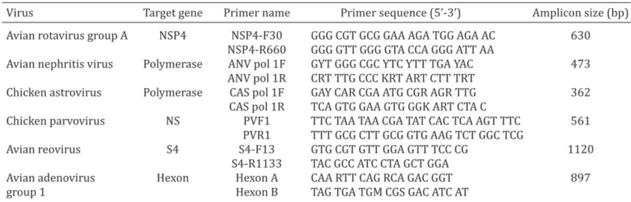
Valencia, CA, USA) according to the manufacturer’s instructions. PCR and RT-PCR detection of viral agents. The screen-ing procedures performed to detect the most common enteric viruses in Brazilian flocks (Moura-Alvarez et al. 2013) included polymerase chain reaction (PCR) assays for the detection of avian adenovirus group 1 (FAdV-1) (Meulemans et al. 2001) and chicken parvovirus (ChPV) (Zsak et al. 2009) and RT-PCR analysis for chicken astrovirus (CAstV), avian nephritis virus (ANV), avian rotavirus group A (ART-A) (Day et al. 2007) and Avian reovirus (AReo) (Pantin-Jackwood et al. 2008). Target genes, primers and amplicon sizes are listed in Table 1. ChPV, FAdV-1, CAstV and ANV strains previously isolated from Brazilian flocks and whose iden -tity was confirmed by sequencing were used as positive controls in the molecular assays. Additionally, Nebraska calf diarrhoea virus and the S1133 strain were used as controls for rotavirus and reovirus, respectively.
RESULTS
Table 2 shows the detection rates of the screened viruses. All flocks that presented enteric problems were positive for at least one of these pathogens. Avian nephritis virus (ANV) and avian rotavirus group A (ART-A) were present in more than 60% of the flocks (75% and 68.75%, respectively). Both chicken astrovirus (CAstV) and chicken parvovirus (ChPV)
Table 1. Primer sequences, amplicon sizes for RT-PCR and PCR assays performed to detect enteric viruses in chickens with RSS in Brazil
Virus Target gene Primer name Primer sequence (5’-3’) Amplicon size (bp) Avian rotavirus group A NSP4 NSP4-F30 GGG CGT GCG GAA AGA TGG AGA AC 630
NSP4-R660 GGG GTT GGG GTA CCA GGG ATT AA
Avian nephritis virus Polymerase ANV pol 1F GYT GGG CGC YTC YTT TGA YAC 473
ANV pol 1R CRT TTG CCC KRT ART CTT TRT
Chicken astrovirus Polymerase CAS pol 1F GAY CAR CGA ATG CGR AGR TTG 362
CAS pol 1R TCA GTG GAA GTG GGK ART CTA C
Chicken parvovirus NS PVF1 TTC TAA TAA CGA TAT CAC TCA AGT TTC 561
PVR1 TTT GCG CTT GCG GTG AAG TCT GGC TCG
Avian reovirus S4 S4-F13 GTG CGT GTT GGA GTT TCC CG 1120
S4-R1133 TAC GCC ATC CTA GCT GGA
Avian adenovirus Hexon Hexon A CAA RTT CAG RCA GAC GGT 897
were both detected in 43.75% of the samples; while no avian reovirus (AReo) or avian adenovirus group 1 (FAdV-1) was detected. In addition, as expected, concomitant infection with two or more viruses were observed in most flocks (75%), and single viral infections were observed in only 25% (n=4) of the flocks. Notably, in the 4 flocks with single viral infections, the main organ in which virus was detected was the pancreas. In general, the pancreas was the organ in which viruses were detected at the highest rates; 33 out of 37 (89.1%) pancreas
samples were positive, by contrast only 6 of 37 (16.2%) pooled intestine samples were positive for the presence of at least one enteric virus. Enteric viruses were also detected at high rates in spleen samples from the different flocks; 77.3% of all spleen samples were positive for at least one virus (Table 3). Finally, a regression analysis (Minitab® software) showed no relationship between the age of birds (8-28 days) and the molecular detection of one or more enteric pathogens (Fig.1).
DISCUSSION
In this case of severe duodenitis and RSS in 16 broiler flo -cks, molecular analysis showed that concomitant infection with 2 or 4 viruses were detected in 75% of the flocks, whi -le sing-le viral infections were detected in the remaining 25% of the flocks. Analysis revealed that the prevalence of viruses such as avian nephritis virus (ANV), avian rotavirus group A (ART-A), chicken astrovirus (CAstV) and chicken parvovirus (ChPV) were high among broilers with digesti -ve disorders. These results are consistent with previous re -ports from Brazil and from several other poultry-producing countries, where enteric problems and growth retardation are often linked to the detection of one or more of these enteric viruses (Otto et al. 2012, Koo et al. 2013, Villarre -al et -al. 2006) and support the hypothesis that RSS has a multifactorial aetiology. Despite of a strong relationship between pathogen detection and the development of the disease the diagnoses has not been clearly established (Koo et al. 2013). Notwithstanding, the diversity of viruses detected during outbreaks, variations in virulence among
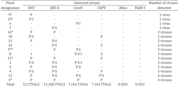
Table 2. Viral detection in each flock and the presence of concomitant infections
Flock Detected viruses Number of viruses
designation ANV ART-A CAstV ChPV AReo FAdV-I detected
9* P - - - 1 virus
15* P-I - - - 1 virus
6 - - P-I - - - 1 virus
7 - P-S - - - - 1 virus
14* P P - - - - 2 viruses
10 P-S - - P - - 2 viruses
11 P P-S - - - - 2 viruses
16 - P-S - S - - 2 viruses
5** - P P-I - - - 3 viruses
8 I - P-S-I S - - 3 viruses
13* P P - P - - 3 viruses
1 P-S P-S P-S-I - - - 3 viruses
2 P P-S P-S - - - 3 viruses
4 P-S P-S - P - - 3 viruses
12 P P-S P-S P-S - - 4 viruses
3* P P P P - - 4 viruses
Total 12 (75%)1 11 (68.75%)1 7 (43.75%)1 7 (43.75%)1 0 (0)1 0 (0)1
*Percentages (in parentheses) were calculated based on the number of positives vs. the number of samples for each virus. P = pancreas; S = spleen; I = intestine. ** Samples sent without spleen.
Fig.1. Regression analysis showing the relationship between the ages of birds and the molecular detection of viral agents. A result of p > 0.05 indicated that there was no relation between the ages of birds and the molecular detection of enteric patho-gens in these sampled flocks.
Table 3. Distribution of enteric viruses in tissues of naturally infected broiler chickens Organs Number of positive samples for each virus (%)* Total positive samples
ART-A ANV CAstV ChPV for each tissue**
Pancreas 11/11 (100.0) 11/12 (91.7) 7/7 (100.0) 4/7 (57.14) 33/37 (89.1) Spleen 7/7 (100.0) 3/5 (60.0) 4/5 (80.0) 3/5 (60.0) 17/22 (77.3) Intestine 0/11 (0.0) 2/12 (16.7) 4/7 (57.14) 0/7 (0.0) 06/37 (16.2) *Percentages (in parentheses) were calculated based on the number of positive tissue samples vs. the number of
serotypes or strains, or similarities in the signs and lesions are observed (Saif 2013). Thereby, concomitant infections are more likely to have a detrimental, synergistic effect on birds (Nuñez & Ferreira 2013, Mettifogo et al. 2014, Nuñez et al. 2015b). Nevertheless, these results show a high pre -valence of enteric viruses circulating in Brazilian chicken flocks located in Minas Gerais.
The result that ANV was detected in 75% of the flocks differs from the results of previous reports for Brazil, whe -re the same virus was found in 35.5 % of turkey flocks Mou -ra-Alvarez et al. 2013) and in 29.6% of chicken flocks tested (Mettifogo et al. 2014). This difference in results could be due to the different sources of diagnostic material used in these studies. While only faecal samples were analysed in previous works, the present study analysed homogenates from the intestine, pancreas and spleen from chickens sho -wing signs of enteric diseases for the detection of viruses, based on the intracellular nature of the agent. This metho -dological difference could explain the higher detection rate in the present study for this and other viruses, including ART-A, CAstV and ChPV. However, despite the high inciden -ce of ANV, it may not be the causative agent of the elevated mortality in these flocks because no visceral urate depo -sition or renal disturbances, which are classical lesions caused by pathogenic ANV, were noticed during necropsy (Siller et al. 1981). On the other hand, ART-A was detected in 68.75% of flocks (11/16), and by contrast with what may have been expected, the pancreas was found to have the highest rate of infection, followed by the spleen; virus was detected in 100% and 91.7% of positive flocks, respec -tively. No virus was detected in the intestine. Studies in rats have shown the capacity of avian rotavirus to disseminate and replicate in different organs, such as the liver, spleen and pancreas; however, the mechanism by which rotavirus escapes the gastrointestinal tract and reaches other organs remains unknown (Crawford et al. 2006).
Affected RSS flocks were positive for CAstV and ChPV at equal rates (43.75% for both viruses). Previous surveys have also found that these pathogens circulate both in birds showing signs suggestive of RSS and in apparently healthy birds in Brazil (Moura-Alvarez et al. 2013, Mettifogo et al. 2014), United States (Pantin-Jackwood et al. 2008), and Po -land (Domanska et al. 2014). Chicken astrovirus was also de -tected in one-day-old breeder chicks, indicating that vertical transmission may occur (Mettifogo et al. 2014). Moreover, parvoviruses have also been identified in day-old broiler chickens showing neurological signs, impaired mobility and diarrhoea, suggesting that these viruses may cause neuro -logical disturbances in young chickens. The neuro-logical disturbances that have been observed were characterized by cerebellar hypoplasia and hydrocephalus (Marusak et al. 2010). These findings suggest that both CAstV and ChPV may potentially be implicated in the aetiology and perpetu -ation of enteric disease and that pathogenicity and clinical signs may vary with the infecting strains.
No AReo and FAdV-1 were detected. These findings are in agreement with other results that show a low preval -ence of these viruses (Moura-Alvarez et al. 2013, Mettifogo et al. 2014). These findings also support the hypothesis of
Decaesstecker et al. (1989) that AReo may not cause en -teric disease, as these viruses do not exhibit pathogenicity when inoculated alone.
Viruses were most frequently detected in the pancreas, among the organs screened for enteric viruses. In general, the pancreas is an accessory organ of the gastrointestinal tract, and it has both endocrine and exocrine functions. The exocrine function of the pancreas is to produce and secrete several digestive enzymes, including trypsinogen, amylase, lipase and procarboxypeptidase, among others (Denbow 2000). In healthy birds, these enzymes are released into the ascending loop of the duodenum, thereby ensuring the di -gestion of lipids, carbohydrates and proteins in the diet (Os -man et al. 1982, Horváthová et al. 2000). By contrast, flocks with characteristic signs of malabsorption syndrome may present alterations in the physiology of the exocrine pan -creas. Previous studies have shown that birds with this syn -drome exhibited diminished pancreatic amylase activity and changes in pancreatic protease levels (Szabo et al. 1989). Histological studies of pancreatic lesions in broilers with RSS revealed fibrosis, inflammation, vacuolar changes in the exo-crine compartment, degeneration of acinar cells and loss of zymogen granules (Qamar et al. 2013). In pigeons, exocrine pancreas disease produced voluminous, coloured faeces and weight loss (Amann et al. 2006), and when aflatoxins are present in the diet, histological changes in the pancreas are observed, together with effects on performance and nutrient absorption (Marchioro et al. 2013). Together, these findings establish a relationship between pancreatic damage and signs of malabsorption, and, more importantly, a relation -ship between pancreatic damage and RSS.
Acknowledgements.- This sudy was supported by grants received from
CNPq no 453920/2014-4, and FAPESP grant no 2013/08560-5. Antonio J. Piantino Ferreira is a recipient of CNPq fellowships.
REFERENCES
Amann O., Visschers M.J.M., Dorrestein G.M., WesterhoI F. & Lumeij J.T. 2006. Exocrine pancreatic insufficiency in pigeons. Avian Pathol. 35:58-62. Crawford S.E., Patel D.G., Cheng E., Hyser J.M., Ciarlet M., Finegold M.J., Con
-ner M.E., Estes M.K. & Berkova Z. 2006. Rotavirus Viremia and Extrain -testinal Viral Infection in the Neonatal Rat Model. J. Virol. 80:4820-4832. Day J.M., Spackman E. & Pantin-Jackwood M.J. 2007. A multiplex RT-PCR
test for the differential identification of turkey astrovirus type 1, turkey astrovirus type 2, chicken astrovirus, avian nephritis virus, and avian rotavirus. Avian Dis. 51:681-684.
Denbow M.D. 2000. Gastrointestinal anatomy and physiology, p.299-325. In: Whittow G.C. (Ed.), Sturkie’s Avian Physiology. Academic Press, San Diego, CA.
Decaesstecker M., Charlier G., Peeters J. & Meulemans G. 1989. Pathogeni -city of fowl enteroviruses. Avian Pathol. 18:697-713.
Domanska K., Jacukowicz A., Bocian L. & Minta Z. 2014. Astroviruses in Polish Commercial Turkey Farms in 2009-2012. Avian Dis. 58:158-164. Giordano S., Serra G., Dones P., Di Gangi M., Failla M.C., Iaria C., Ricciardi F.,
Pernice L.M., Pantaleo D. & Cascio A. 2013. Acute pancreatitis in children and rotavirus infection. New Microbiol. 36:97-101.
Horváthová V., Janeček Š. & Šturdík E. 2000. Amylolytic enzymes: their specificities, origins and properties. Biol. Bratislava 55:605-615. Koo B., Lee H., Jeon E., Jang H., Han M. & Mo I. 2013. An unusual case of con
-comitant infection with chicken astrovirus and group A avian rotavirus in broilers with a history of severe clinical signs. J. Vet. Sci. 14:231-233. Marchioro A., Mallmann O., Diel A., Dilkin P., Rauber R.H., Blazquez F.J.H.,
Oliveira M.G.A. & Mallmann C.A. 2013. Effects of aflatoxins on perfor -mance and exocrine pancreas of broiler chickens. Avian Dis. 57:280-284. Marusak R.A., Guy J.S., Abdul-Aziz T.A., West M.A., Fletcher O.J., Day J.M., Zsak
L. & Barnes H.J. 2010. PARTovirus-Associated Cerebellar Hypoplasia and Hydrocephalus in day old broiler chickens. Avian Dis. 54:156-160. Mettifogo E., Nuñez L.F.N., Chacón J.L., Santander Parra S.H., Astolfi-Ferreira
C.S., Jerez J.A., Jones R.C. & Piantino Ferreira A.J. 2014. Emergence of en -teric viruses in production chickens is a concern for avian health. Scient. World J. 2014:450423. <http://dx.doi.org/10.1155/2014/450423> Meulemans G., Boschmans M., Berg T.P. & Decaesstecker M. 2001. Polymera
-se chain reaction combined with restriction enzyme analysis for detection and differentiation of fowl adenoviruses. Avian Pathol. 30:655-660. Moura-Alvarez J., Chacon J.L., Scanavini L.S., Nuñez L.F.N., Astolfi-Ferreira
C.S., Jones R.C. & Piantino Ferreira A.J. 2013. Enteric viruses in Brazilian turkey flocks: single and multiple virus infection frequency according to age and clinical signs of intestinal disease. Poult. Sci. 92:945-955. Moura-Alvarez J., Nuñez L.F., Astolfi-Ferreira C.S., Knöbl T., Chacón J.L., Mo
-reno A.M., Jones R.C. & Ferreira A.J. 2014. Detection of enteric pathogens in Turkey flocks affected with severe enteritis, in Brazil. Trop. Anim. He -alth Prod. 46:1051-1058.
Nuñez L.F.N. & Ferreira A.J.P. 2013. Viral agents related to enteric disease in commercial chicken flocks, with special reference to Latin America. World´s Poult. Sci. J. 69:853-864.
Nuñez L.F., Parra S.H., Mettifogo E., Catroxo M.H., Astolfi-Ferreira C.S. & Piantino Ferreira A.J. 2015a. Isolation of chicken astrovirus from spe -cific pathogen-free chicken embryonated eggs. Poult Sci. 94:947-954. Nuñez L.F., Santander Parra S.H., Mettifogo E., Astolfi-Ferreira C.S. & Pian
-tino Ferreira A.J. 2015b. Isolation and molecular characterisation of chicken pARTovirus from Brazilian flocks with enteric disorders. Brit. Poult. Sci. 56:39-47.
Nuñez L.F., Sá L.R.M., Parra S.H.S., Astolfi-Ferreira C.S., Carranza C. & Piantino Ferreira A.J. 2016a. Molecular detection of chicken pARTovi -rus in broilers with enteric disorders presenting curving of duodenal loop, pancreatic atrophy, and mesenteritis. Poult. Sci. <http://dx.doi. org/10.3382/ps/pev439>
Nuñez L.F., Santander Parra S.H., Carranza C., Astolfi-Ferreira C.S., Buim M.R. & Piantino Ferreira A.J. 2016b. Detection and molecular characteri -zation of chicken astrovirus associated with chicks that have an unusual condition known as “white chicks” in Brazil. Poult. Sci. <http://dx.doi. org/10.3382/ps/pew062>
Otto P.H., Ahmed M.U., Hotzel H., Machnowska P., Reetz J., Roth B., Trojnar E. & Johne R. 2012. Detection of avian rotaviruses of groups A, D, F and G in diseased chickens and turkeys from Europe and Bangladesh. Vet. Microbiol. 156:8-15.
Osman A.M. 1982. Amylase in chicken intestine and pancreas. Comp. Bio -chem. Physiol. 73:571-574.
Pantin-Jackwood M.J., Day J.M., Jackwood M.W. & Spackman E. 2008. En -teric viruses detected by molecular methods in commercial chicken and turkey flocks in the United States between 2005 and 2006. Avian Dis. 52:235-244.
Qamar M.F., Aslam H. & Jahan N. 2013. Histopathological studies on stun -ting syndrome in broilers, Lahore, Pakistan. Vet. Med. Int. 2013:1-6. DOI: 10.3382/ps/pev439
Rebel J.M., Balk F.R., Post J., Van Hemert S., Zekarias B. & Stockhofe N. 2006. Malabsorption syndrome in broilers. World´s Poult. Sci. J. 62:17-29. Saif Y.M. 2013. Viral Enteric Infections, p.375-376. In: Swaine D.E., Glisson
J.R., McDougald N.L.K., Suarez D.L. & Nair V. (Eds), Diseases of Poultry. Wiley-Blackwell, Oxford, UK.
Siller W.G. 1981. Renal pathology of the fowl - a review. Avian Pathol. 10:187-262.
Szabo J., Salyi G. & Rudas P. 1989. Effect of malabsorption syndrome on pancreatic function in broilers. Poult. Sci. 68:1553-1560.
Villarreal L., Uliana G., Valenzuela G., Chacon J.L., Saidenberg A., Sanches A., Brandão P.E., Jerez J.A. & Ferreira A.J.P. 2006. Rotavirus detection and isola -tion from chickens with or without symptoms. Braz. J. Poult. Sci. 8:187-191. Zsak L., Strother K.O. & Day J.M. 2009. Development of a polymerase chain reaction procedure for detection of chicken and turkey parvoviruses. Avian Dis. 53:83-88.