240
INTRODUCTION Revista da Sociedade Brasileira de Medicina Tropical 43(3):240-243, mai-jun, 2010
Article/Artigo
Gastroenteric virus detection in fecal samples from women in Goiânia,
State of Goiás, Brazil
Detecção de vírus gastroentéricos em amostras fecais de mulheres em Goiânia, Estado de
Goiás, Brasil
Rui Gilberto Ferreira
1, Ana Maria Tavares Borges
2, Fabiola Souza Fiaccadori
2, Menira Borges de Lima Dias e Souza
2,
Rodrigo Alessandro Togo Santos
2and Divina das Dôres de Paula Cardoso
2ABSTACT
Introduction: his was a prospective study that included women seen in the obstetrics and gynecology sector of Hospital das Clínicas, Federal University of Goiás, in Goiânia, State of Goiás, with the aim of detecting rotaviruses, adenoviruses, caliciviruses and astroviruses. Eighty-four women participated in the study and from these, 314 fecal samples were collected. Out of all of the women, 29 were seropositive for HIV and 55 were seronegative, and 45 and 39 were pregnant and non-pregnant, respectively. Methods: Fecal samples were collected from each woman once every two months over the period from July 2006 to June 2007, and they were screened for rotaviruses by means of polyacrylamide gel electrophoresis and immunoenzymatic assays, for caliciviruses and astroviruses by means of RT-PCR and for adenovirus by means of immunoenzymatic assays. he astroviruses were genotyped using nested PCR. Results: Among the 84 patients, 19 (22.6%) were positive for either calicivirus (14/19) or astrovirus (6/19), while one women was positive for both viruses in fecal samples collected on diferent occasions. Most of the positive samples were collected during the months of July and August (astrovirus) and September and October (calicivirus). None of the samples analyzed was positive for rotavirus or adenovirus. Gastroenteric viruses were detected in 13/19 (68.4%) of the pregnant women, whether HIV-seropositive or not. Conclusions: he results from the present study showed that neither pregnancy nor HIV-seropositive status among the women increased the risk of infection by any of the gastroenteric viruses studied. his study presents data on gastroenteric virus detection among pregnant and/or HIV-positive women. Key-words: Gastroenteric viruses. Calicivirus. Astrovirus. HIV-positive. Pregnant women.
RESUMO
Introdução: Este foi um estudo prospectivo que incluiu mulheres atendidas no setor de obstetrícia e ginecologia do Hospital das Clínicas da Universidade Federal de Goiás, em Goiânia, Estado de Goiás com o objetivo de detectar rotavírus, adenovírus, calicivírus e astrovírus. Oitenta e quatro mulheres participaram no estudo e destas, 314 amostras fecais foram coletadas. Do total de mulheres, 29 eram soropositivas para HIV, 55 soronegativas, 45 e 39 estavam grávidas e não-grávidas, respectivamente. Métodos: Amostras fecais foram coletadas de cada mulher uma vez a cada dois meses pelo período de Julho-2006 a Junho-2007, foram triadas para rotavírus pela metodologia de eletroforese em gel de poliacrilamida (EGPA) e através de ensaio imunoenzimático (EIE), para calicivírus e astrovírus por RT-PCR e por EIE para adenovírus. Os astrovírus foram genotipados por Nested-PCR. Resultados: De 84 pacientes, 19 (22,6%) foram positivas para calicivírus (14/19) ou astrovírus (6/19), sendo que uma mulher foi positiva para ambos os vírus em amostras fecais coletadas em diferentes ocasiões. A maioria das amostras positivas foi coletada no período de Julho a Agosto (astrovírus) e de Setembro a Outubro (calicivírus). Nenhuma das amostras analisadas foi positiva para rotavírus ou adenovírus. Os vírus gastroentéricos foram detectados em 13/19 (68,4%) mulheres grávidas, as quais eram HIV-soropositivas ou não. Conclusões: Os resultados do presente estudo mostram que nem o estado gravídico das mulheres nem a soropositividade para HIV aumentaram o risco para a infecção por nenhum dos vírus gastroentéricos estudados. Este estudo apresenta dados sobre a detecção de vírus gastroentéricos entre mulheres grávidas e/ou HIV-positivas.
Palavras-chaves: Virus gastroentéricos. Calicivírus. Astrovírus. HIV-positivo. Mulheres grávidas.
1. Departamento de Ginecologia e Obstetrícia, Faculdade de Medicina, Universidade Federal de Goiás, Goiânia GO. 2. Laboratório de Virologia Humana, Instituto de Patologia Tropical e Saúde Pública, Universidade Federal de Goiás, Goiânia GO
Address to: Dr. Rui Gilberto Ferreira. Laboratório de Virologia Humana/IPTSP/UFG. Rua 235 S/N,
sala 420, Setor Universitário, 74605050 Goiânia, GO, Brasil. Phone/Fax: 55 62 32096122/62 32096363
e-mail: [email protected]
Received in 29/09/2009
Accepted in 25/03/2010
Acute gastroenteritis is a major public health issue around the world, and is associated with high morbidity-mortality rates, especially among children. Gastroenteric viruses constitute the single most important cause of acute gastroenteritis in children up to ive years of age, worldwide1,2. Among
the more than twenty known gastroenteric viruses, the most important etiological agents of acute gastroenteritis are rotaviruses, enteric adenoviruses, astroviruses and human caliciviruses3-5.
Elderly and immunosuppressed individuals are among those with the highest susceptibility to infection and reinfection by gastroenteric viruses6-9.
It is also believed that pregnant women and HIV-seropositive individuals are more vulnerable to viral infections than is the general population7,10.
We decided to conduct this study because of the epidemiological importance of viral gastroenteritis and the assumption that pregnant women, whether HIV-seropositive or not, could constitute a group potentially at risk of viral infection and the development of gastroenteric disease, and also due to the scarcity of information on the incidence of gastroenteric viruses among the adult female population
Our study presents novel data on gastroenteric viruses (astroviruses and caliciviruses) among adult pregnant women (either HIV-seropositive or not) from the central-western region of Brazil.
METHODS
241
Ferreira RG et al - Gastroenteric viruses in women in Goiânia, GoiásRESULTS
From sample size analysis, the appropriate sample size for this study was estimated to be 84 women (p = 0.24; signiicance level of 5%, and 9% error). herefore, 84 women were selected to participate in this study, out of all of the women who were seen at the ObGyn service of HC-UFG over a one-year period. he women had spontaneously sought assistance or had been referred for consultation at the selected services. In accordance with the predeined criteria for inclusion and continuation in the groups, women with chronicle gastrointestinal conditions that could progress to diarrhea episodes (Crohn’s disease, Lupus or irritable bowel syndrome) were excluded from the study. All the participants conirmed their participation in the study by signing a writen consent form, and the study was approved by the Ethics Commitee for Human and Animal Medical Research of Hospital das Clínicas, Federal University of Goiás.
A data collection system provided the database for identifying the patients (age, obstetric history and present pregnancy) and for information about previous diarrhea episodes and HIV viral load. he CD4+ and CD8+ T cell counts of the HIV-positive women who were positive for any of the gastroenteric viruses studied were also recorded. Fecal samples were collected every two months over a 12-month period, and at the time of every diarrhea episode, in appropriate plastic receptacles, at HC-UFG or in patients’ homes when necessary. he samples were transported in ice and delivered to the virology laboratory of the Institute of Tropical Pathology and Public Health of the Federal University of Goiás, where the material was stored at -20°C until further processing.
The fecal samples were initially processed to obtain a 20% solution in phosphate-bufered saline (PBS; pH 7.4). he fecal suspensions were then homogenized and clariied by centrifugation at 9,000rpm for 10 minutes. he supernatant were then stored at -20°C, until further testing.
To detect rotavirus A (RVA), polyacrylamide gel electrophoresis (PAGE) was performed as described by Pereira et al11, and the gels
were stained using silver nitrate12. he samples were also screened for
RVA using a commercial immunoenzymatic assay (RIDASCREEN®
Rotavirus), following the manufacturer’s instructions (R-Biopharm, Germany). To detect adenovirus, a commercial immunoenzymatic assay (RIDASCREEN®
Adenovirus) was used in accordance with the manufacturer’s instructions (R-Biopharm). To detect calicivirus and astrovirus, the viral RNA was irst extracted as described by Boom et al13, with modiications14, and the samples were screened for calicivirus
by means of reverse-transcriptase polymerase chain reaction (RT-PCR) using the primer pairs Ni/E315 and 289/29016, and for astroviruses
using the primer pair Mon 269/27017. Astrovirus-positive samples were
further genotyped by means of nested PCR, as described by Sakamoto et al18. To determine possible associations between positive indings of
gastroenteric viruses, pregnancy and/or HIV-seropositivity, Fisher’s exact test was applied at a signiicance level of 0.05.
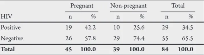
A total of 314 fecal samples were collected from 84 women who were seen at the Ob-Gyn service of HC-UFG, between July 2006 and June 2007. hese women were distributed into four groups characterized as follows: HIV-seropositive women (pregnant and non-pregnant) and HIV-seronegative women (pregnant and non-pregnant). Table 1 shows the distribution of the women regarding clinical characteristics. It was found that out of the 84 women, 45 were pregnant and HIV-positive.
TABLE 1- Distribution of the women seen at the Ob-Gyn service of HC-UFG between June 2006 and July 2007, according to the sampling groups.
Pregnant Non-pregnant Total HIV n % n % n % Positive 19 42.2 10 25.6 29 34.5 Negative 26 57.8 29 74.4 55 65.5
Total 45 100.0 39 100.0 84 100.0
P = 0.053
TABLE 2 - Distribution of the women seen at the Ob-Gyn service of HC-UFG between July 2006 and June 2007, according to immunological status (pregnant and/or HIV-seropositive) and positive indings of gastroenteric viruses.
Gastroenteric viruses Pregnant/HIV-positive yes No
n % n % Yes 13 68.4 41 63.1 No 6 31.6 24 36.9 Total 19 100.0 65 100.0 p = 0.199
In terms of age distribution, 43.7% of the women were up to 30 years of age, ive (7.8%) were considered teenagers (up to 19 years of age), 32.8% were aged between 20 and 30 years and 23.5% were more than 40 years old. No statistical diference (p = 0.053) was found between the groups of seropositive women and HIV-seronegative women, in relation to their pregnancy status.
Nineteen (22.6%) of the 84 women participating in the study had at least one fecal sample that was positive for either astroviruses or caliciviruses. hus, out of the total of 314 fecal samples collected, 33 (10.5%) were positive for either calicivirus or astrovirus. Caliciviruses were detected in 14 (16.6%) women and astrovirus in six (7.1%) women. One woman was positive for astrovirus and calicivirus in diferent samples collected on diferent occasions. RVA and adenovirus were not detected in any of the fecal samples collected.
he results in Table 2 show that 68.4% (13/19) of the women who were positive for gastroenteric viruses were pregnant (six were HIV-seropositive and seven were seronegative), although no statistical diference was observed when this group was compared with the group of women who were positive for gastroenteric viruses and were HIV-seronegative but were not pregnant (p = 0.199).
Among the 19 women that were positive for gastroenteric viruses, 13 had only one positive fecal sample and six had more than one positive fecal sample collected at diferent times, as observed in
Table 3. One of these women had three fecal samples, collected with two-month intervals between them, which were positive for gastroenteric viruses, and another woman had two positive fecal samples, with a six-month interval between them.
The circulation patterns of calicivirus and astrovirus were characterized by detection peaks during the months of June and August, for astrovirus, and during the months of September and October for calicivirus.
Genotyping of the six astrovirus-positive samples revealed predominance of genotype 1 (66.6%), followed by genotypes 3 and 8 (16.7% each).
242
Rev Soc Bras Med Trop 43(3):240-243, mai-jun, 2010
DISCUSSION
TABLE 3 - Distribution of the gastroenteric viruses in the various samples collected from the women seen at the Ob-Gyn service of HC-UFG between July 2006 and June 2007.
Gastroenteric viruses
calicivirus astrovirus Total Sample n % n % n % First 1 7.1 4 66.7 5 25.0 Second 3 21.4 0 0,0 3 15.0 hird 1 7.1 2 33.3 3 15.0 Fourth 2 14.3 0 0.0 2 10.0 Fith 1 7.1 0 0.0 1 5.0 First and second 1 7.1 0 0.0 1 5.0 First and third 2 14.3 0 0.0 2 10.0 Second and ith 2 14.3 0 0.0 2 10.0 First/second/third 1 7.1 0 0.0 1 5.0
Total 14 100.0 6 100.0 20 100.0
HIV-seronegative and pregnant. Among the 65 women who did not have any positive fecal samples for gastroenteric viruses, four (6.2%) had sporadic diarrhea episodes (three were HIV-seropositive, of whom two were pregnant and one was non-pregnant, and the fourth patient was non-pregnant and HIV-seronegative).
he HIV viral load was accessed at the time of the irst fecal sample collection, and was found to be undetectable (< 50 copies/ cell) in ive (71.4%) of the women who were HIV-seropositive (either positive or negative for gastroenteric viruses). From analysis on the cell immune status data of the 19 women for whom at least one fecal sample was positive for gastroenteric viruses, it was observed that the CD4+ T cell counts were low in only ive (26.3%) of them.
In the present study, calicivirus and astrovirus were detected in fecal samples collected from 84 adult women. Data from the literature show that the detection rates for these viruses among children with gastroenteritis in Brazil vary between 12 and 47%4,14,19-21. However,
there is no information available on the prevalence of these agents among the adult female population in Brazil.
It is known that in developing countries, infection by gastroenteric viruses, especially rotavirus, adenovirus and astrovirus, usually occurs during childhood, and mainly among children under five years of age3,2,21-24. Caliciviruses infect people of all ages and constitute
important etiological agents for diarrhea, while noroviruses are the main causative agents of non-bacterial epidemic acute gastroenteritis, worldwide5. Although uncommon, infection by gastroenteric
viruses can generally also occur among adults, especially elderly and immunosuppressed individuals6-8.Acute gastroenteritis due to
astrovirus has been reported in immunosuppressed (transplanted or HIV-seropositive) adults and children9,25. Walter & Mitchell also
reported positive indings of these viruses in nurseries23.
In our study, the only gastroenteric viruses found in the adult female population were calicivirus and astrovirus, while rotavirus or adenoviruses were not detected in any of the samples collected. Additionally, none of the fecal samples collected from the youngest group of women (teenagers) were positive for any of the viruses studied. he reasons for this epidemiological patern have not yet been elucidated, and require further investigation. From analysis
on virus positivity according to the women’s ages, we observed that the women with positive indings were predominantly aged 20-30 years, followed by 40-50 years, with prevalence rates a litle higher than those detected for calicivirus4.20 and astrovirus14,19 in children
with acute gastroenteritis under ive years of age.
Regarding virus seasonality, caliciviruses and astroviruses were mainly detected during the rainy season in our region. his circulation pattern corroborates the seasonality data obtained from other studies in the region, which have shown higher incidence rates for astroviruses and caliciviruses during periods with elevated relative air humidity19,20.
he focus on public health and preventive measures (even when treatment is not available) has given greater visibility to pregnant women (whether HIV-seropositive or not), because of the greater vulnerability of their immune system10. The inclusion of
HIV-seropositive women in this study was justiied by the assumption that this condition is associated with impaired immune status that could consequently predispose such individuals to infection by other pathogens, such as gastroenteric agents.
Although our results suggested that there was an association between HIV-seropositivity and/or pregnancy and positive indings of calicivirus and astrovirus (68.4%), especially astroviruses, this was not statistically signiicant. We hypothesize that the immunological impairment of these HIV-positive women may have been atenuated by the antiretroviral therapy and clinical follow-up, considering that more than one third of these women presented undetectable viral load and CD4+ cell counts higher than 250 cells/mm3. he fecal
samples were obtained at two-month intervals (up to ive samples per women), and all of them were screened for rotavirus, adenovirus, calicivirus and astrovirus.
For some women, more than one sample was positive for the same agent, while for one woman, more than one sample collected at diferent times was positive for calicivirus and astrovirus. It is known that gastroenteric viruses such as noroviruses can be excreted by immunocompetent individuals for more than two weeks ater infection24. Additionally, the immunosuppressive state induced
during pregnancy may contribute towards higher susceptibility or even reactivation of viral infection10.
In addition, positive indings of calicivirus in diferent samples from the same women, collected up to six months apart, could be due to reinfection by a diferent calicivirus genotype. Genotyping and sequencing of the samples might have helped elucidate this mater, but this could not be performed in the present study because of to the low RNA concentration in the samples.
he genomic characterization of the astrovirus detected in this study revealed predominance of genotype 1, which corroborates with data from the literature showing that serotypes 1 and 2 are the most prevalent worldwide18,21,23,25. In a study conducted in Goiânia in
2002, Cardoso et al. observed that the overall prevalence of genotype 1 astrovirus in children with acute diarrhea, was 2.8%14. In a more
recent study, Silva et al detected genotype 1 in 42.8% of the positive samples, followed by genotype 2 (23.2%) and genotype 4 (14.3%)14.
Santos et al reported similar astrovirus detection rates (3%) among children less than ive years of age who were hospitalized due to acute gastroenteritis in Goiânia and Brasília (7%)19.In another study,
Andreasi et al4 reported a detection rate of 3.1% for astrovirus, among
243
CONFLICT OF INTEREST
he authors declare that there is no conlict of interest.
REFERENCES
although serotype 1 was the most prevalent in our study, samples of serotypes 3 and 8 were also detected.
he clinical follow-up on the women enrolled in our study did not reveal any clinical peculiarities of relevance when we compared the women who were positive for gastroenteric viruses with those who were not. However, a higher frequency of diarrhea episodes was observed among the women who were positive for gastroenteric viruses (15.8%), compared with those who were negative (6.2%).
Considering the close contact between the mothers and their newborns, it is recommended that mothers should maintain good personal hygiene and handle the baby carefully, especially among women who are infected with gastroenteric viruses, so that virus transmission to the baby is prevented.
In analyzing positive indings of gastroenteric viruses among pregnant women, not only the immunological vulnerability of the gestational process but also the possibility of vertical transmission of these agents should be taken into consideration. We believe that our indings and will also help to elucidate some aspects of the epidemiological features of these agents, which are important causes of morbidity-mortality worldwide, especially among children.
1. Linhares AC, Bresee JS. Rotavirus vaccines and vaccination in Latin America. Rev Panam Salud Publica 2000; 8:305-331.
2. Wilhelmi I, Roman E, Sánchez-Fauquier A. Viruses causing gastroenteritis. Clin Microbiol Infect 2003; 9:247-262.
3. Cardoso DDP, Soares CMA, Souza MBLD, Azevedo MSP, Martins RMB, Queiróz DAO, et al. Epidemiological features of rotavirus infection in Goiânia, Goiás, Brazil, from 1986 to 2000. Mem Inst Oswaldo Cruz 2003; 98:25-29. 4. Andreasi MS, Cardoso DDP, Fernandes SM, Tozeti IA, Borges AM, Fiaccadori
FS, et. al. Adenovirus, calicivirus and astrovirus detection in fecal samples of hospitalized children with acute gastroenteritis from Campo Grande, MS, Brazil. Mem Inst Oswaldo Cruz 2008; 103:741-744.
5. Glass RI, Noel J, Ando T, Frankhauser R, Belliot G, Mounts A, et al. The epidemiology of enteric caliciviruses from human: Reassessment using new diagnostic. J Infect Dis 2000;181(S2):254-261.
6. Anderson EJ, Weber SG. Rotavirus infection in adults. Lancet Infec Dis 2004; 4:91-99.
7. Rodríguez-Guillén L, Viaai E, Alcalá AC, Pujol FH, Liprandi F, Ludert JE. Calicivirus infection in human immunodeiciency vírus seropositive children and adults. J Clin Virol 2005; 33:104-109.
8. Sebire NJ, Malone M, Shah N, Anderson G, Gaspar HB, Cubit WD. Pathology of astrovirus associated diarrhea in a paediatric bone marrow transplant recipient. J Clin Pathol 2004; 57:1001-1003.
9. Trevino M, Prieto E, Peíialver D, Aguilera A, García-Zarbate A, García-Riestra C, et al. Diarrea por adenovirus y astrovirus en pacientes imunodeicientes hospitalizados. Enferm Infecc MicrobiolClin 2001;19:7-10.
10. Andosova LD, Kontorshchikova KN, Katkova Niu, Mikhaleva OV, Demina VA. Role of immunological factors in the diagnosis and prediction of intrauterine fetal infections. Clin Lab Diagn 2008; 6:44-46.
11. Pereira HG, Azeredo RS, Leite JPG, Candeias JAN, Rácz ML, Linhares AC, et al. Electrophoretic study of the genome of human rotaviruses from Rio de Janeiro, São Paulo and Pará, Brazil. J Hyg Camb 1983; 90:117-125.
12. Herring AJ, lnus NF, Ojen CK, Snodgrass DR, Menzies JD. Rapid diagnosis of rotavirus infection by direct detection of viral nucleic acid in silver-stained polyacrylamide gels. J Clin Microbiol 1992; 16:473-477.
13. Boom R, Sol CJA, Salimans MMM, Jansen CL, Werthem-Ven-Dillean PME, Noordaa JVD. Rapid and simple method for puritication of nucleic acids. J Clin Microbiol 1990; 28:495-503.
14. Cardoso DDP, Fiaccadori FS, Souza MBLD, Martins RMB, Leite JPG. Detection and genotyping of astroviruses from children with acute gastroenteritis from Goiânia, Goiás, Brazil. Med Sci Monit 2002; 8:CR624-628.
15. Green J, Gallimore CI, Norcot JP, Lewis D, Brown DWG. Broadly reactive reverse transcriptase polymerase chain reaction for the diagnosis of SRSV–associated gastroenteritis. J Med Virol 1995; 47:392-398.
16. Jiang X, Espul C, Zhong WM, Cuello H, Matson DO. Characterization of a novel human calicivirus that may be a naturally occurring recombinant. Arch Virol 1999; 144:2377-2387.
17. Noel JS, Lee TW, Kurtz JB, Glass RI, Monroe SS. Typing of human astrovirus from clinical isolates by enzyme immunoassay and nucleotide sequencing. J Clin Microbiol 1995; 33:797-780.
18. Sakamoto T, Negishi H, Wang QH, Akihara S, Kim B, Nishimura S, et al. Molecular epidemiology of astroviruses in Japan from 1995 to 1998 by reverse transcription-polymerase chain reaction with serotype-speciic primers (1 to 8). J Med Virol 2000; 61: 326-331.
19. Santos AT, Borges AMT, Costa PSS, Teixeira JMS, Giugliano LG, Leite JPG, et al. Astrovirus infection in children living in the Central West region of Brazil. Mem Inst Osvaldo Cruz 2007;102: 209-213.
20. Borges AMT, Teixeira JMS, Costa PSS, Giugliano LG, Fiaccadori FS, Franco RC, et al. Detection of calicivirus from fecal samples from children with acute gastrenteritis in the west Central Region of Brazil. Mem Inst Osvaldo Cruz
2006;101: 721-724.
21. Silva PA, Santos AT, Costa PSS, Teixeira JMS, Giugliano LG, Andreasi MAS, et al. he circulation of human astrovirus genotypes in the Central West Region of Brazil. Mem Inst Oswaldo Cruz 2009; 104:655-658.
22. Okitsu-Negishi S, Nguyen TA, Phan TG, Ushijima H. Molecular epidemiology of viral gastroenteritis in Asia. Ped lnt 2004; 46:245-252.
23. Walter JE, Mitchell DK. Astrovirus infection in children. Curr Opin Infect Dis 2003; 16:247-253.
24. Murata T, Katsushima N, Mizuta K, Muraki Y, Hongo S, Matsuzaki Y. Prolongued norovirus shedding in infants ≤ 6 months of age with gastroenteritis. Ped Infect Dis J 2007; 26:46-49.
25. De Grazia S, Giammanco GM, Colomba C, Cascio AM, Arista S. Molecular Epidemiology of astrovirus infection in italian children with gastroenteritis. Clin Microbiol Infect 2004; 10:1025-1029.