UNIVERSIDADE DA BEIRA INTERIOR
Ciências da Saúde
Desenvolvimento de novas abordagens terapêuticas
para a regeneração da pele
Daniela Sofia Rodrigues Figueira
Dissertação para obtenção do Grau de Mestre em
Ciências Biomédicas
(2º ciclo de estudos)
Orientador: Prof. Doutor Ilídio Joaquim Sobreira Correia
Co-orientador: Mestre Sónia Alexandra Pereira Miguel
Mestre Tiago Ruivo Correia
iii
List of publications
Publication in a peer-reviewed international journal:
Fradique, R., Correia, T. R., Miguel, S. P., de Sá, K. D., Figueira, D. R., Mendonça, A. G., & Correia, I. J., Production of new 3D scaffolds for bone tissue regeneration by rapid prototyping. Journal of Materials Science: Materials in Medicine, 20016. 27(4), 1-14.
Correia, T. R., Figueira, D. R., de Sá, K. D., Miguel, S. P., Fradique, R., Mendonça, A. G. & Correia, I.J., 3D printed scaffolds with bactericidal activity aimed for bone tissue regeneration. International Journal of Biological Macromolecules, 2016. In press.
iv
Ser digna na partida, na despedida, Dizer adeus com jeito, Não chorar para não enfraquecer o emigrante, Mesmo que o emigrante seja o nosso irmão mais novo.
Dobrar-lhe as camisas, Limpar-lhe as sapatilhas com um pano húmido, Ajudá-lo a pesar a mala que não pode levar mais de 20 kg. Quanto pesará o coração dele?
E o meu? (...) Nunca chorar! Mesmo que o pai esteja a chorar, Mesmo que estejam todos a chorar. Acordar mais cedo para lhe fazer torradas antes da viagem, Com manteiga, com doce de mirtilo, Com tudo o que houver no frigorífico, E não pensar que nunca mais seremos pequenos outra vez,
Cheios de mãe e de pai no quarto ao lado, Cheios de emprego no quarto ao lado, Quando ainda existia Portugal. É tanto o que se pede a um ser humano no século XXI. Que morra de medo e de saudade no aeroporto Francisco Sá Carneiro. Mas que não chore.
vi
À minha mãe Bela que sempre acreditou; Ao meu pai Jorge que ainda não acredita.
viii
Acknowledgments
A wise man once said that we should cultivate the habit of being grateful for every good thing that comes to our lives, and to give thanks continuously. Therefore, I wish to offer my most heartfelt thanks to the following people that somehow contributed for the work present in this dissertation.
First and foremost, I would like to express my deepest gratitude to my supervisor Professor Ilídio Correia, whose expertise, critical thinking, advices, and exigency made me grow up professionally. Without his guidance and constant feedback, this M.Sc. thesis would not have been achievable. It was a real privilege and honor to work with him.
Secondly, I must express my gratitude to Sónia Miguel, my co-supervisor, who is one of these unique people that “walk two miles when asked to walk one”. I thank her for the all the knowledge she shared with me, for the companionship, constant support, and availability. Girl, there are no words for your kindness. I also would like to thank Tiago Correia for all his encouragement and co-supervision since my B.Sc. project.
I would also like to show my appreciation to Professor Abílio Silva for his help with the mechanical assays.
I must acknowledge all of my group colleagues for being always helpful and for the funny moments, either discussing Game of Thrones or the birthday cake Marco did not brought.
I have to offer my special thanks to my dearest friends who accompanied me throughout my academic life. To Marco Carvalho, Luís Xavier and Pedro Sousa I thank for the endless talks about endless subjects, for the laughs and for making me feel “one of the guys” during the long nights of study or fun in Travessa Escura. It was the closest I have ever been from a Friends episode and that is priceless.
Words cannot express how thankful I am to Kevin Sá, a.k.a. my part-time lover and full-time friend, for his endless love, enthusiasm and support. Most of all, I thank him for being such an adventurous partner who makes me happy and embraces every single challenge with me. We are growing up together distractedly, my love. “Grow old with me; the best is yet to be”.
Finally, but by no means least, my gratitude goes to my family members who looked closely at my progress, kept encouraging me towards success and supported me financially. I am so grateful to my beautiful mom, Anabela Rodrigues, for his unwavering faith in me and in my abilities. Her humor has kept me sane while her optimism and persistence have pushed me in ways I could never have done for myself. Thank you for everything, but mostly for never once having said, “I
ix told you so”. I also owe a special thank you to my father, Jorge Figueira, who showed me the true worth of hard work and the life that is not written in books.
To my gorgeous sister Márcia I thank for everything she taught me and for the inspiration she brings to my life. Her rebellion turned into poems and her heightened sensitivity to the promises of life are one the most beautiful realities of my life. To my brother Duarte, that little thing that keeps my eyes brilliant since the day he was born 5 years ago, I thank for all the strength he gives me, for surprising me with his shrewd eye on the world and for do not let dream came out of my life.
And I want to thank you, Lord, for life and all that is in it. Thank you for the day and for the hour, and the minute.
xi
Resumo
Todos os anos, milhões de pessoas em todo o mundo são vítimas de queimaduras, feridas crónicas ou feridas agudas que comprometem a integridade da pele. Tendo em vista a recuperação da pele lesada, uma das abordagens terapêuticas mais utilizadas consiste no uso de autoenxertos. Estes, no entanto, apresentam algumas limitações tais como a escassa disponibilidade de tecido dador e longos períodos de internamento hospitalar. Neste contexto, a engenharia de tecidos tem-se focado no desenvolvimento de matrizes sintéticas capazes de reproduzir a estrutura nativa da pele e promover a sua regeneração. Esta tese de mestrado apresenta o trabalho realizado ao longo dos últimos 9 meses onde foi produzida uma membrana com duas camadas através da técnica de eletrofiação que se pretende usar como substituto de pele. A camada superior da membrana era composta por ácido hialurónico e policaprolactona e foi concebida com o objetivo de atuar como barreira física contra agentes externos e ainda proporcionar o suporte mecânico necessário. Por outro lado, a camada inferior da membrana foi produzida com quitosano e zeína e posteriormente funcionalizada com ácido salicílico com o intuito de lhe conferir propriedades anti-inflamatórias e antimicrobianas. Os resultados obtidos revelaram que a membrana produzida possui porosidade, propriedades mecânicas e biocompatibilidade adequadas. Por outro lado, verificou-se a ausência de formação de biofilme durante, pelo menos, 5 dias. Os resultados obtidos revelam que a membrana possui as propriedades adequadas para ser usada na regeneração de feridas cutâneas.
Palavras-Chave
xiii
Resumo alargado
A pele é o maior órgão do corpo humano, constituindo cerca de 8% da sua massa total. As principais funções da pele incluem proteção contra agentes externos, regulação da temperatura corporal e manutenção da homeostasia de líquidos. No entanto, a integridade deste órgão é frequentemente afetada por doenças ou trauma (ex. queimaduras). Geralmente, as lesões cutâneas têm uma elevada morbilidade associada e, em último caso, podem causar a morte do paciente.
Consoante o tipo e a duração do processo de cicatrização de feridas, as lesões cutâneas podem ser classificadas como agudas ou crónicas. A cicatrização de feridas é realizada através de uma cascata de eventos complexos e dinâmicos, que incluem a coagulação, inflamação, síntese e deposição de matriz extracelular, angiogénese, fibroplasia, epitelização, contração e remodelação. Tendo em vista a recuperação do tecido lesado, uma das abordagens clinicas mais recorrentes consiste na utilização de autoenxertos. No entanto, este tipo de terapia é limitado pela parca disponibilidade de tecido dador, particularmente em casos de pacientes com elevada percentagem do corpo queimada ou comprometida. Alternativamente, alguns cirurgiões recorrem ao uso de aloenxertos e xenoenxertos para o tratamento de lesões cutâneas. Porém, estas têm associado o risco de infeções e rejeições imunológicas. Para colmatar estas limitações, nas últimas décadas, a engenharia de tecidos tem desenvolvido vários substitutos de pele com o objetivo de acelerar o processo de cicatrização de feridas e restabelecer as funções da pele. De acordo com a camada da pele que se tem que substituir, os substitutos de pele podem ser classificados como epidérmicos, dérmicos e dermo-epidérmicos. Os substitutos de pele podem ser produzidos com polímeros naturais e/ou sintéticos, sendo que alguns têm células incorporadas. Contudo, nenhum dos substitutos de pele produzidos até agora é capaz de reestabelecer, na íntegra, as propriedades funcionais e anatómicas da pele.
Entre uma diversidade de sistemas usados na promoção da cicatrização de feridas cutâneas, as membranas produzidas pelo processo de eletrofiação têm sido objetivo de intensa investigação nos últimos anos. A técnica de eletrofiação utiliza forças eletrostáticas para produzir malhas nanofibrosas com diâmetros entre 50-500 nm, que são semelhantes às dimensões das fibras de colagénio presentes na matriz extracelular. Esta semelhança estrutural torna as membranas produzidas por eletrofiação excelentes substratos para a adesão celular. Adicionalmente, a elevada porosidade e o baixo tamanho de poro permitem as trocas gasosas e conferem ainda proteção contra infeção bacteriana no local da ferida, respetivamente. Os exsudados das feridas são também eficazmente absorvidos pelas membranas devido ao seu elevado rácio área/volume, ajudando a manter um ambiente húmido no local da ferida. Por outro lado, estas estruturas nanofibrosas são conformáveis e adaptáveis a feridas com arquitetura irregular e podem ser facilmente funcionalizadas com moléculas que promovem o processo de cicatrização.
xiv Desta forma, o presente estudo teve como objetivo a produção de uma membrana com duas camadas pelo processo de electrofiação para aplicação na regeneração de pele lesada. A camada superior da membrana era composta por ácido hialurónico e policaprolactona e foi desenvolvida com o intuito de proporcionar integridade estrutural ao sistema e simultaneamente constituir uma barreira física contra agentes externos. Por outro lado, a camada inferior foi concebida de forma a estimular o processo de cicatrização e ainda possuir propriedades antimicrobianas e anti-inflamatórias. Para tal, foi produzida uma membrana nanofibrosa composta por quitosano e zeína com ácido salicílico incorporado.
Após otimizado o processo produção, procedeu-se à caracterização físico-química e biológica da membrana. Os resultados obtidos revelaram que a membrana produzida possui porosidade, propriedades mecânicas e biocompatibilidade adequadas. Por outro lado, verificou-se a ausência de formação de biofilme na superfície da membrana durante pelo menos 5 dias. Os resultados obtidos revelam que a membrana possui as propriedades adequadas para ser usada na regeneração de feridas cutâneas.
xvi
Abstract
Every year, millions of patients suffer burns, chronic or surgical-related wounds. Autografts are still the gold standard used for the treatment of these injuries, although they have several drawbacks like limited availability of donor sites, patient morbidity, and long periods of hospitalization. To surpass such drawbacks, several studies have been focused on the development of polymeric matrices that are able to reproduce the skin native structure and also improve its regeneration. Herein, a bilayer membrane was produced by electrospinning and its properties have been characterized through in vitro assays. The upper layer of the membrane was comprised of hyaluronic acid and polycaprolactone in order to provide mechanical support and also to act as a physical barrier against external threats. Chitosan and zein were used to produce the bottom layer. Furthermore, salicylic acid was also incorporated in this layer for conferring anti-inflammatory and antimicrobial properties to the membrane. The obtained results showed that the produced electrospun meshes display an ideal porosity, appropriate mechanical properties, controlled evaporative water loss and an initial burst release of SA. Moreover, the membranes did not exhibit any toxic effects for human fibroblast cells and promoted their adhesion, spread, and proliferation. In addition, no biofilm formation was noticed on their surface along the experiments. The obtained data reveal that this electrospun membrane possesses the required properties to be used in wound healing.
Keywords
xviii
Contents
1. Introduction ... 2
1.1. Skin ... 2
1.1.1. Functions and structure ... 2
1.1.1.1. Epidermis ... 3 1.1.1.2. Basement Membrane ... 4 1.1.1.3. Dermis... 5 1.1.1.4. Hypodermis ... 5 1.1.1.5. Skin appendages... 5 1.2. Wounds... 6 1.2.1. Wound Healing ... 8
1.2.1.1. Normal Wound Healing ... 9
1.2.1.2. Non-healing Wounds ... 11
1.2.2. Wound Healing Therapies ... 11
1.2.2.1. Autografts, allografts, and xenografts ... 11
1.2.2.2. Skin Tissue Engineering ... 12
1.3. Nanofibrous membranes produced by electrospinning ... 15
1.3.1. Electrospun nanofibrous membranes produced with natural polymers ... 17
1.3.1.1. Chitosan ... 18
1.3.1.2. Hyaluronic Acid ... 18
1.3.1.3. Zein ... 19
1.3.2. Electrospun nanofibrous membranes produced with synthetic polymers ... 19
1.3.2.1. Polycaprolactone ... 20
1.4. Antimicrobial Agents ... 20
1.4.1. Salicylic Acid ... 21
1.5. Aims ... 21
2. Materials and Methods... 23
2.1. Materials ... 23
2.2. Methods ... 23
xix
2.2.2. Production of the electrospun bilayer membrane ... 23
2.2.4. Characterization of the mechanical properties of EBM ... 25
2.2.5. Evaluation of the porosity of EBM ... 25
2.2.6. Contact angle analysis of EBM ... 26
2.2.7 Water vapor transmission rate (WVTR) of EBM ... 26
2.2.8. Characterization of EBM biodegradation profile ... 26
2.2.9. Characterization of the release profile of SA ... 26
2.2.10. Characterization of the biological properties of EBM ... 27
2.2.10.1. Evaluation of cell viability and proliferation in the presence of EBM ... 27
2.2.10.2. Confocal microscopic analysis ... 27
2.2.10.3. Evaluation of the antimicrobial properties of EBM ... 27
2.2.11. Characterization of the morphology and biological performance of the EBM by SEM analysis ... 28
2.2.12. Statistical Analysis ... 28
3. Results and discussion ... 30
3.1. Deacetylation of Chitosan ... 30
3.2. Morphological characterization of the scaffolds ... 30
3.3. Attenuated Total Reflectance-Fourier Transform Infrared Spectroscopic analysis ... 32
3.4. Characterization of the mechanical properties of the EBM ... 33
3.5. Evaluation of the porosity of EBM ... 35
3.6. Determination of contact angle at the surfaces of EBM ... 36
3.7. Water vapor transmission rate... 37
3.8. Characterization of the biodegradation profile of EBM ... 38
3.8. Determination of SA release from EBM ... 40
3.9. Evaluation of cell viability and proliferation in the presence of EBM ... 41
3.10. Characterization of the antimicrobial properties of the produced membranes ... 44
4. Conclusion ... 48
5. Bibliography ... 50
xxi
List of figures
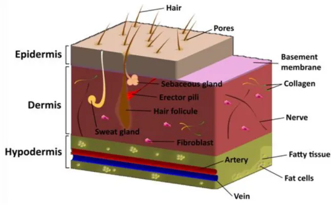
Figure 1. Schematic representation of the structure of normal skin tissue. ... 2
Figure 2. Representation of the structure of the skin. ... 4
Figure 3. Representation of the different type of skin wounds... 7
Figure 4. Representation of the healing process ... 8
Figure 5. Schematic representation of the five stages of wound healing process ... 10
Figure 6. Representation of a tissue engineering strategy used to regenerate a functional tissue12 Figure 7. Representation of a conventional electrospinning apparatus ... 16
Figure 8. Chemical structure of natural and synthetic polymers ... 17
Figure 9. Schematic representation of the setup used to produce the EBM... 24
Figure 10. Representative macroscopic images of the top and bottom view of the produced EBM. ... 31
Figure 11. Macroscopic and SEM cross-section images of the EBM. ... 32
Figure 12. ATR-FTIR analysis of the EBM and raw materials ... 33
Figure 13. Representative stress–strain curve and macroscopic images of the EBM during the tensile test. ... 34
Figure 14. Characterization of the porosity of the EBM ... 36
Figure 15. Characterization of the water contact angles of EBM... 37
Figure 16. Characterization of the water vapor transmission profile of EBM ... 37
Figure 17. Evaluation of weight loss of EBM ... 39
Figure 18. SEM images of EBM after 1, 3 and 7 days of being incubated with PBS. ... 40
Figure 19. Release profile of SA from electrospun EBM ... 41
Figure 20. Microscopic images of NHDF in the presence of EBM ... 42
Figure 21. Evaluation of NHDF viability when cultured in the presence of EBM ... 43
Figure 22. SEM micrographs of fibroblasts morphology at surface of EBM. ... 43
Figure 23. CLSM images of fibroblasts in contact with EBM ... 44
Figure 24. Evaluation of the bactericidal activity of EBM ... 45
xxiii
List of tables
Table 1. Tissue-engineered skin constructs commercially available ... 14 Table 2. Degree of deacetylation of the different CS ... 30 Table 3. Mechanical parameters of EBM and native human skin ... 35 Table 4. Water vapor transmission profile of EBM ... 38
xxv
Acronyms
1DUVS 3D ATR-FTIR bFGF CFU COX CS CSLM DD DETCs DMEM-F12 DMF DMSO EBM ECM EDTA EGF EtOH FBS FGF HA HB-EGF HIV LMW MMPs MPa MTT NHDF PBS PCL PDGF PEG PFA PLA RGD RTFirst Derivative UV-spectroscopy Three-Dimensional
Attenuated Total Reflectance-Fourier Transform Infrared Spectroscopy Basic Fibroblast Growth Factor
Colony Forming Units Cyclooxygenase Chitosan
Confocal Laser Scanning Microscopy Deacetylation
Dendritic γδ Epidermal T Cells Dulbecco’s Modified Eagle’s Medium Dimethylformamide
Dimethyl Sulfoxide
Electrospun Bilayer Membrane Extracellular Matrix
Ethylenediaminetetraacetic Acid Epidermal Growth Factor
Ethanol
Fetal Bovine Serum Fibroblast Growth Factor Hyaluronic Acid
Heparin-Binding Epidermal Growth Factor Human Immunodeficiency Virus
Low Molecular Weight Matrix Metalloproteinases Megapascal
3-(4,5-dimethylthiazol-2-yl)-2,5-diphenyltetrazolium bromide Normal Human Dermal Fibroblasts
Phosphate-Buffered Saline Solution Polycaprolactone
Platelet-Derived Growth Factor Poly(ethylene glycol)
Paraformaldehyde Poly(lactic acid)
Arginine-Glycine-Aspartic Acid Room Temperature
xxvi SA SEM TFE TFG-β TIMP TNF–α UV VEGF WVTR ZN ZnO Salicylic Acid
Scanning Electron Microscopy Trifluoroethanol
Transforming Growth Factor-β Tissue inhibitor of Metalloproteinase Tumor Necrosis Factor-α
Ultraviolet
Vascular Endothelial Cell Growth Factor Water Vapor Transmission Rate
Zein Zinc Oxide
Development of new therapeutic approaches for skin regeneration
2
1. Introduction
1.1. Skin
1.1.1. Functions and structure
Skin is the largest and heaviest organ of the human body, with an area of 1.5–2.0 m2 and a weight
of 3.5–10 kg [1]. This complex organ is the outermost barrier of the body that protects inner organs from microbial pathogens, mechanical and chemical insults, regulates the body temperature, gives support to blood vessels and nerves, and prevents dehydration. Furthermore, it is also involved in the immune surveillance and sensory detection process [2].
Anatomically and functionally, the skin is formed by three connected layers, the epidermis, the dermis and the hypodermis (figure 1). The basement membrane separates physically epidermis from the dermis, acting as a consistent and dynamic interface [3]. Associated with the skin layers are various appendages such as hair follicles, nails, sweat and sebaceous glands that play different functions [4]. A more detailed description of the structure of the human skin is given in the following sections.
Development of new therapeutic approaches for skin regeneration
3
1.1.1.1. Epidermis
Epidermis is the most superficial layer of the skin and it is composed of stratified squamous epithelium. Epidermis thickness varies according to body location [4]. The main function of the epidermis is to protect the skin from potential threats and also to act as an efficient barrier at the top of the skin [5]. As can be observed in figure 2A, the epidermis is composed of five distinct cell layers, according to the different stages of keratinocyte maturation. From the deepest to the most superficial, these layers are known as strata basale, spinosum, granulosum, lucidum, and
corneum [6].
The stratum basale consists in several layers containing cuboidal nucleated epithelial cells, also known as skin stem cells. These stem cells have a high self-renewal capacity and move upwards while they differentiate [3]. Initially, they are in the stratum basale and then they migrate to the
stratum spinosum where the keratinization process begins. At this stage, cells start to lose their
cytoplasm, suffer shape variation and start to synthesize keratin [7]. In the stratum granulosum, differentiated keratinocytes are highly active and accumulate lipid granules that are critical for the maintenance of a water barrier due to its hydrophobic nature. In fact, while delivering the content of the secretory lamellar granules to the intercellular spaces, the keratinocytes present in this layer become flat and spread. In strata lucida and corneum, cells enter into the programmed cell death process and all of the cytoplasmic organelles are degraded [4]. The
stratum corneum is the most external layer of the epidermis and represents the final stage of
keratinocyte differentiation [8]. It is composed of 10-20 layers of completely differentiated dead keratinocytes, known as corneocytes, that are interspersed with intercellular lipids, mainly ceramides and sphingolipids [5]. The keratinized stratum corneum is in direct contact with the environment and provides the main barrier to prevent water loss and penetration of hazard agents. Keratinocytes migration from the stratum basale to the outer stratum corneum takes approximately 28 days [9].
In addition to keratinocytes, which account for about 80% of epidermal cells, the epidermis is also composed by melanocytes, responsible for the pigmentation, Langerhans cells, which play a sensory role, and Merkel cells that are involved in the skin immune defense system [6, 7].
Development of new therapeutic approaches for skin regeneration
4
Figure 2. Representation of the structure of the skin. The outermost layer of the epidermis, the stratum corneum, is fundamental for avoiding microorganisms penetration and also protect the skin from environmental insults. The other strata (lucidum, granulosum, spinosum and basale) are fundamental for the remodeling of the stratum corneum. The epidermis also contains Langerhans cells and melanocytes (A). The dermis has as mainly cellular components the fibroblasts but also contains cells of the immune system (neutrophils and macrophages), blood vessels, and nerve fibers (B).
1.1.1.2. Basement Membrane
In the basal surface of the epithelium, it is found the basement membrane that is responsible for establishing a functional link between the epidermis and the dermis. Skin basement membrane has a thickness of 50-100 nm and it is composed of two main regions: a) lamina lucida, the layer closer to the epidermis, composed of laminin, entactins, and dystroglycans and b) lamina densa that is located above the papillary dermis and is mostly formed by collagen type IV [10,11]. This junction regulates the permeability to substances that migrate from the dermis to epidermis and vice versa [12]. Epidermis attachment to the basement membrane is ensured by hemidesmosomes that bind to the keratin filaments of keratinocytes. In turn, the basement membrane is bound to the dermal layer through fibrils of collagen VII that are interspersed into the papillary dermis [13].
Development of new therapeutic approaches for skin regeneration
5
1.1.1.3. Dermis
The dermis is located below the basement membrane and is the major component of human skin, with 3-5 mm thick. This layer supplies energy and nutrition to the overlying epidermis and also provides mechanical integrity to the skin due to the arrangement of collagen fibers that are deposited by local fibroblasts [7].
The dermis is composed of two distinct layers: the upper papillary dermis and the deeper reticular dermis [3]. The papillary dermis is essentially composed of loose connective tissue, namely elastin fibers interspersed with collagen fibers. It also holds numerous blood vessels that assure nutrients delivery, remove waste products and also allow the regulation of the body temperature. In turn, the reticular dermis is denser and composed by larger collagen fibers that confer flexibility to the skin [14]. Fibroblasts are the main cell type found in the dermis and they are responsible for the production and deposition of extracellular matrix (ECM) components. The dermal layer also contains cells of the immune system (neutrophils and macrophages), lymphatic vessels, nerve fibers, sweat and sebaceous glands, the deep portion of hair follicles and endothelial cells (figure 2B) [4].
1.1.1.4. Hypodermis
Hypodermis is the deepest skin layer and it is mainly composed of adipose tissue. This layer insulates the body and provides mechanical protection against physical shock [15]. Structurally, hypodermis is divided into lobules containing adipose cells separated by fibro-vascular septa. The septa are composed of collagen and reticulin fibers, blood and lymphatic vessels. In addition to adipose cells, hypodermis also contains fibroblasts and macrophages, that have an important role in the stimulation of thermogenesis during cold exposure or exercise [3].
1.1.1.5. Skin appendages
Skin has a variety of appendages, such as hair, nails, sebaceous and sweat glands, which maintain and protect the skin and their functions as explained in the following topics.
Hair
Hair, a unique mammalian trait, has important functions in thermoregulation, physical protection, sensory activity, and social interactions. Each hair arises through a tubular invagination of the epidermis into the dermis [6]. Histologically, hair is arranged in three concentric layers - the inner layer or medulla, the middle layer or the cortex and the outer layer or cuticle – that are composed of keratin [4, 16]. Hair follicles are distributed over the entire surface of the body, although there are specific regions of the body where they are absent (soles of the feet, palms of the hands, glans penis, clitoris, labia minora, and mucocutaneous junctions). At the proximal end of the hair follicle is the hair bulb that contains a population of hair stem cells [6].
Development of new therapeutic approaches for skin regeneration
6
Nail
The nail is a highly keratinized structure that grows on the dorsal tip of the fingers [17]. Nail functions include physical protection of the end of the fingers and toes, assistance in manipulation and scratching [18]. The nail unit is composed of the proximal nail fold, the nail matrix, the nail bed and hyponychium, which together form and support the nail plate, which is a keratinized structure that continuously grows throughout life [18, 19]. The nail is also composed of water, lipids and trace elements like iron, zinc, and calcium [16].
Sebaceous Glands
Sebaceous glands are holocrine glands that are widely distributed throughout the skin but are mainly associated with hair and, therefore, concentrated on the face and scalp. Furthermore, they are absent from the palms, soles, and dorsum of feet [20]. Sebaceous glands often open directly into the hair follicle and produce sebum, an oily complex of triglycerides, fatty acids and their breakdown products (wax esters, squalene and cholesterol esters). Due to its lipid-hydrophobic composition, sebum acts as a natural lubricant that protects the skin against friction and avoids its dehydration [6].
Sweat Glands
Sweat glands are located within the dermis. They are composed of coiled tubes and found all over the skin, but in abundance on palms, soles, axillae and forehead [6, 16, 17]. These glands secrete sweat, which is fundamental for the thermoregulation and excretion of metabolites [17]. There are two types of sweat glands – eccrine and apocrine – according to their secretory mechanism. The eccrine sweat glands secrete high amounts of an aqueous liquid following a merocrine mechanism. In turn, the apocrine glands secrete low amounts of a lipid-rich liquid and join up into the hair canal instead of the skin surface [21].
1.2. Wounds
Due to its exposition to environmental conditions, the skin suffers different types of lesions and diseases that compromise its structure and functions. According to the US Wound Healing Society, wounds are defined as the result of disruption of normal anatomic structure and functions of the skin [22].
Wounds - particularly surgical incisions, thermal injuries and chronic ulcers - are a major medical issue, since they lead to physical incapacity and ultimately to the death of the patient [23,24]. In fact, every year, more than 70 million surgical procedures are performed just in the United States of America with more than one-third resulting in hypertrophic scarring or keloid formation [25]. It is also estimated that burn injuries affect more than 11 million people worldwide annually [25, 26]. On the other hand, the number of patients suffering from chronic wounds, mainly pressure
Development of new therapeutic approaches for skin regeneration
7 ulcers, and diabetic foot ulcers, is increasing and affects nowadays more than 1% of the worldwide population [27].
According to the number of skin layers that are damaged, wounds can be divided into superficial, superficial partial-thickness, deep partial thickness, and full thickness, as depicted in figure 3 [28, 29].
Superficial injuries, typically caused by sunburns, light scalds and grazing. They are
usually able to self-renewal within few days, due to the presence of epidermal stem cells. Such injuries do not require specific surgical treatment since only the epidermis is affected. Moreover, this type of lesions regenerates rapidly without scarring, since no ECM deposition occurs.
Superficial partial-thickness injuries affect the epidermis and the upper part of the
dermis. This type of injury is followed by epidermal blistering and severe pain to the patient. They usually heal rapidly (≈2 weeks) in a healing process that involves the epithelialization from the margins of the wound through basal keratinocytes that migrate from the wound edge, hair follicles or sweat glands. In fact, the rate of skin regeneration depends on the density of these skin appendages. For example, thin hairless skin (e.g. inner arm, eyelids) heals more slowly than hairy skin (e.g. back, scalp).
Deep partial-thickness injuries involve a dermal damage, where the skin appendages are
destroyed and they are characterized by a slow healing. Scarring is pronounced in this type of injuries since fibroplasia is more intensive when compared with superficial partial-thickness wounds.
Full-thickness injuries involve the complete destruction of the epidermis and dermis.Sometimes, this type of injury can also affect the hypodermis, muscle, and even bone. Skin appendages are destroyed and, therefore, there is no potential source to trigger epithelial regeneration. Full-thickness skin wounds lead to extensive scarring, cause mobility restrictions and cosmetic issues to the patients. If not properly treated, the full thickness damaged tissue will start to break down providing a perfect nutrient source and environment for invading microorganisms.
Development of new therapeutic approaches for skin regeneration
8
1.2.1. Wound Healing
Wound healing is a complex biological process that allows the restoration of skin integrity. Based on the nature of the healing process, wounds can be divided in acute and chronic. Acute wounds (figure 4A) occurs as a consequence of mechanical injuries (abrasions, superficial, surgical or traumatic wounds) [27]. In this type of injury, the normal wound healing process occurs (see section 1.2.1.1. for further details) [30]. In turn, chronic wounds (figure 4B) are usually associated with comorbidity conditions, such as diabetes, obesity, tumor or microbial infection. In this type of wound, the healing process is not effective and a state of pathologic inflammation occurs (see section 1.2.1.2.) [30, 31].
Figure 4. Representation of the healing process that occurs in acute and chronic wounds. Acute wound healing (A) is characterized by cell migration, proliferation and also by the secretion of high levels of growth factors that are involved in the stimulation of the synthesis of new ECM; Chronic wound healing (B) is characterized by an exuberant inflammation, increased MMPs production that impairs cell proliferation and hence delays the formation of the new skin tissue.
Development of new therapeutic approaches for skin regeneration
9
1.2.1.1. Normal Wound Healing
The normal wound healing process involves a series of coordinated events including bleeding and coagulation, acute inflammation, cell migration, proliferation, differentiation, angiogenesis, re-epithelialization, synthesis and remodeling of ECM [24]. These complex events can be divided into five overlapping steps: wounding (A), hemostasis (B), inflammation (C), proliferation (D) and remodeling (E) (see figure 4 for further details).
Wounding
The wounding phase (figure 5A) is the first stage of the wound healing process. After the skin damage occurs, the wound site is filled with fluids from injured blood and lymphatic vessels. At this point, bacteria start to invade the open and unprotected wound [32].
Hemostasis
Hemostasis begins immediately after the injury occurs in order to prevent bleeding. This process involves the formation of a provisional matrix at the wound site (figure 5B). The extrinsic and intrinsic coagulation cascades are activated, leading to platelet aggregation. Such event promotes the vasoconstriction of the injured vessels, reducing blood loss and simultaneously filling the tissue gap with a blood clot comprised of fibrin, fibronectin, vitronectin, and thrombospondin. The clot also acts as a provisional matrix for the migration of leukocytes, keratinocytes, fibroblasts, endothelial cells and growth factors during the wound healing process [32, 33].
Inflammation
The inflammatory phase of the wound healing is represented on figure 5C. It occurs hemostasis and can last up for 6 days. This phase is characterized by the sequential influx of immune cells. Specifically, monocytes that migrate to the wound site, where they differentiate into macrophages and remove bacteria and foreign material through phagocytosis. Monocytes are attracted to the wound site by several chemoattractive agents, such as clotting factors, platelet-derived growth factor (PDGF), transforming growth factor-β (TGF-β), elastin and collagen breakdown products. In the wound, macrophages produce numerous growth factors, such as TGF-β, tumor necrosis factor-α (TNF-α), heparin-binding epidermal growth factor (HB-EGF), and fibroblast growth factor (FGF) that are involved in the activation of keratinocytes, fibroblasts, and endothelial cells. Mast cells are also active in this phase and release granules filled with enzymes, histamine, and other active amines. These mediators are responsible for the characteristic symptoms associated with inflammation, i.e. rubor (redness), calor (warmth),
tumor (swelling), dolor (pain) and functio laesa (loss of function). Lymphocytes, particularly
CD4+, CD8+, and dendritic γδ epidermal T cells (DETCs), arrive approximately 6 days after the
Development of new therapeutic approaches for skin regeneration
10
Proliferation
The proliferative phase starts on the 6th day after the injury occurs and lasts for about 3 weeks
(figure 5D). It is characterized by the re-epithelization of epidermis, repair of the dermal layer and neovascularization. At this stage, keratinocytes migrate from the surrounding tissue to the wound, where they became organized in stratified layers. This migration is stimulated by connexins and by the contact established between the keratinocytes and fibrin molecules. It is noteworthy that the migration, proliferation, and differentiation of the keratinocyte cells lead to the closure of the epithelial gap and to the restoration of the epithelium. On the other hand, the dermis is restored by fibroblasts that synthesize and secrete new ECM. The migration of fibroblasts into the wound is facilitated by ECM-cleaving matrix metalloproteinases (MMPs) that are produced by these cells. Furthermore, fibroblasts attach to the fibrin matrix and produce collagen (predominantly type I). As the collagen matures, intramolecular and intermolecular cross-links are created, increasing the strength of the new skin tissue. Simultaneously, the wound microenvironment, which is characterized by low pH, reduced oxygen content, and high concentration of lactate contribute for triggering the angiogenic process. This process is also stimulated by vascular endothelial cell growth factor (VEGF), basic fibroblast growth factor (bFGF), and TGF-β [21, 34, 36].
Remodeling
The remodeling of the wound is the final phase of the normal wound healing process and may last for 1–2 years or even longer (figure 5E). In this phase, all the processes that were activated after injury occurrence cease. Specifically, it is verified the degradation and replacement of immature ECM, the return to a normal vascular density and the apoptosis of inflammatory cells at the wound site. All of these changes lead to the contraction of the wound and to the formation of acellular scar tissue. Although the remodeling process continues for a long period of time, the scar tissue does not achieve the strength of the native skin [32, 34, 36].
Figure 5. Schematic representation of the five stages that characterize the normal wound healing process. During wounding damaged skin is vulnerable to bacteria invasion (A). The hemostasis is characterized by the
Development of new therapeutic approaches for skin regeneration
11
formation of a clot that becomes a provisional ECM (B). Inflammatory phase is characterized by inflammatory cells infiltration (C). In the proliferative phase, new fibroblasts and keratinocytes migrate into the wound, in order to perform the re-establishment of the dermal layer and the re-epithelization of the epidermis, respectively (D). In remodeling, the wound contracts and an acellular scar tissue is formed. The chronologic duration of each phase of the wound healing is also represented in the figure.
1.2.1.2. Non-healing Wounds
Non-healing wounds occur in situations where the normal healing process is delayed, incomplete, and does not follow the proper order [30]. A chronic wound is continuously subjected to inflammation that is caused by an excessive infiltration of neutrophils and, therefore, is unable to progress through the normal stages of the wound healing [34]. This up-regulation of the inflammatory cascade can be caused by pressure, bacterial overgrowth, leukocyte trapping or ischemic injury. Neutrophils release the enzyme elastase, which is capable of destroying important healing factors, such as PDGF and TGF-β that promote proliferation and ECM deposition. Moreover, the mitogenic activity of cells is suppressed in chronic wounds and they cannot enter into the proliferative phase, unlike what happens in normal wound healing process. It is noteworthy that the time required for chronic wound healing is increased and, in some cases, the wound size increases over time [31].
1.2.2. Wound Healing Therapies
1.2.2.1. Autografts, allografts, and xenografts
Autologous skin grafts are still the gold standard used in the clinic for the treatment of full-thickness skin wounds [37]. Before the autografting process, an early excision of the damaged skin tissue is performed [38]. Subsequently, the autograft procedure is performed with a dermatome that cuts thin slices of the epidermis and initial part of the dermis of the patient uninjured skin, which are subsequently transplanted to the wound site [39]. Such grafting procedure provides sufficient coverage without risk of rejection but presents serious drawbacks like limited availability of donor sites, induction of scar formation, patient morbidity, and long periods of hospitalization [2].
Alternatively, allograft and xenograft procedures have also been used for the treatment of severe wounds. In this case, implantable skin is collected from human and non-human cadavers, respectively. However, these procedures cannot be seen as efficient therapeutic alternatives to autografts since they present risks of viral transmission (e.g., hepatitis B and C or HIV), immune rejection and their use are dependent on tissue availability on skin banks [2, 39].
Hence, in the last few years, researchers from Tissue Engineering field attained significant progress in the development and clinical use of improved skin substitutes that are able to surpass some of the limitations associated with auto-, allo-, and xenografts.
Development of new therapeutic approaches for skin regeneration
12
1.2.2.2. Skin Tissue Engineering
Tissue engineering is an interdisciplinary field that combines the principles and methods of engineering and life sciences toward the development of biologic substitutes that are able to restore, maintain, or improve the functions of native tissues. The main strategy used in the area of tissue engineering involves cell seeding in contact with biomaterials, which serve as temporary scaffolds, in order to restore the structure and functions of a tissue (see figure 6) [40].
Figure 6. Representation of a tissue engineering strategy used to regenerate a functional tissue. Such therapeutic approach involves cell seeding in a porous scaffold. Cells are isolated from the patient (1) and expanded in vitro (2). Subsequently, cells are seeded in a scaffold (3). Ultimately, the construct is transplanted to the damaged tissue in order to restore its structure and function.
Skin tissue engineering revolutionized the therapy of extensive acute or chronic wounds. An ideal skin substitute has to fill certain requirements, such as biocompatibility, biodegradability, non-toxicity, and non-immunogenicity. It may also protect against bacterial infection, be nonadherent to the wound site, allow gaseous and fluid exchanges, absorb the excess exudates, and support the handling during application and regeneration of the new tissue. Furthermore, it should also be capable of improving the healing process, mimic the native skin environment, be comfortable and cost effective [2, 41].
Today, as a result of intense research, a myriad of skin substitutes exists and some of them are already available to be used in the clinic. Depending on wound severity, skin substitutes can be classified as epidermal, dermal and bilayered/dermo-epidermal substitutes (see table 1) [29]:
Development of new therapeutic approaches for skin regeneration
13 ● Epidermal Skin Substitutes
Epidermal skin substitutes aim to restore the epidermis by promoting the reepithelialization of the wound. The majority of the epidermal skin substitutes are created by isolation of keratinocytes from the patient, followed by its subsequent in vitro cultivation on biocompatible substrates (e.g. bovine collagen, hyaluronic acid, etc.). When a suitable cell mass has grown, it is transferred to the wound bed for restoring the barrier function and enhancing the wound healing [42, 43]. In general, the available epidermal substitutes are expensive, difficult to handle due to their thin and fragile nature, cannot be used to treat full-thickness skin wounds and their production is time-consuming [44].
MySkin® is an example of an epidermal skin substitute that uses subconfluent autologous
keratinocytes grown on a silicon support layer [29]. Contrariwise, Cryoskin® and Celaderm® are
epidermal replacements containing allogeneic keratinocytes [45]. Suprathel® represents a reliable
acellular epidermal skin substitute, based on polylactide, trimethylene carbonate and ε-caprolactone [45, 46].
● Dermal Skin Substitutes
Dermal skin substitutes are able to restore the injured dermis and are usually acellular. They display great structural integrity, prevent the wound from contracting and are able to mimic the basic properties of ECM [11]. However, they cannot efficiently replace the epidermal layer of the skin.
Alloderm®, Integra®, MatriDerm®, and Oasis® are examples of acellular dermal replacements.
Integra® consists of a porous scaffold made of bovine collagen type I and shark
chondroitin-6-sulphate glycosaminoglycan bonded to a silicone membrane. MatriDerm® is made up of bovine
collagen and elastin hydrolysate. Cellular dermal substitutes, such as TransCyte® or Dermagraft®,
use autologous dermal fibroblasts or neonatal human foreskin fibroblasts [42, 45]. ● Bilayered/ Dermo-Epidermal Skin Substitutes
Bilayered/ Dermo-epidermal skin substitutes are the most advanced skin substitutes, which are conceived to mimic the histological structure of normal skin with both epidermal and dermal layers [29]. These bilayers provide biological stimulus for skin regeneration and at the same time act as temporary wound coverage. Keratinocytes and fibroblasts, either autologous or allogenic, are commonly used to prepare these bilayered structures [44]. Nevertheless, the bilayered skin substitutes that are available in the market are very expensive, present risk of infection to the patient and have low mechanical properties.
Apligraf® consists of viable allogeneic neonatal fibroblasts grown in a bovine collagen type I gel
Development of new therapeutic approaches for skin regeneration
14 engineered skin construct that includes fibroblasts seeded into a bovine type I collagen sponge, on top of which keratinocytes are seeded to form a confluent layer [42]. Additionally, Mediskin® is
an example of an acellular dermo-epidermal skin substitute that is based on porcine xenografts [48].
Although some of the different skin substitutes are very complex and effective, they are not able to completely mimic the skin’s natural environment. Therefore, further studies are needed to improve these products.
Table 1. Skin substitutes available in the market.
Company Application Cells Biomaterials
Epide
rm
al
Celaderm® Inc., Connecticut, USA Advanced BioHealing, Chronic wounds Frozen cultured allogeneic
keratinocytes -
Cryoskin® Cryo Diffusion, Lery, France Chronic wounds keratinocytes from Allogenic
newborn foreskin Silicon backing matrix
Myskin® CellTran Ltd, Sheffield, UK
Diabetic foot; venous leg ulcers;
burns
Cultured autologous
human keratinocytes Polyacrylic acid membrane
Suprathel® Bochum, Germany Stapleline GmbH,
Acute superficial partial-thickness injuries - Copolymer of polylactide and ε-caprolactone De rm al
Alloderm® LifeCell Corporation, New Jersey, USA thickness wounds Full- and partial- - lyophilized dermis Human acellular
Integra® Inc., New Jersey, USA Integra Life Sciences, thickness wounds Full- and partial- - glycosaminoglycan Bovine collagen I; matrix
Matriderm® Billerbeck, Germany Medskin Solutions, thickness wounds Full- and partial- -
Bovine non-crosslinked lyophilized dermis, coated with α-elastin
hydrolysate
Oasis® Cook Biotech Inc., Indiana, USA thickness wounds Full- and partial- - Acellular porcine small intestine submucosa matrix
Transcyte® Inc., Connecticut, USA Advanced BioHealing, Full-thickness burns allogeneic fibroblasts Neonatal foreskin Nylon mesh covered with silicone
Dermagraft® Inc., Connecticut, USA Advanced BioHealing, Full-thickness burns allogeneic fibroblasts Neonatal foreskin Polyglactin scaffold
De rm o-Epid er m al Apligraf ® Organogenesis, Inc.,
California, USA Chronic wounds
Allogenic keratinocytes;
fibroblasts Bovine collagen
OrCel® Inc., New York, USA Ortec Internation, Chronic wounds keratinocytes; Allogenic
fibroblasts Bovine Collagen
Mediskin® Brennen Medical, Minnesota, USA Acute and chronic wounds - xenograft chemically Frozen porcine crosslinked
Development of new therapeutic approaches for skin regeneration
15
1.3. Nanofibrous membranes produced by electrospinning
Electrospinning is a technology that uses electrostatic forces to produce polymeric nanofibrous meshes with diameters ranging from 2 nm to several micrometers [49]. Electrospinning was for the first time described in literature more than a century ago, in 1902, when Cooley and Morton separately patented methods and apparatuses for electrically dispersing fluids. However, only recently electrospun polymeric nanofibers became a topic of huge research [50].
A conventional electrospinning setup (figure 7) is composed of a syringe pump, a capillary needle connected to a syringe, a high-voltage power supply, and a metal collector. In this technique two electrodes are used - one is connected to the needle and the other is fixed to the collector - in order to complete a circuit that produces an electric field. The syringe pump is used to force the solution to pass through the needle with a controlled flow rate. When a high voltage is applied at the tip of the capillary needle, the polymeric solution remains at the tip of the needle due to surface tension. With the increasing of the intensity of the electrical field, the charge causes the elongation of the droplet into a cone, known as the Taylor Cone. An additional increase in the electric field will generate a critical value with which the repulsive electrostatic forces overcome the surface tension and a polymer jet is ejected from the tip of the Taylor Cone. Before reaching the collector, the jet undergoes a series of electrically driven instabilities and gradually thins in the air due to solvent evaporation [50, 51]. If the jet is collected in a stationary collect, as shown in figure 8, non-woven meshes composed of randomly oriented nanofibers are obtained. However, aligned nanofibers can also be produced by this technique using rotary collectors [52].
The production of electrospun meshes is dependent on specific parameters such as the precursor solution (e.g. conductivity, surface tension, viscosity and solvent selection), processing variables, and environmental conditions. The control of these particular features has a direct impact on the mean diameter and arrangement of the accumulated fibers. Particularly, solutions with high viscosity lead to the production of fibers with a high diameter, while an increase in the surface tension or conductivity of the polymer solution results in a decrease in nanofiber diameter. Furthermore, an increase in voltage or a decrease in feed rate results in a reduction of the fiber diameter. However, a high electrical potential is not desired since it may lead to the formation of beads or to the formation of defective nanofibers. Moreover, the distance between the needle and the collector needs to be sufficient to allow solvent evaporation but close enough to allow production of fibers with the required morphology. Regarding the environmental parameters, an increase in temperature is responsible for decreasing the average fiber diameter, due to a decrease in solution viscosity, while an increase in humidity increases the average fiber diameter due to polymer swelling [32, 50].
Development of new therapeutic approaches for skin regeneration
16
Figure 7. Representation of a conventional electrospinning apparatus that is used to produce non-woven meshes composed of randomly oriented nanofibers. Bioactive molecules that may enhance the wound healing process have been incorporated in electrospun meshes.
In the last years, electrospun nanofibrous membranes become one of the most studied platforms for skin tissue engineering applications. According to Augustine and Abrigo, the main advantages of the electrospun membranes to be used as skin substitutes are [52, 53].
Mimic the ECM
The nanofibers produced by electrospinning have diameters ranging from 50-500 nm, which is within the diameter range presented by the collagen fibrils found in the natural ECM. The structural similarity is very important in cell adhesion, proliferation, migration, and differentiation.
Exudate uptake capacity
The high surface area presented by electrospun nanofibrous meshes allows them to absorb the wound exudates. The great absorptive properties of this type of skin substitute help in the maintenance of a moist environment at the wound site.
Semi-permeable
The porous structure of electrospun meshes allows gas exchange and confers protection against bacteria invasion.
Conformability
Conformability of the material is a crucial issue when materials are aimed for skin tissue engineering. Electrospun nanofibrous membranes are easy to fit in a wound with irregular architecture, thus providing a great conformability.
Development of new therapeutic approaches for skin regeneration
17
Functionality
The nanofibers produced by electrospinning can be easily functionalized with bioactive compounds in order to improve skin regeneration. Depending on the stage of treatment and the intended functionality, drug molecules (e.g. antiseptics, antifungal, and vasodilators), genes, antimicrobial agents, growth factors (e.g. FGF, EGF, and TGF) and even cells (e.g. fibroblasts and keratinocytes) can be delivered to the wound, in a controlled manner, for improving the wound healing process. Blend, coaxial and emulsion electrospinning are strategies employed to fabricate biofunctional nanofibrous scaffolds (figure 7). To produce blended electrospun meshes, active agents are mixed with the polymeric solution before performing the electrospinning process [54]. In coaxial electrospinning, a syringe containing a core and an outer compartment is used. Through this technique, the active agents are incorporated in the core and the polymeric solution in the outer compartment. Subsequently, both polymer and active agent are coaxially and simultaneously electrospun to produce fibers with a core-shell structure [32]. Another route to obtain core-shell nanofibers is by performing emulsion electrospinning, which is a technique that does not require a particular needle setup. In this type of electrospinning, an aqueous solution, with active agents is emulsified into an organic phase containing a polymer that forms the shell [55].
1.3.1. Electrospun nanofibrous membranes produced with natural polymers
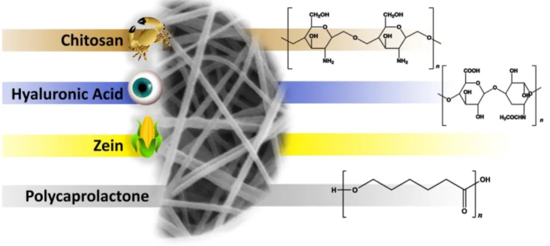
Natural polymers are usually biocompatible and present low immunogenicity. Moreover, natural polymers have an inherent capacity to promote cell adhesion since they have specific aminoacids sequences, such as RGD (arginine/glycine/aspartic acid). Cellulose acetate, chitosan, collagen, elastin, gelatin, hyaluronic acid, silk fibroin, and zein are examples of natural polymers successfully electrospun and applied for skin regenerative purposes. The main drawback of natural polymers is associated to its poor mechanical properties [54]. In the following sections, some of the natural polymers are described in further detail.
Figure 8. Chemical structure of some examples of natural and synthetic polymers used in skin tissue engineering.
Development of new therapeutic approaches for skin regeneration
18
1.3.1.1. Chitosan
Chitosan (CS) is obtained from chitin, which is the second most abundant polysaccharide found in nature. This polymer is usually obtained from the shells of crustaceans, the exoskeleton of insects and cell walls of fungi [54]. CS polysaccharide is composed of glucosamine and N-acetyl-glucosamine [56]. CS has many advantages like biocompatibility, biodegradability, antimicrobial and hemostatic activity. Moreover, CS can stimulate collagen synthesis, a feature that is fundamental for enhancing the wound healing [2].
CS is easily incorporated into films, gels, or sponges, but very hard to electrospun in its pure form since it forms highly viscous solutions. Hence, this polymer is usually modified or combined with other polymers in order to be electrospun. Zhou et al produced the first CS-based electrospun membranes aimed for skin regeneration. Their electrospun membranes were produced with water soluble CS and poly(vinyl alcohol) that promoted the adhesion and proliferation of mouse fibroblast cells [57]. The antimicrobial properties of CS are also reported in the literature by several authors. Cai et al produced CS/silk fibroin electrospun membranes that inhibit the growth of the Gram-negative bacteria Escherichia Coli (E.Coli) [58]. Antunes and collaborators produced a deacetylated/arginine-modified chitosan electrospun membrane with enhanced bactericidal activity against both gram-positive Staphylococcus Aureus (S. Aureus) and gram-negative E.Coli [59].
1.3.1.2. Hyaluronic Acid
Hyaluronic acid (HA), also known as hyaluronan or hyaluronate, is a linear polysaccharide composed of repeating units of glucuronic acid and N-acetylglucosamine that is found in the ECM of connective tissues [60]. It can be obtained from bacteria or from the enzymatic digestion of connective tissues [56]. HA has excellent biocompatibility and biodegradability. Furthermore, as a natural polysaccharide present in the skin ECM, HA presents a high water retention capacity, and as a component of granulation tissue, it facilitates the migration of inflammatory cells and fibroblasts into the healing wounds through the interaction with CD44 receptors present in the plasma membrane [54].
The application of HA electrospun meshes in the wound healing process has been limited by its high viscosity and surface tension [61]. Hsu and colleagues produced HA/collagen electrospun membranes cultured with foreskin fibroblast cells. In their study, they verified that HA promotes a scarless wound healing since it decreases the ratio of tissue inhibitor of metallopeptidase 1 (TIMP1) to matrix metalloproteinase-1 (MMP1) characteristic of scarless wounds [62]. Later on, HA/ Polycaprolactone (PCL) /Silk fibroin electrospun membranes were produced to be used in skin tissue engineering. The addition of HA gave hydrophilic properties to the membranes, which caused the suppression of non-specific protein adsorption, leading to a reduction of fibrosis thickness and macrophages adhesion in vivo. Further, this study showed that HA-based membranes present a significant increase in fibroblasts proliferation and adhesion. These findings suggested that HA-based electrospun membranes are excellent candidates to be used as wound
Development of new therapeutic approaches for skin regeneration
19 dressings [63]. Recently, HA electrospun meshes were used as growth factors delivery systems in order to promote wound healing [64, 65]. A recent study reported the efficient encapsulation of EGF in HA/PCL electrospun membranes [64]. Moreover, Lai and collaborators developed an HA/collagen electrospun membrane capable of performing a controlled release of several angiogenic growth factors, such as PDGF, VEGF, bFGF, and EGF, either directly embedded in the nanofibers or encapsulate in gelatin nanoparticles according to the wound healing stage required. Moreover, this study also showed that the produced membranes possess mechanical properties similar to those of human native skin [65].
1.3.1.3. Zein
Zein (ZN) is a protein found in corn or maize. It is mainly composed of nonessential amino acids such as glutamic acid (21–26%), leucine (20%), alanine (10%) and proline (10%). ZN is biodegradable, biocompatible, moderately hydrophobic, highly elastic and easily electrospinable [66, 67].
The study of ZN nanofibers for biomedical applications began in 2009. Yao and co-workers reported the production of electrospun blends composed of ZN/silk fibroin with improved mechanical properties and biocompatibility [68]. In 2013, ZN nanofibers were used as drug delivery system for wound healing applications. Huang et al. produced ZN nanofibers loaded with Ibuprofen through coaxial electrospinning technique. The in vitro release experiments showed that the drug-loaded fibers provided sustained drug release for 10 hours [69]. Another study reported the production of ZN/polyurethane/cellulose acetate electrospun membranes blended with an antimicrobial agent, streptomycin sulfate. These electrospun membranes presented features, such as hydrophilicity, excellent cell adhesion capacity, and blood clotting activity [70]. Moreover, ZN/silver nanoparticles electrospun membranes were also produced for being applied as wound dressings by Dashdorj and collaborators. The results obtained demonstrated that membranes had a good cytocompatibility and fibroblasts were able to adhere on the composite nanofibers and also displayed bactericidal activity against S.aureus and E.coli [71].
1.3.2. Electrospun nanofibrous membranes produced with synthetic polymers
Synthetic polymers are often characterized by having improved mechanical properties (viscoelasticity and strength), and slow degradation rates. Typical synthetic polymers used in biomedical applications are hydrophobic biodegradable polyesters, such as polyglycolide, polylactide, polyurethane, and PCL, which were already used as nanofibrous scaffolds [54]. In the following section, the use of PCL to produce electrospun mats for wound healing purposes is reviewed in further detail.
Development of new therapeutic approaches for skin regeneration
20
1.3.2.1. Polycaprolactone
PCL is an aliphatic linear polyester, synthesized through ring-opening polymerization of ε-caprolactone. It is characterized by being biocompatible, bioresorbable, inexpensive and degrades through the hydrolysis of its ester linkages under physiological conditions. Therefore, it has been used as a valuable material for tissue engineering applications. However, the use of PCL in tissue engineering is compromised by its hydrophobic nature that limits cell adhesion and leads to uncontrolled biological interaction [72].
In order to overcome such drawbacks, PCL has been blended with other polymers such as chitosan or gelatin. Duan et al investigated the feasibility of using gelatin/PCL electrospun membranes to produce an epidermal skin substitute. These membranes showed good mechanical and biological properties that are compatible with their use in epidermis regeneration [73]. Bonvallet and co-workers synthesized PCL/collagen electrospun membranes with micropores to be used as dermal skin substitutes. These nanofibers accelerated the wound closure and stimulated the regeneration of healthy dermal tissue [74]. Another study, performed by Augustine and colleagues, reports the development of a PCL electrospun membrane loaded with zinc oxide (ZnO) nanoparticles. This was the first study that evaluated the ability of biomaterials containing ZnO nanoparticles to enhance the mammalian cell proliferation. The authors noticed that fibroblasts adhered and proliferated when seeded on top of these membranes, a process that is crucial for enhancing the wound healing process [53].
1.4. Antimicrobial Agents
Bacterial infections are regarded as the most severe and devastating complications associated with the implantation of biomaterials in the human body [75]. Nowadays, it is estimated that 65-80% of bacterial infections are caused by organisms that form biofilms on implants surface, compromising their successful application [76]. In this context, the development of biomaterials with antimicrobial activity is fundamental to avoid infections related to materials implantation. So far, different approaches have already been used to confer antimicrobial properties to tissue engineering constructs. Some authors reported the prevention of bacterial adhesion via through modification of surface charges or incorporation of adhesion-resistant coatings (e.g. polymer brushes or diamond-like carbon coatings) [59, 77-80]. Other antimicrobial strategies include the release of antimicrobial agents (e.g. antibiotics, drugs, metallic nanoparticles, nitric oxide or quaternary ammonium compounds) to the surrounding areas in order to avoid contaminations by bacteria [81-84].
However, the commonly used antimicrobial agents display cytotoxicity and induce microbial resistance [85]. Therefore, there is a huge demand to develop of new and effective antibacterial tissue engineering constructs that do not trigger any adverse effect on the host.