Mariana Cabral Monteiro Matos Santos
Characterization of microRNAs expression
during zebrafish skeletal development
Mariana Cabral Monteiro Matos Santos
Characterization of microRNAs expression
during zebrafish skeletal development
Mestrado em Biologia Marinha
Trabalho efetuado sob a orientação de:
Professor Doutor Paulo Gavaia
Doutor Daniel Tiago
Characterization of microRNAs expression
during zebrafish skeletal development
Declaração de autoria do trabalho
Declaro ser a autora deste trabalho, que é original e inédito. Autores e trabalhos consultados estão devidamente citados no texto e constam da listagem de referências incluída. Esta publicação não poderá ser reproduzida de forma parcial ou integral, nem poderá ser copiada de alguma maneira, incluindo eletronicamente, mecanicamente, fotocopiada, gravada ou digitalizada, sem autorização escrita do autor.
© Mariana Santos
"A Universidade do Algarve tem o direito, perpétuo e sem limites geográficos, de arquivar e publicitar este trabalho através de exemplares impressos reproduzidos em papel ou de forma digital, ou por qualquer outro meio conhecido ou que venha a ser inventado, de o divulgar através de repositórios científicos e de admitir a sua cópia e distribuição com objetivos educacionais ou de investigação, não comerciais, desde que seja dado crédito ao autor e editor."
i
Agradecimentos
Durante o último ano dediquei-me à realização desta tese de mestrado. Quem me conhece sabe o quanto gosto de desafios e este foi mais um superado. Após inúmeros altos e baixos, que só as pessoas que me acompanharam os conhecem, sinto-me finalmente aliviada por conseguir concluir mais uma etapa da minha vida. Este espaço é, então, dedicado àqueles que deram a sua contribuição, dos quais dependi tanto pelo seu importante apoio e incentivo e sem os quais esta tese não se teria tornado uma realidade. Ficarei eternamente grata a todos eles.
Aos meus orientadores, Doutor Daniel Tiago e Professor Doutor Paulo Gavaia agradeço o apoio, a disponibilidade, pelo saber que transmitiram, pelas opiniões valiosas e pelas críticas (necessárias!) bem como pela valiosa colaboração para solucionar todos os entraves que surgiram ao longo da realização deste trabalho.
A todos os colegas com os quais tive o prazer de partilhar o espaço de trabalho, em especial à Vânia Roberto pelas inúmeras dúvidas resolvidas, pela ajuda fundamental prestada e sobretudo pela paciência, e ao Gil Martins por me introduzir ao fantástico mundo do peixe-zebra, um enorme obrigado! Nada disto teria sido possível sem vocês!
Aos meus colegas de casa e amigos, Francisca Ribeiro e Teófilo Sales, um enorme obrigado pelo companheirismo e boa disposição constantes que proporcionaram momentos de descontração e animação, mesmo quando o mesmo não parecia possível. À Joana Firmino, Matilde Costa, Marisa Magalhães obrigada por todas as confidências e todos os cafés partilhados. São fantásticas!
Um especialíssimo obrigado à de sempre, Rita Coutinho, pela amizade incondicional que transcende muitos laços familiares. Ficarei para sempre agradecida pela ajuda a solucionar os meus problemas, mesmo quando falo de coisas que em nada se relacionam com Direito.
Também não poderia deixar de fazer uma pequena nota para a ‘’pessoa’’ que em mais contribuiu para a minha ‘’sanidade mental’’ ao longo deste percurso todo, ao meu cão Porto, que me proporcionou o apoio mais incondicional de todos e que me obrigava a desligar do trabalho para poder lhe dedicar tempo e passear (quantos problemas não tiveram solução durante esses passeios!).
Por último e mais importante, aos meus pais, pelo seu apoio incansável, pela paciência constante e pelo incentivo, agradeço-vos do fundo do coração.
ii
Abstract
Skeletogenesis is a highly conserved developmental process that involves osteogenic and chondrogenic mechanisms, and vast number molecular determinants. High conservation of skeletogenesis and the numerous technical advantages of zebrafish, make this organism a suitable scientific model for the study of this process. In this context, miRNAs recently emerged as important and highly conserved regulators of skeleton formation in vertebrates. MiRNAs are a sub-class of ncRNAs with ~22 nucleotides that function as negative or positive regulators of gene expression and are involved in crucial biological mechanisms. Not surprisingly, miRNAs have been implicated in several physiological and pathological processes, including in skeleton formation. Nevertheless, their in vivo effects in zebrafish have now started to be demonstrated. In our lab, three miRNAs, miR-214, miR-20a, and miR-29a, were recently implicated in both fish and mammalian skeletogenesis. However, there is lack of information regarding the effects of these miRNAs in vivo. Therefore, the purpose of this study was to investigate the skeletogenic effects of these miRNAs using zebrafish as model. We started by investigating levels and sites of expression of miR-20a and miR-29a (already performed for miR-214) throughout zebrafish development, using qPCR and in situ hybridization techniques. Simultaneously, we proceeded to 1) the creation of transgenic zebrafish lines overexpressing miR-214, miR-20a, and miR-29a, in a constitutive manner (using constructs containing cmv promoters); and 2) the creation of transgenic zebrafish lines overexpressing miR-20a, in skeleton tissues (using cartilage-specific collagenXa1 promoter and bone-specific
osteocalcin promoter). So far, only one founder specimen, for cmv-miR-29a construct,
was obtained. As an alternative approach, we forced the overexpression of miR-29a in zebrafish larvae by microinjecting embryos with up to 9 μM of miRNA mimic of dre-miR-29a in eggs, and investigated skeleton phenotypes at 6 dpf.
iii
Resumo
Os reguladores fundamentais para uma correta formação óssea são semelhantes entre os vertebrados superiores, como é o caso dos mamíferos, e os teleósteos. Comparativamente, os peixes possuem uma elevada conservação dos eventos que ocorrem durante o desenvolvimento e dos mecanismos moleculares encarregues da sua regulação, nomeadamente durante a formação da cartilagem e do osso durante os estágios larvares. Esta conservação dos processos resulta numa partilha das características anatómicas e do desenvolvimento entre o esqueleto dos vertebrados superiores e dos peixes. Assim sendo, o peixe-zebra emerge como um modelo científico apropriado para o estudo da esqueletogenese. As vantagens deste pequeno vertebrado consistem na sua fácil e pouco dispendiosa manutenção, no facto dos seus primeiros estágios larvares serem translúcidos, o que permite o acompanhamento pormenorizado do desenvolvimento sem recorrer a técnicas invasivas de observação, a sua elevada utilização pela comunidade científica, que ajuda na validação dos resultados obtidos e na obtenção fácil de informação acerca do organismo em estudo. Neste contexto, os miRNAs surgiram recentemente como importantes reguladores da esqueletogénese altamente conservados entre vertebrados. Os miRNAs caracterizam-se por serem uma sub-classe de RNAs não codificantes com aproximadamente 22 nucleótidos e que podem funcionar como reguladores positivos ou negativos durante a expressão génica. Foram documentadas diversas funções associadas a esta classe de moléculas desde o seu envolvimento em processos biológicos cruciais, como processos celulares tipo apoptose e diferenciação celular, inativação do cromossoma X e durante o desenvolvimento. Cada miRNA pode ser responsável pela regulação de centenas de genes, levando à conclusão de que uma larga maioria dos genes humanos serão controlados por miRNAs. Por essa mesma razão, foi também reportado o seu envolvimento em diversos processos patológicos, como é o caso do cancro, em que diversos miRNAs possuem uma expressão desregulada. Apesar da informação referente ao modo de ação deste tipo de moléculas ter aumentado nos últimos anos, pouco ainda se sabe acerca do seu envolvimento na regulação dos genes responsáveis por um correto desenvolvimento ósseo. Os efeitos in vivo destas moléculas no peixe-zebra começam agora a ser estudadas, tal como é o caso desta dissertação. Neste trabalho pretendeu-se investigar as funções putativas de três miRNAs, o 214, miR-20a e o miR-29a, no desenvolvimento do esqueleto do peixe-zebra. Estes miRNAs foram
iv
recentemente estudados no nosso laboratório utilizando linhas celulares derivadas do osso de peixe, tendo-se observado genericamente uma conservação entre peixes e mamíferos dos seus efeitos osteogénicos. Começámos por investigar os níveis de expressão de do miR-20a e o miR-29a (a expressão do miR-214 foi já estudada) ao longo do desenvolvimento do peixe-zebra, desde a fase de 1000 células até aos noventa dias pós fertilização, altura em que o peixe se encontra maturado sexualmente e é possível proceder à diferenciação entre os sexos. A quantificação dos miRNAs foi efetuada recorrendo a uma técnica de PCR quantitativo, que revelou picos de expressão: aos 2 dias pós fertilização no caso do miR-20a e dos 6-12 dias pós fertilização no caso do miR-29a. Estes picos de expressão poderão estar ligados à formação óssea em peixe-zebra, cujo desenvolvimento sofre profundas alterações durante esses períodos. Simultaneamente, foram efetuadas recolhas de animais para a avaliação do local da expressão, através da técnica de hibridizações in situ em secções histológicas de peixe-zebra, ou ainda recorrendo a uma técnica de whole mount. As amostras escolhidas para a realização da hibridização in situ foram selecionadas consoante os valores obtidos durante a quantificação dos miRNAs e apenas foram processadas as amostras que revelaram maior quantidade de miRNA expresso, como foi o caso dos dias 2, 6, 12, 15, 22 pós-fertilização. Apesar das sondas de LNA aumentarem a sensibilidade da técnica de hibridização in situ, os miRNAs pouco abundantes continuam a ser de difícil visualização. Após diversas alterações ao protocolo, nomeadamente ao nível da temperatura de hibridização, recurso ao uso de proteinase K e concentração da sonda, a técnica continuou a revelar-se infrutífera. Possíveis explicações para a não detecção dos miRNAs analisados poderão estar relacionadas com: degradação das sondas (uma vez que nem o controlo positivo, o miR-199, detetado em estudos anteriores, foi aqui identificado); ou a presença de RNAses em algum dos reagentes utilizados. Outro tipo de abordagem adotada, para o estudo funcional destes miRNAs, consistiu na tentativa de criação de cinco linhas transgénicas distintas de peixe-zebra, com sobre-expressão destes miRNAs: três linhas transgénicas que sobre-expressavam os miR-214, miR-20a e miR-29a (respetivamente) de um modo constitutivo, através do uso de um promotor cmv; e duas linhas transgénica que sobre-expressavam o miR-20a de um modo mais específico, associado à cartilagem e ao osso e recorrendo ao uso dos promotores de colagénio 10 e osteocalcina, respetivamente. Até ao momento foi apenas encontrado um founder entre os peixes-transgénicos injetados com o construct cmv-miR-29a. Finalmente, numa alternativa para compreender os efeitos miR-29a, foi realizado um estudo recorrendo a uma molécula mimetizadora (miRNA
v
mimic) do dre-miRNA-29a (estudo realizado de forma semelhante aquele anteriormente realizado para o miR-214 por V. Roberto). Esta técnica visa a simulação da presença exacerbada dos miRNAs num sistema, neste caso durante o início do desenvolvimento larvar do peixe-zebra. O objetivo era perceber os efeitos do miR-29a no desenvolvimento inicial do esqueleto através das alterações fenotípicas resultantes. Diversos estudos preliminares foram necessários, uma vez que concentrações previamente usadas no estudo semelhante realizado no nosso laboratório para o miR-214 (18 µM) revelaram ser tóxicas, induzindo uma elevada taxa de mortalidade e de malformações ao fim de três dias após a microinjeção. Durante o decorrer da experiência a mortalidade foi registada e decorreu-se à captura de imagens dos embriões. Findados os 6 dias após microinjeção, 11 larvas foram recolhidas peixe-zebra wild type, 16 para a concentração de 0 µM e 20 larvas para cada uma das concentrações mais elevadas testadas (2.5 µM e 5 µM). Posteriormente procedeu-se ao processamento histológico das mesmas para recorrer à observação de possíveis fenótipos.
Palavras-chave: Danio rerio, osso, cartilagem, sobre-expressão, transgénico, miRNA mimics
vi
Index
Agradecimentos . . . i
Abstract . . . ii
Resumo . . . iii
Index . . . vi
Figure index . . . vii
List of abbreviations . . . .viii
Chapter 1. Introduction . . . 1
1.1) Zebrafish as a scientific model to investigate skeletogenesis 1
1.2) Skeletogenesis 2
1.3) Bone formation 3
1.4) RNA interference process 5
1.5) MicroRNAs as posttranscriptional regulators 6
1.6) MicroRNAs involvement in skeleton formation 8
1.7) miR-214 10
1.8) miR-29a 11
1.9) miR-20a 12
Chapter 2. Materials and methods. . . 14
2.1) RNA extraction 14
2.2) Quantitative real time PCR (qPCR) 14
2.3) In Situ Hybridization (ISH) 15
2.4) Producing transgenic zebrafish overexpressing miRNAs 15
2.5) miRNA mimics effect on development 17
Chapter 3. Results and discussion. . . 18
3.1) Study of skeleton-related miRNAs expression patterns and sites of expression throughout zebrafish development 18
3.2) Transgenic lines establishment 23
3.3) miRNA mimics effect on zebrafish development 28
vii
Figure index
Figure 1.1 Biogenesis of miRNA (found in Kloosterman and Plasterk, 2006) ………...…………7 Figure 1.2 miRNAs and their target genes that regulate osteoblast differentiation (Adapted from Vimalraj
and Selvamurugan, 2011) ……….9
Figure 2.1 Chien Lab construct representation...………16 Figure 3.1 Analysis of mature miR-20a expression levels during development of zebrafish, measured by
qPCR. The values were normalized to levels of zebrafish U6 small RNA, and the mean of all the samples was used as reference. Values are the mean of at least 3 independent replicates; #: significantly different; hpf: hours post fertilization; dpf: days post fertilization………...………...………19
Figure 3.2 Analysis of mature miR-20a expression levels during development of zebrafish, measured by
qPCR. The values were normalized to levels of zebrafish U6 small RNA, and brain was used as reference. Values are the mean of at least 3 independent replicates ………..…..……20
Figure 3.3 Analysis of mature miR-29a expression levels during development of zebrafish, measured by
qPCR. The values were normalized to levels of zebrafish U6 small RNA and using the mean of all the samples as reference. Values are the mean of at least 3 independent replicates; *: significantly different; hpf: hours post fertilization; dpf: days post fertilization …...………...………22
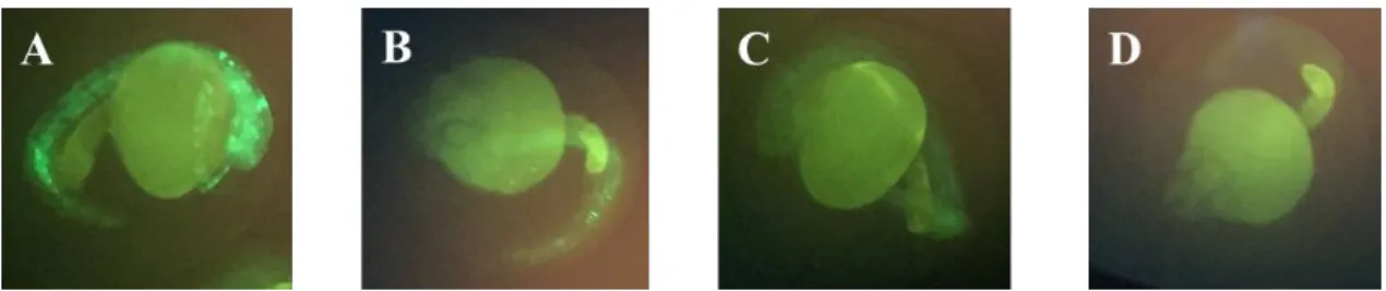
Figure 3.4 GFP fluorescence of zebrafish larvae (2 dpf) microinjected with cmv-miR-29a. Frames A, B
and C represent GFP positive transgenic carriers and frame D represents a wild-type specimen. Images were obtained in IX-81 fluorescence microscope (Olympus) ……….………..…..25
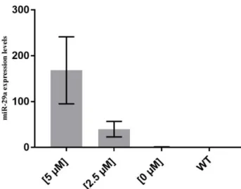
Figure 3.5 miRCURY LNA miRNA mimics for dre-miRNA-29a expression levels to confirm the
overexpression of this miRNA, measured by qPCR. The values were normalized to levels of zebrafish U6 small RNA and using wild type zebrafish as reference. Values are the mean of at least 3 independent replicates. This analysis was performed at 3 dpi. Dpi: days post injection; WT: wild type zebrafish …...28
Figure 3.6 Effects of miRCURY LNA miRNA mimics for dre-miRNA-29a [5 µM] in 6 dpi zebrafish
larvae. The red arrow shows an asymmetry between the otoliths position. The yellow arrow shows malformations in meckels cartilage, in the palatoquadrate, in the hyoid arch and in the symplectic bone. The third image represents an aberrant larvae with no discernable cranial structures. Dpi: Days post injection ………..…29
Figure 3.7 Effects of miRCURY LNA miRNA mimics for dre-miRNA-29a [2.5 µM] in 6 dpf zebrafish
larvae. The black arrow shows a compression in the posterior part of the notocord. The yellow arrow shows the presence of only one otolith. The red arrows show an assimetry between the otholits position and size. Dpf: Days post fertilization ………..30
viii
List of abbreviations
alpl - alkaline phosphatase
BMMs - Bone Marrow Monocytes
BM-MSCs - Bone Marrow Mesenchymal Stem cells BMP - Bone Morphogenetic Protein
DGCR8 - DiGregory Syndrome Critical Region DIG - Digoxigenin
dpf - days post fertilization dpi - days post injection dsRNA - double stranded RNA ECM - Extracellular Matrix
EDTA - Ethylenediaminetetraacetic Acid ESC - Embryonic Stem Cell
GFP - Green Fluorescent Protein hpf - hours post fertilization IR - Ischemia / Reperfusion ISH - In Situ Hybridization LNA - Locked Nucleic Acid mgp - matrix Gla protein miRNAs - microRNAs mRNA - messenger RNAs ncRNA - non-coding RNA nt - nucleotides
OC - Osteocalcin PFA - Paraformaldehyde
piRNAs - piwi-interacting RNAs PMCs - Palatal Mesenchymal Cells pre-miRNA - precursor miRNA pri-miRNA - primary miRNA
PTGS - Posttranscriptional Gene Silencing qPCR - quantitative PCR
rasiRNAs - repeat associated small interfering RNAs RISC - RNA-Induced Silencing Complex
RNAi - RNA interference SB - Sleeping Beauty
siRNAs - small interfering RNAs SMC - Skeletal Muscle Cells
TGFB - Transforming Growth Factor Beta VEGF - Vascular Endothelial Growth Factor
1
Chapter 1. Introduction
1.1) Zebrafish as a scientific model to investigate skeletogenesis
The zebrafish (Danio rerio) is a small tropical fish from the order Cypriniformes representative of the family Cyprinidae (Laale 1977) and is an excellent model to investigate vertebrate biology, in particular the development (Spoorendonk et al. 2010). Zebrafish can reproduce prolifically and reach adulthood within 3 months, producing hundreds of eggs throughout the year (Bilotta et al. 1999; Eaton and Farley 1974; Lawrence 2007). The eggs have a rapid development, are small, non-adherent, emersible and practically transparent, as well as the larvae, allowing the examination of developmental stages without interfering with the process (Laale 1977). This last feature allows the easy identification of many cell types throughout the development (Kimmel and Warga 1987). Some other advantages are that it is an easily and cheaply maintained species that allow for high densities in the laboratory (Lawrence and Mason 2012; Stuart et al. 1990). This resulted in a large scale use of this fish as a model, and thus reinforcing its advantages by the amount of information and lab tools that were developed and became available for this species.
Regarding skeletogenesis, key regulators of bone formation are highly conserved between mammals and teleosts thus making the zebrafish an adequate scientific model to study this process (Spoorendonk et al. 2010). Despite some specific differences, anatomic and developmental features of fish and mammalian skeletons share a high similarity, with most structures in the skull, the axial and the appendicular skeleton formed by identical bones. In addition fish have a high conservation of developmental events and of the molecular mechanisms regulating skeletogenesis, including the early formation of a cartilaginous precursor followed by bone formation through endochondral and intramembranous ossification (Laizé, Gavaia, and Cancela 2014). The first stable lines of transgenic zebrafish were developed in 1988 (Stuart, Mcmurray, and Westerfield 1988) and since then many others have been created, some with the purpose to emulate human diseases (Dodd et al. 2000). In that sense, in our laboratory, many cell lines and other fish derived systems (e.g. zebrafish caudal fin regeneration) were established in the past years, and have been used to investigate the role of molecular determinants of fish skeleton formation (Laizé et al. 2014). The availability of all these systems makes zebrafish a
2
serious alternative to mammalian models, particularly concerning the investigation of skeleton formation.
1.2) Skeletogenesis
Two main processes occur during the skeleton development, the histogenesis and morphogenesis. As the name suggests, the histogenesis consists on the differentiation of tissues and morphogenesis comprises the location, shape, and size of the resulting tissues of the previous event through time (Eames et al. 2012a).
The skeleton is present in several structures and locations in the vertebrate body, with many shapes and sizes and is mainly constituted of two distinct tissues, the bone and cartilages. The cells that comprise these tissues are osteoblasts, osteocytes and osteoclasts in bone, and chondroblasts and chondrocytes in cartilage. Osteoblasts and chondroblasts share a common progenitor and derive from mesenchymal cells while the osteoclasts derive from a myelomonocytic lineage.
Bone morphogenetic protein (BMP) is one of the main signaling molecules responsible for the regulation in higher vertebrates of, among others, bone formation, and it is partly responsible for the differentiation of bone marrow mesenchymal stem cells (BM-MSCs) into the osteogenic lineage (Pittenger et al. 1999).
Unlike the classification used for the mammalian skeletal tissues, fishes include several types of bone and cartilage, some intermediates between the bone and cartilage, and even many intermediary tissues between connective tissue and bone (as reviewd in Boglione et al., 2013; Hartmann, 2006 and Witten, Huysseune, & Hall, 2010) .
Some other differences observed between fish and higher vertebrates are that the calcium present in the bones of the later is only mobilized in case of extreme calcium deficiency (Lewis-mcCrea and Lall 2010), the space of the bone marrow present in higher vertebrates is mainly filled with adipose tissue in fish and the main hematopoietic organ is the head kidney (Witten and Huysseune 2009).
3
1.3) Bone formation
Bone formation occurs by two distinct processes, endochondral ossification and intramembranous (or dermal) ossification (Hall 1978). In both cases the skeleton starts to form by the condensation of the mesenchymal cells, also referred to as “membranous skeleton”, that dictate the shape and location for the future bone formation. Condensations act as the primary resource to build the skeleton and later it is modified onto and phylogenetically (Hall and Miyake 2000).
In intramembranous (or dermal) ossification the cells comprising the tissue are osteoblasts, responsible for the mineralization of the bone matrix, and the resulting flat bones have no intermediary cartilaginous structures (Hall and Miyake 1992). This process is responsible, among others, for the formation of scales (Sire and Akimenko 2004), the fins rays dermal skeleton (Haas 1962), the cleithrum and the operculum (Grandel and Schulte-Merker 1998).
The most common process for higher vertebrates and teleosts, is the endochondral ossification, responsible for the formation of long bones in vertebrates, in which the bone slowly replaces the pre-existing cartilage template (Hall and Miyake 2000). In zebrafish, most of the head skeleton, for instance, the bones that constitute the neurocranium and pharyngeal skeleton, develop first as cartilage (C. Cubbage and Mabee 1996), as well as the vertebral arches (Morin-kensicki, Melancon, and Eisen 2002).
In endochondral ossification the intermediate cartilage template starts to form by a process called chondrogenesis. The mesenchymal cells that aggregate give rise to chondroblasts that start to express specific molecular markers such as the proteoglycan
aggrecan and collagenIIa1 that allow for the distinction between these and the
undifferentiated mesenchymal cells that remain in the perichondrium, located in the periphery of the forming tissue (Horton 1993). Chondrocytes that are able to deposit a calcified extracellular matrix (ECM) rich in collagenXα1, undergo unidirectional proliferation thus resulting in the elongation of the bone, and, when the cycle is complete, they became hypertrophic and die through a process of apoptosis.
Proliferating and reserve chondrocytes are called non-hypertrophic chondrocytes. The hypertrophic chondrocytes can be subdivided into two distinct populations, the prehypertrophic, expressing collagenIIα1 and the hypertrophic chondrocytes proper
4
which start to express collagenXα1, a specific molecular hypertrophy marker in higher vertebrates (Poole 1991; Reichenberger et al. 1991). In zebrafish, collagenXα1 is also expressed during larval development in the cranial intramembranous bones, such as the
cleithrum and the opercular bones, in cartilaginous structures, such as the ethmoid plate
(Simões et al. 2006) and during the fin regeneration process in both scleroblasts, that are osteoblasts-like cells responsible for the secretion of fin ray bone matrix and developing scales, and in the basal epithelial cell layer adjacent to the bone matrix of the ray (Avaron et al. 2006).
Around the time when hypertrophy starts to take place, in the perichondral tissue some cells start to differentiate into osteoblasts expressing runx2 that will be responsible for the formation of the bone collar, a mineralized structure around the cartilaginous core (Caplan 1987).
The vascularization of the tissue favoured by vascular endothelial growth factor (VEGF)-dependent pathway will result in the presence of chondroclasts, responsible for the degradation of this ECM (Vu et al. 1998), and progenitors of the osteoblasts from the bone collar. The cartilaginous ECM will slowly be replaced by a bone ECM, rich in
collagenIa1 and collagenIIa1 (Benjamin and Ralphs 1991). The ossification, in higher
vertebrates, occurs centrifugally and once the hypertrophic area starts to reach the periphery of the future bone, chondrocytes will start to move distally forming the growth plate, an avascular structure at the ends of the expanding bone (Caplan 1987).
In zebrafish the process of endochondral ossification is absent, instead a persisting cartilage rod remains in the bone shaft and if cartilage is removed, it is replaced by adipose tissue (reviewed in Boglione et al., 2013). Although, the process of endochondral ossification typically gives rise to the vertebrae in higher vertebrates, in the zebrafish the vertebral centra ossify without any cartilaginous template (Huxley 1859). The most common ossification process in teleosts is, therefore, the perichondral ossification and larvae only have, essentially, this type of ossification (Boglione et al. 2013).
The perichondral bone is formed by cells that were previously part of the perichondrium, that start to secrete the bone matrix, and is located in contact with a cartilaginous template. Cartilage can, therefore, became enclosed in perichondral bone,
5
start to hypertrophy and be replaced by adipose tissue (such as it is the case of the splanchnocranium) (Witten et al. 2010).
The chondrogenesis is, thus, an important and complex process in the beginning of the skeletogenesis and throughout the skeleton development, being important for osteogenesis, bone elongation and skeleton mobility by the presence of cartilage in the joints. Several complex processes are involved in the regulation of chondrogenesis and include signalling pathways, growth and transcription factors and also being subject to post-transcriptional control, the main subject of this work.
1.4) RNA interference process
RNA-silencing systems evolved before multicellularity and were present in primitive eukaryotic cells (Molnár et al. 2007). In these systems, this mechanism is important to avoid the replication of virus and transposons and to keep the genome integrity (Jensen, Gassama, and Heidmann 1999).
It was first observed in E. coli the presence of some molecules with the capacity of inhibiting the mRNA mediated processes by an antisense control mechanism (Eckhardt and Lührmann 1979; Jayaraman et al. 1981). The first demonstration of a process of RNA interference (RNAi) in complex systems was performed in the nematode Caenorhabditis
elegans, after the injection of double stranded RNA (dsRNA) and consequent silencing
of genes with homologous complementary sequences (Fire et al. 1998). Since then, several types of non-coding RNA (ncRNA) molecules with RNA interference capacity have been found, including piwi-interacting RNAs (piRNAs), small interfering RNAs (siRNAs), repeat associated small interfering RNAs (rasiRNAs) and microRNAs (miRNAs) (Bartel and Chen 2004), the topic of focus of this work. Many different essential functions have been ascribed to ncRNAs (Berezikov and Plasterk 2005; Goodrich and Kugel 2006; Morey and Avner 2004). At the molecular level, these molecules perform posttranscriptional gene silencing (PTGS) and regulate gene expression either by mediating the repression of mRNA translation (Moss 2000) or promoting mRNA degradation (Elbashir 2001).
6
1.5)
MicroRNAs as posttranscriptional regulatorsmiRNAs are a sub-class of ncRNAs with ~22 nucleotides (nt) (Kloosterman and Plasterk 2006) negative regulators of gene expression that have been described in plants, animals, virus (Griffiths-Jones et al. 2008) and in one unicellular eukaryotic organism (Molnár et al. 2007).
The first miRNA, so called lin-4 was discovered in C. elegans in 1993 (Lee, Feinbaum, and Ambros 1993; Wightman, Ha, and Ruvkun 1993), and since then, many more have been found by combining bioinformatics predictions with experimental (mostly RNA sequencing) approaches (Berezikov, Cuppen, and Plasterk 2006). These miRNAs have been deposited in several online databases, being the miRBase currently the most important one. The 21st release (June, 2014) contains 28645 entries, expressing 35828 mature miRNA products, in 223 different species.
The formation of fully mature miRNAs (Figure 1.1) begins with the transcription of a primary miRNA (pri-miRNA), mostly mediated by RNA polymerase II (Cai, Hagedorn, and Cullen 2004). Pri-miRNAs are then processed by a protein complex containing the Drosha enzyme and the DiGregory syndrome critical region gene B (DGCR8) cofactor (needed for the enzyme to locate the pri-miRNA), which form the precursor miRNA (pre-miRNA) still inside the cellular nucleus (Lee et al. 2003). Then, Exportin-5 transports the pre-miRNAs out of the nucleus, into the cytoplasm, where further processing takes place (Bohnsack, Czaplinski, and Görlich 2004). Cytoplasmic enzyme Dicer will then originate an imperfect dsRNA (Ketting et al. 2001). In theory, the strands with lower 5’-end stability (determined by G/C occurrence) are loaded into RNA-induced silencing complex (RISC), becoming a mature miRNA, while the other strand is degraded (Schwarz et al. 2003). However, in some cases, the opposite strands are loaded into RISC, thus originating star miRNAs. Regardless of that, mature miRNAs direct the RISC to target messenger RNAs (mRNA) by their complementarities to specific matching regions in the 3’UTRs. Then, miRNAs/RISC interactions with target mRNAs can promote 2 different effects: 1) translation repression or 2) direct mRNA cleavage (Dalmay 2013). The amount of complementary might be the variable in charge of the fate of the mRNA molecules (Brodersen and Voinnet 2009; Pillai 2005). This process can be used as an alternative route to degradation since it can impose a translational block to mRNA (Kloosterman and Plasterk 2006).
7
Figure 2.1 Biogenesis of miRNA (found in Kloosterman and Plasterk, 2006)
Since it is very rare for the mRNA to have a perfect complementarity with the miRNA, being the only exception the HOXB8 mRNA with miR-196 (Yekta, Shih, and Bartel 2012), by allowing several mismatching opportunities in a database search, hundreds of potential binding sites will arise. Usually nucleotides from the positions 2-8 of the miRNA are perfectly complementary to the mRNA (Dalmay 2008)and comprise the seed region in the 5’end of mature miRNAs that will, therefore, assume great importance in this process.
Several groups of target sites exist (reviewd in Dalmay, 2013), the 5’-dominant canonical target sites, 5’-dominant seed-only target sites and 3’-compensatory target sites. The first two include no mismatches on the seed but the first has several matches in the rest miRNA, contrary to the second. The last one present some mismatches in the seed region but also show extensive base pairing in the other regions.
miRNAs can have two mechanisms of translational repression by either suppression the initiation of the translation, in the ribosome-free fraction (Pillai 2005), or act at the post-initiation stage, in the polysome fraction (Petersen et al. 2006).
Since mRNA silencing comprise translation repression and its degradation, the question remains as to which is more diffuse. Two studies tried to answer this question (Baek et al. 2008; Selbach et al. 2008), but much remains to be discovered. It might be the case that it could start with one of the processes and then change the method during development, or that different tissues will present different approaches (Dalmay 2013)
8
Since their early discovery, miRNAs have been shown to be involved in crucial biological processes, including cellular processes (e.g. apoptosis and cellular proliferation (Rana 2007), genomic imprinting (Sleutels, Zwart, and Barlow 2002), X-chromosome inactivation (Chureau et al. 2002; Lanz et al. 1999), and development (Kloosterman and Plasterk 2006). Not surprisingly, in the last years miRNAs dysregulation have also been implicated in several pathological processes, including oncogenesis (Zhang et al. 2007).
1.6) MicroRNAs involvement in skeleton formation
In recent years, miRNAs were also shown to play an essential role in skeleton formation. For instance, several miRNAs were shown to be modulated by the BMP signaling, which in turns promotes osteogenesis (Phimphilai et al. 2006). On another example, Runx2, one of the main orchestrators of osteogenic differentiation, was shown to be targeted by at least 11 miRNAs (reviewd in Kapinas & Delany, 2011). In general, the regulation of osteoblast differentiation can be negatively or positively influenced by miRNAs, as represented in Figure 2 (present in the next page).
In two very important studies, where Dicer was knocked-out in the cartilage (directed by collagen II α1 promoter; Kobayashi et al., 2008) and bone (directed by
collagen I α1 and Osteocalcin promoters; Gaur et al., 2010) of mice models, partially
unveiled the importance miRNAs in skeleton formation. In cartilage, absence of mature miRNAs, due to lack of Dicer enzyme, revealed the importance of these molecules in the regulation of chondrocyte proliferation and inhibition of premature differentiation. The number of proliferating chondrocytes was shown to be lower, which resulted in a rapid and increased differentiation into postmitotic hypertrophic chondrocytes (Kobayashi et al. 2008). The functional role of miRNAs in osteoblast and bone formation was shown firstly by inactivating Dicer in early osteoblasts (Dicer knockout directed by Collagen Ia1 promoter). This knockout led to 100% foetal lethality at E15.5 stage, the embryos displayed a deformed cartilaginous skeleton and completely lacked bone formation. Dicer inactivation in mature osteoblasts resulted in viable adult animals, but induced two distinct phenotypes: firstly, Dicer knockout lead to delayed bone formation at perinatal stage; later, Dicer inactivation strongly increased mineralized cortical bone formation. These results suggest that Dicer generated miRNAs are essential for early osteoblast differentiation, and later in development decisive for proper calcification, probably
9
through an unexplained increment on the production of bone matrix proteins (Gaur et al. 2010).
Interestingly, the in vivo effects of miRNAs have now started to be also demonstrated using zebrafish. Thus, miR-182 osteogenic effect, which was initially investigated in a mouse osteoblast cell line where it was shown to be responsible for an increased cell apoptosis and osteoblast differentiation inhibition, was finally demonstrated in zebrafish where it was shown that the overexpression of miR-182 had a negative effect on osteogenesis (Kim et al. 2012).
In the last years, our laboratory has been investigating miRNAs effects on skeleton formation using fish models (in vivo and in vitro). This work was focused on the particular skeletogenic effects of three miRNAs: miR-214 (Roberto et al., manuscript under preparation), miR-29a (Roberto et al. 2014) and miR-20a (Tiago et al., 2014), that will be described next.
Figure 1.2 miRNAs and their target genes that regulate osteoblast differentiation (Adapted from
10
1.7) miR-214
miR-214 is a miRNA that is highly conserved across vertebrates (Watanabe et al. 2008). In the last years, miR-214 was shown to be involved in several physiological and pathological processes, including: angiogenesis in mice (Van Mil et al. 2012), myogenesis in zebrafish (through regulation of the Hedgehog signalling; Flynt, Li, Thatcher, Solnica-Krezel, & Patton, 2007), cardiac and liver fibrosis in mice (Aurora et al. 2012; Knabel 2013), cardiac stress by ischemia/ reperfusion (IR) injury (Aurora et al., 2012) and during cardiac hypertrophy and heart failure (Rooij et al. 2006), and cardiomyocytes protection from apoptosis during myocardial infarction (Boon and Dimmeler 2014). In vitro studies with mir-214 have also showed its relevance in the commitment of embryonic stem cell (ESC) into specific cell lineages, such as skeletal muscle cells (SMC) or specific neural cell populations, (Juan et al. 2009; Lee et al. 2009), myogenic differentiation (Feng et al. 2011) and tumorigenesis (Penna et al. 2011; Yin et al. 2010).
Regarding skeletal development, miR-214 essential role started to be demonstrated in mice models lacking Dnm3os, the transcript in which miR-214 is inserted, that is derived of an intronic region in the gene Dnm3. In this mice models, several skeletal abnormalities were observed, being shorter and having hypoplastic dorsal neural arches of the cervical vertebrae, revealing the possible importance of this miRNA for the normal skeletal development (Watanabe et al., 2008). More recently, higher levels of miR-214 expression were correlated with lower levels of bone formation in mice. In that study, miR-214 was also shown to inhibit osteoblast differentiation and matrix mineralization
in vitro and in vivo, confirming its particular role in skeleton formation (Wang et al.
2013). Apparently, miR-214 promoted these effects mainly through repression of ATF4, a co-factor of RunX2 transcription factor and a main orchestrator of osteogenesis. This, in turn, resulted in decreased expression of bone markers genes such as oc (osteocalcin) and alpl (alkaline phosphatase) (Wang et al. 2013; Yang et al. 2004). At that time authors proposed that miR-214 could be involved in skeletal disorders, such as osteoporosis (Wang et al. 2013). In last year, miR-214 was shown to participate also in osteoclastogenesis by promoting osteoclast differentiation from bone marrow monocytes (BMMs) (Zhao et al. 2015). In our lab, the miR-214 involvement in chondrogenesis is in the process of being demonstrated. It was shown that the promotor of Dnm3os where this miRNA is inserted were active in chondrocyte cells and that the miR-214 overexpression
11
dulled the chondrocyte differentiation, probably due to the targeting of Atf4 in chondrocytes.
1.8) miR-29a
miR-29a is a miRNA that was also previously implicated in many physiological and pathological processes, being down-regulated during liver fibrosis in mice hepatocytes (Knabel 2013) and its upregulation was shown to mediate differentiation of cardiac stem cells into cardiomyocytes (De Pauw et al. 2014). The miR-29 family is also involved in the regulation of cell proliferation, differentiation and apoptosis (Aluru et al. 2013) and might perform a neuroprotective function and be correlated with Alzheimer’s disease (Hébert et al. 2008; Kole, Swahari, and Hammond 2011). In humans, miR29c was shown to be a tumour suppressor factor and for that reason it was created a cancer patient score to discriminate between good/bad prognosis using this miRNA and miR233 (Stamatopoulos et al. 2009).
Regarding skeleton formation, the miR-29 family was shown to play an essential role mainly through regulation of osteoblast function and differentiation (Kapinas and Delany 2011). During the first stages of osteoblastogenesis miR-29a expression is low, allowing higher levels of osteonectin expression and the consequent formation of collagen fibril (van Rooij et al. 2008). As the matrix and the osteoblast reach their final maturation stages, the expression of this miRNA increases, which is mediated by Wnt signalling (Hartmann 2006). Consequently, miR-29a suppresses the deposition of collagen, necessary for natural bone turnover (van Rooij et al. 2008). To promote osteoblast differentiation miR-29a will modulate Wnt signalling in a positive feedback loop (Hartmann 2006; Kapinas, Kessler, and Delany 2009). More recently, in a study developed in our laboratory (Roberto et al. 2014), the expression pattern of miR-29a was shown to be high during cell differentiation and extracellular matrix (ECM) mineralization of a fish (gilthead seabream) bone-derived cell line (ABSa15) by the targeting of sparc protein. The overexpression of this miRNA promoted early differentiation and consequent premature mineralization. It was also shown that the miR-29 family is highly conserved among vertebrates. More specifically, this miRNA induced higher b-catenin protein levels, possibly due to the stimulation of canonical Wnt signaling.
12
1.9) miR-20a
miR-20a belongs to the miRNA-17-92 cluster (previously identified as a human oncogene), and its overexpression was linked to angiogenesis in different human tumours (Fish and Srivastava 2009; Quintavalle et al. 2011) and could enhance the proliferation and invasion capacities in an ovarian cancer cell line (Fan et al. 2010). In humans, it was also shown to be related to G1 transition in diploid cells, and thus being responsible for a form of cell cycle timing progression (Pickering, Stadler, and Kowalik 2009). This cluster was also associated to the proliferation of cardiomyocytes in mouse (Chen et al. 2013), and cardiac regeneration in zebrafish (Poss, Wilson, and Keating 2002). Specifically, miR-20a was identified as a cardioprotective miRNA and could be used for its therapeutic properties to prevent cardiac remodelling (Frank et al. 2012). This cluster is also responsible for the regulation of hematopoiesis, some immune functions, and is involved in the cardiopulmonary system development (reviewd by Bonauer & Dimmeler, 2009). In primary neurons, the BMP-2 stimulation lead to an increase in this cluster transcription, leading to the upregulation of mature miR-20a, that further repressed BMPRII expression which became part of a negative feedback loop responsible for the stabilization of the BMP signalling and therefore is responsible for the maintenance of cellular homeostasis and the prevention of apoptosis (Sun et al. 2013).
Recent studies have shown the putative involvement of miR-20a in bone cell differentiation in vitro. In mouse palatal mesenchymal cells (PMCs) miR-20a was shown to repress the transforming growth factor beta (TGFB) pathway, thus resulting in the stimulation of proliferation, inhibition of collagen synthesis and regulation of palatal shelf elongation and elevation (Li et al. 2012). In another study, the expression of endogenous miR-20a was shown to increase during the course of osteogenic differentiation in human BM-MSC, and to specifically target several molecules responsible for the downregulation of the osteogenic agent BMP, which resulted in the upregulation of BMP/Runx2 signalling (Zhang et al. 2011).
More recently, in a different study performed in our laboratory using fish bone-derived cell lines (ABSa15 cells), miR-20a was shown to be poorly expressed in undifferentiated cells and to increase in later stages of osteogenic differentiation. Interestingly, miR-20a overexpression in these cells was clearly shown to repress BMP-2, by the upregulation of matrix Gla protein (mgp) transcript, and to sustain cells in
13
undifferentiated state. This resulted in a significant delay in extracellular matrix mineralization. It was thought that, by default, the low expression of this miRNA in undifferentiated cells may result in a higher BMP-2 signalling activity, leading to osteogenic differentiation (Tiago et al. 2014). The BMP signalling pathway was also implicated in bone formation in a treatment with dorsomorphin, an inhibitor of BMP signalling (Hao et al. 2008), during 2–3 dpf zebrafish that lead to a clear reduction in calcification of all the bone elements (Windhausen et al. 2015).
14
Chapter 2. Materials and methods
2.1) RNA extraction
Zebrafish eggs were obtained from a natural spawning broodstock of wild type fish (AB) maintained in-house. Eggs were collected and incubated in 1 L aquariums with system water until hatching. Larvae and adult fish were maintained under standard conditions (Westerfield 2000). Total RNA was extracted from different stages throughout the development (1k cell, 26 and 36 hours post fertilization (hpf), 2, 3, 4, 5, 6, 12, 15, 22, 29, 35, 40, 51, 61, 69, 81 and 90 days post fertilization (dpf)), and from adult tissues, using a protocol adapted from the guanidium-phenol-chloroform extraction method, previously described by Chomcyzynski and Sacchi (1987). To eliminate contaminant genomic DNA, total RNA was digested with RQ1 RNAse-free DNAse, according to manufacturer’s instructions. Then, total RNA was further purified using phenol-chloroform-isoamyl alcohol mixture (25:24:1) added to equivalent volume of RNA sample. The RNA integrity was analyzed by electrophoresis in an agarose gel (1%) and its quantity and purity was assessed by UV spectrophotometry (NanoDrop ND-1000, Thermo Scientific, Madison, WI, USA).
2.2) Quantitative real time PCR (qPCR)
Total RNA (450 ng) were polyadenylated and reverse-transcribed using the NCode™ miRNA First-Strand cDNA Synthesis kit (Invitrogen) and an oligo dT primer, according to manufacturer’s instructions. Then, 0.8 ng of cDNA were used template to amplify miR-20a and miR-29a mature miRNAs using zebrafish specific (TAAAGTGCTTATAGTGCAGGTAG for miR-20a and TAGCACCATTTGAAATCG CTTA for miR-29a) and universal primers, and the Platinum® SYBR® Green qPCR SuperMix-UDG, according to the manufacturer’s protocol. The following cycling program was used in the StepOnePlus system (Applied Biosystems, Invitrogen, Grand Island, NY, USA) thermocycler: 50ºC for 2 minutes, 95ºC for 2 minutes, 40 cycles of: 95ºC, 15 seconds and 63ºC, 30 seconds and finally the melt curve: 95ºC for 15 seconds, 60ºC for one minute increasing up until 85ºC, 0.5ºC at the time for 15 seconds. Relative expression of miRNAs was calculated by the ΔΔCt method (Livak and Schmittgen 2001), normalized using expression levels of U6 small nuclear RNA (U6) and using the mean of
15
all the stages as a reference sample, in the case of the study of the miRNAs expression levels during the development, and the brain in the case of the study of the zebrafish tissues. Values are the mean of at least 3 independent replicates. To access significant differences in miRNA expression throughout the zebrafish development and in the adult tissues, a One-way ANOVA followed by Tukey’s multiple comparisons test was performed using GraphPad Prism version 6.00 for Windows, GraphPad Software, La Jolla California USA, www.graphpad.com.
2.3) In Situ Hybridization (ISH)
In parallel with the analysis of miR expression, samples from the same developmental stages were collected and fixed in paraformaldehyde 4% (PFA) for 24 hours. The specimens were decalcified with a 0.1M ethylenediaminetetraacetic acid (EDTA) in PFA (1%) and pH 7.4. Depending on the larval stage, decalcification time varied from one week (in 22 dpf larvae) up to 2 months (in adults). Samples were stored in methanol (100%) at -20ᵒC until further use.
The in situ hybridization protocol was performed according to Kloosterman, Wienholds, Bruijn, Kauppinen, & Plasterk (2006), using LNA (Locked Nucleic Acid)-modified oligonucleotide 5’-Digoxigenin (DIG)-labelled specific probes (Exiqon), a scrambled probe as a negative control. For the first stages of development (up to 2dpf) a whole-mount ISH was performed, while larger specimens were embedded in paraffin and sectioned (5-7 µm thick sections). Several protocol adaptations were performed in order to optimize results, including changing the deparaffination process, by switching from ethanol to methanol, testing different hybridization temperatures (between 47-50-54ºC), and testing different proteinase K concentrations (0 g/ml, 5g/ml and 10g/ml).
2.4) Producing transgenic zebrafish overexpressing miRNAs
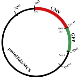
The constructs used in this study were previously prepared in the lab using the backbone of pMinitol2 vector (simplified structure in Figure 2.1) from Chien Lab (Kwan et al. 2007). Briefly, the oligonucleotide sequences of zebrafish miRNA-214, pri-miRNA-20a and pri-miRNA-29a were obtained from Sigma, hybridized and cloned into pcDNA6.2. The same sequences were amplified from the vector using specific primers
16
containing the XbaI restriction site. Both these sequences and the pMinitol2 vector were digested with XbaI. Afterwards, these fragments suffered a purification process, and were then ligated by DNA ligase, cloned and sequenced.
Figure 2.1 Chien Lab construct representation
In order to prepare oc-miR-20a and col-miR-20a constructs, the sequences of the osteocalcin promoter (sequence available in the gene code by the accession AY526532; cloned in pGEM-T-Easy) and collagen Xa1 promoter (Simões et al., 2006; cloned in pGL2) were amplified using specific primers containing the restriction sites SalI and EcoRI. Both the fragments and the vector were digested with SalI and EcoRI. Once again, the resulting fragments were purified and ligated using DNA ligase, cloned and sequenced.
Wild type zebrafish (AB) were crossed and fertilized eggs were prepared for microinjection as previously described (Bill et al. 2009; Clark et al. 2011). Eggs in the zygote stage were kept in embryo medium and fixed in a 2% agarose gel, and injected using a Nanoliter 2010 microinjector (World Precision Instruments) coupled to the MMJR micromanipulator (World Precision Instruments), and using the following injection mix: 1.25 ng of DNA and 1.25 ng of transposase in 1X Dannieu. A volume of 4.6 nl was microinjected into each embryo at 1 cell stage. For zebrafish eggs microinjected with the CMV promoter constructs, incorporation success was first evaluated 24 hours after the procedure and tracked along time. GFP positive fish,
17
independently of the tissue, were further used to obtain progeny. Regarding zebrafish eggs microinjected with zebrafish osteocalcin and collagen Xa1 promoter constructs, all specimens were further used to cross and obtain progeny for screening. The fish were then kept under standard conditions until they reached the adult stage (Westerfield 2000) and afterwards crossed with a wild type AB zebrafish in order to establish new transgenic lines by the screening of the F0 larvae. The second microinjection session was performed under the same conditions and the plasmids used were the colX-miR20a and the oc-miR20a.
2.5) miRNA mimics effect on development
Wild type zebrafish eggs were microinjected with miRCURY LNA miRNA mimics for dre-miRNA-29a (Exiqon). Several microinjection sessions were performed using different concentrations, since the concentrations previously used (18µM) in the lab for other miRNAs revealed to be toxic for the developing embryos. The concentrations tested were of 9 µM, 5 µM, 2.5 µM and 0 µM diluted in DEPC treated water, with a 1X Dannieu. The eggs in zygote stage were injected using the Nanoliter 2010 microinjector (World
Precision Instruments) coupled to the MMJR micromanipulator (World Precision Instruments) and each egg was injected with 4.6nl. The embryos were kept in embryo
medium in the first 3 days after injection in petri dishes and were then transferred to 250 ml tanks in autoclaved system water. After 5 days post injection (dpi), the fish were fed with Artemia sp. up until the 6DPI, when the experiment ended. At 3 dpi, 10 embryos from each treatment were collected for RNA extraction, as previously described. At the 6 dpi, several (11-20) fish were collected for histological analysis from each treatment, as previously described. Fish were stored in ethanol 100% at -20ºC until they were histologically processed for staining cartilaginous structures using Alcian blue staining (0.1% m/v) for 20 minutes and then stored in glycerol 75%. During this experiment several pictures were daily taken, mortality registered and hatching tracked.
18
Chapter 3. Results and discussion
This dissertation follows the work line of previous works studied in our lab. The miR-214 involvement in chondrogenesis is in the process of being demonstrated, since it’s overexpression lead to a decrease in the chondrocytes differentiation (Roberto et al., manuscript under preparation). Regarding miR-29a, its overexpression lead to an early differentiation and consequent premature mineralization (Roberto et al. 2014). Finally, miR-20a was implicated in osteogenic differentiation, since its overexpression lead to the sustaining of cells in an undifferentiated state. As shown, the implication of these miRNAs in skeletogenesis has been shown recently and this work’s objective is to continue with the gathering of information regarding this subject and to further conclude about the implications of the gene regulation properties of these miRNAs in bone formation events.
3.1) Study of skeleton-related miRNAs expression patterns and sites of expression throughout zebrafish development
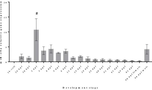
In order to investigate a possible association/contribution of miR-20a and miR-29a to specific stages of zebrafish development, and in particular to skeletogenesis, the expression levels of these two miRNAs throughout zebrafish development were determined in RNA samples collected from 1-k cell stage until sex differentiated adult zebrafish (90 dpf). The results of miR-20a quantification throughout the zebrafish development are presented in Figure 3.1. From the values observed in 1k-cell stage, it is possible to conclude that miR-20a is most likely not maternally inherited. The expression levels from 1k-cell stage until the 2nd dpf show no statistically different values (p-value > 0.05), and therefore it is likely to assume that this miRNA only starts to be expressed on the 2nd dpf. In zebrafish, the chondrification of cranio-facial elements from mesenchymal condensations commences at 2 dpf (Kimmel et al. 1995) and ossification starts at approximately 3 dpf, when the cleithrum, the fifth branchial arch, and the opercle are being formed (Cubbage and Mabee 1996). Here, we show that miR-20a is up regulated around that period, being statistically different of the other developmental stages (p-value < 0.0001), suggesting a possible involvement in early ossification. This result is consistent with previously demonstrated miR-20a involvement in early
19
osteogenic differentiation in vitro (Tiago et al. 2014; Zhang et al. 2011). Apparently, after this peak of expression, a downward slope is observed, until it reaches a stabilization at the 35th day. This is highly consistent with miR-20a inhibitory role on osteogenic
differentiation in fish bone-derived cells (Tiago et al. 2014). As previously suggested, it seems that miR-20a expression needs to decrease in order for ossification to proceed. However, miR-20a expression could also be related with many other organs being formed around the 2nd dpf.
In the adults, there is also an interesting observation: there is a statistically difference (p-value > 0.05) between male and female miR-20a expression, where males show up to fivefold higher expression than the females. Male zebrafish also show differences between the observed and the values obtained after the 35th dpf. Without the results from the in situ hybridization one can only speculate about the possible tissues and organs that might be responsible for the values of the miRNAs observed, but this could be related to the gametogenesis or other structural differences present between male and female zebrafish. 1k -c el l 26 h pf 36 h pf 2 d pf 3 d pf 4 d pf 5 d pf 6 d pf 12 d pf 15 d pf 29 d pf 35 d pf 40 d pf 51 d pf 61 d pf 69 d pf 82 d pf 90 d pf fe m al e 90 d pf m al e 0 5 1 0 1 5 2 0 D e v e l o p m e n t s t a g e m i R -2 0 a r e l a t i v e g e n e e x p r e s s i o n
Figure 3.1 Analysis of mature miR-20a expression levels during development of zebrafish, measured by
qPCR. The values were normalized to levels of zebrafish U6 small RNA, and the mean of all the samples was used as reference. Values are the mean of at least 3 independent replicates; #: significantly different; hpf: hours post fertilization; dpf: days post fertilization
20
In an attempt to understand the specific involvement of miR-20a in specific processes in zebrafish, we analysed its expression in different organs collected from adult specimens (Figure 3.2). Our results showed a higher miR-20a expression in the brain, being x-y-fold higher than remaining tissues, i.e. muscle, heart, branchial arches (lowest expression), vertebrae and skull. Although the expression of miR-20a was not significantly different in later tissues, the brain showed statistical differences between the remaining tissues (p-value < 0.02), except when compared with the heart, where no clear differences were shown (p-value > 0.06). Sun et al. (2013) showed the importance of this miRNA in the maintenance of the cellular homeostasis and prevention of apoptosis in primary neurons, by acting in a negative feedback loop to control BMP-2. This could potentially explain the higher values observed in the zebrafish brain. Another organ that might be interesting analyse in the future is the gonads, since a 5-fold difference in expression was observed between different genders (Figure 3.1). Once again, our results showed that miR-20a is widely expressed in the skeleton, being expressed in tissues resulting from different types of ossification: intramembranous ossification in skull versus endochondral ossification in vertebrae.
B ra i n H ea rt M us cl e B ra nc hi a l a rc he s V er te br ae Sk ul l 0 . 0 0 . 5 1 . 0 1 . 5 Z e b r a f i s h t i s s u e s m i R -2 0 a r e l a t i v e g e n e e x p r e s s i o n
Figure 3.2 Analysis of mature miR-20a expression levels during development of zebrafish, measured by
qPCR. The values were normalized to levels of zebrafish U6 small RNA, and brain was used as reference. Values are the mean of at least 3 independent replicates.
21
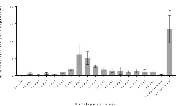
Regarding miR-29a expression throughout development, it is possible to observe 2 different peaks of expression, being the first (Figure 3.3) between the 6th and the 12th day
(p-value < 0.05) and the last (marked as * on Figure 3.3) observed in male adult zebrafish (p-value < 0.0001). As it was the case for miR-20a, the 1k-cell stage levels of miRNA expression were low, revelling that this miRNA are not maternally inherited. Also, these same values were not significantly different from the values observed during the entire zebrafish development except for the 2 peaks of expression previously referred, revealing the presence of low expression levels. Concerning the first peak of expression, i.e. at 6 dpf, Eames et al. (2012) showed that in the ceratohyal there are signs of early chondrocyte hypertrophy, the perichondrium starts to mineralize, and dentaries show abundant bone matrix net in Meckel’s cartilage, suggesting an onset of bone formation in the cranium. It was also shown by Kimmel, Miller, & Moens (2001) that the formation of the larvae cartilaginous cranial skeleton was completed at 6 dpf and endo and perichondral ossification would start to take place after this period (Cubbage and Mabee 1996). Another study showed that around the 6th dpf, the centra of the axial skeleton start to be formed sequentially, initiating with the third and fourth centrum (Bird and Mabee 2003). Since, miR-29a was previously shown to positively regulate genes associated to extracellular matrix (ECM) deposition in bone, specifically SPARC, an important protein for ECM assembly and deposition (Kapinas et al. 2009), it is possible that the higher values observed in the first peak of expression (represented as # in Figure 3.3) are a result of increased ECM mineralization in the cranium. However, we cannot discard a putative involvement of miR-29a in the formation of other organs concomitantly being formed at 6 dpf. In the beginning of zebrafish development, low levels of miRNA were detected but show a sign of increase before the first peak of expression, although not being statistically different (p-value < 0.05). After the peak, it shows a slow decrease in the levels of this miRNA expression, leading to a stabilization to values that show no clear difference (p-value < 0.05) with the first (p-values observed (from 1k-cell stage until the 6 dpf).
As it was the case for miR-20a, also a clear difference is shown for miR-29a between male and female adult zebrafish. In this particular case, the difference observed is even higher and it might lead to some conclusions regarding the involvement of this miRNA involvement during the gametogenesis or regarding some sex specific event.
22 1k -c el l 26 h pf 36 h pf 2 d pf 3 d pf 4 d pf 5 d pf 6 d pf 12 d pf 15 d pf 29 d pf 35 d pf 40 d pf 51 d pf 61 d pf 69 d pf 82 d pf 90 d pf fe m al e 90 d pf m al e 0 5 1 0 1 5 2 0 D e v e l o p m e n t s t a g e m i R -2 9 a r e l a t i v e g e n e e x p r e s s i o n
Figure 3.3 Analysis of mature miR-29a expression levels during development of zebrafish, measured by
qPCR. The values were normalized to levels of zebrafish U6 small RNA and using the mean of all the samples as reference. Values are the mean of at least 3 independent replicates; *: significantly different; hpf: hours post fertilization; dpf: days post fertilization
In order to investigate the specific involvement our miRNAs of interest, i.e. miR-20a and miR-29a (this analysis was already performed for miR-214 within the scope of V. Roberto’s work; manuscript under preparation) in skeleton development, we decided to study their specific sites of expression in zebrafish larvae. Larvae for qPCR analysis and in situ hybridization (ISH) were simultaneously collected, but only the larvae displaying highest levels of miR-20a and miR-29a expression through qPCR were further analysed through ISH: 2, 6, 12, 15, 22 dpf. A procedure similar to that previously applied for miR-214 was tested. We used LNA-modified DNA probes (LNA probes), which are a class of bicyclic high-affinity RNA analogues that result in a higher hybridization affinity regarding complementary DNA and RNA molecules coupled with higher thermal stability (Vester and Wengel 2004), and a protocol previously described by Kloosterman et al. (2006). Although LNA probes allow to increase the sensitivity of the ISH results, the low abundant miRNAs, or those only expressed in a few cells, are still of difficult observation. This might have been the case for the miRNAs that were here tentatively studied, since they presented much different expression values when comparing, with miR-214 (previously investigated in the lab; Roberto et al., manuscript under preapration). Despite several attempts, and after adjusting several ISH parameters,
23
including hybridization temperatures and probes concentrations (up to 60 nM), we could not obtain any signal for miR-20a and miR-29a. However, a lack of signal due to a hypothetical low abundance of miRNAs cannot explain why we could not obtain any signal for our positive control: a probe against miR-199, which is a miRNA that is highly abundant in tested larvae (our previous data from V. Roberto’s work) Therefore, other possible explanations for these negative results are: a problem with the reagents used in the process, which should be RNAse-free; or possible probes degradation, which validity have expired.
Many miRNAs have an organ specific expression pattern and are mostly expressed at later stages of development (Wienholds and Plasterk 2005). A final ISH attempt will be carried in adult zebrafish specimens in order to finally discard all the options and to further conclude about the integrity of the reagents.
3.2) Transgenic lines establishment
In this study we aimed at the creation of 3 zebrafish transgenic lines overexpressing constitutively (through a cmv promoter, in pminitol2 vector) 3 different miRNAs, the miR-214, miR-20a and miR-29a. Of those, only one founder fish was positively identified, belonging to one of the cmv-miR-29a microinjected fish. Because of these results other constructs were used in order to direct the overexpression of the miRNAs over specific skeleton tissues, such as cartilage and bone. This was performed resorting to the use of different promoters (collagen Xa1and osteocalcin) for miR-20a, since some experiences are taking place at our lab regarding the miR-214 (Roberto et al. n.d.).
In order to establish new zebrafish transgenic lines with specific overexpression of miR-20a, miR-29a or miR-214, we prepared different constructs (previously developed in the lab), and microinjected in zebrafish eggs. Microinjection is a powerful technique that allows the manipulation of the expression of specific genes in vertebrate systems (Stuart et al. 1988) and investigate particular developmental processes (Grabher, Joly, and Wittbrodt 2004). In addition, the incorporation of exogenous DNA constructs can be followed by the injection of fluorescent lineage tracer dyes (Rembold et al. 2006; Wittbrodt 2005). The dominant reporter cassette marker can be a fluorescent protein, as it is the case of the green fluorescent protein (GFP), used in this work. The use of this


![Figure 3.6 Effects of miRCURY LNA miRNA mimics for dre-miRNA-29a [5 µM] in 6 dpi zebrafish larvae](https://thumb-eu.123doks.com/thumbv2/123dok_br/18437759.896497/40.892.350.557.590.874/figure-effects-mircury-mirna-mimics-mirna-zebrafish-larvae.webp)
![Figure 3.8 Effects of miRCURY LNA miRNA mimics for dre-miRNA-29a [2.5 µM] in 6 dpf zebrafish larvae](https://thumb-eu.123doks.com/thumbv2/123dok_br/18437759.896497/41.892.299.531.288.518/figure-effects-mircury-mirna-mimics-mirna-zebrafish-larvae.webp)
