UNIVERSIDADE DA BEIRA INTERIOR
Ciências da Saúde
Therapeutic application of retinoic acid-loaded
nanoparticles for neurological and vascular repair
in stroke
Márcia Carvalho da Fonseca
Dissertação para obtenção do Grau de Mestre em
Ciências Biomédicas
(2º ciclo de estudos)
Orientadora: Doutora Raquel Ferreira
Coorientadora: Prof. Doutora Liliana Bernardino
Agradecimentos
Chegado o final desta importante etapa, não posso deixar de agradecer aqueles que de uma forma ou de outra contribuíram para a realização deste trabalho.
Em primeiro lugar quero agradecer há minha orientadora, Doutora Raquel Ferreira, pelo tanto que me ensinou. Não foi só a nível profissional mas pessoal também. Passou de orientadora a amiga! Por isso tudo, agradeço toda a amizade, dedicação, paciência, compreensão, riso e choro. Foi um enorme orgulho para mim. Obrigada do fundo do coração!
Agradeço também há minha co-orientadora, Prof. Doutora Liliana Bernardino, por toda a dedicação e sugestões dadas ao longo deste ano sempre em prol de um melhor resultado. Agradeço todos os conselhos e ensinamentos que sem dúvida levo comigo para o futuro. Patrícia Lindeza, um muito obrigada por todas as conversas e toda a força! Em momento algum deixas-te que me faltasse uma palavra de carinho e conforto. Muito obrigada pela amizade e paciência que foram sem dúvida fundamentais para o meu sucesso na realização deste trabalho. Levo para a vida todos os momentos de partilha!
Tiago Santos, foi um gosto conhecer e conviver com uma pessoa com tanta luz e positivismo. Obrigada por todo o incentivo, a paciência e ensinamentos transmitidos. Levo comigo todos os momentos de riso, diversão e convívio.
Um obrigada ao restante grupo de trabalho com quem tive o prazer de aprender e partilhar ao longo deste ano. Estou grata por tudo o que me ensinaram!
O mais especial agradecimento àquelas que são sem dúvida o meu maior apoio, conforto e a fonte de toda a minha força, a minha mãe e irmã. Muito obrigada por lutarem comigo e acreditarem sempre em mim, mas acima de tudo obrigada por todo amor, carinho, compreensão e por toda a paciência que sei que é preciso comigo. Sem vocês nada disto teria feito sentido. Depois, e como não podia deixar de ser, agradeço há Alcina Amorim, pelo apoio a todos os níveis que sempre me deu, por toda a motivação transmitida, toda a amizade, força e porque sempre acreditou que era eu capaz, muito muito obrigada.
Ao Marco Saldanha, um mega obrigada por ter aparecido na minha vida no início desta difícil etapa. Muito obrigada por toda a paciência, o companheirismo, amor, amizade, compreensão e serenidade transmitida. Agradeço por nunca me ter deixado baixar os braços e me fazer acreditar em mim mesma.
Raquel Faro, Inês Albuquerque e Lais Oliveira, obrigada por todos os conselhos, por todas as horas de conversa a rir e a chorar e por saberem dizer sempre o que preciso de ouvir na hora certa, não só nesta fase, mas em muitas outras. Sei que qualquer coisa que aconteça estarão sempre lá, por isso agradeço do fundo do coração toda a amizade e confiança depositada em mim.
Resumo
O acidente vascular cerebral é a principal causa de morte e incapacidade em Portugal, causando encargos socioeconómicos significativos. Contudo, os tratamentos atuais apenas funcionam numa percentagem reduzida de pacientes e podem comportar efeitos secundários graves. Uma das principais estruturas afetadas pelo acidente vascular cerebral é a vasculatura cerebral, responsável pelo suporte trófico dos tecidos que irriga e por facilitar a migração de células estaminais neurais à zona lesionada. O objetivo principal deste projeto é desenvolver uma terapia, com potencial pró-angiogénico, que recupere a vasculatura cerebral afetada e por outro lado, potencie a sobrevivência e diferenciação das células estaminais neurais. Este trabalho propõe a utilização de nanopartículas contendo ácido retinóico, uma vez que o ácido retinóico apresenta propriedades relevantes para o tratamento eficaz do acidente vascular cerebral: é um agente anti-inflamatório, regulador da função vascular e pró-neurogénico. Esta formulação contorna as limitações do ácido retinóico livre: baixa solubilidade e degradação rápida permitindo que este atinja o seu potencial. Assim, os objetivos deste projeto são determinar a função moduladora do ácido retinóico encapsulado em nanopartículas em parâmetros específicos da função/atividade das células endoteliais, em que estas são sujeitas a um modelo in vitro de privação de oxigénio/glicose para reproduzir a condição isquémica. Definir o papel das células endoteliais tratadas com o ácido retinóico encapsulado em nanopartículas na secreção de fatores que promovam a sobrevivência e diferenciação de células estaminais neurais. Este tratamento mostrou-se promissor na proteção e proliferação de células endoteliais em condições basais e promoveu a sobrevivência de células endoteliais em condições isquémicas. Quando sujeitas a privação de oxigénio/glucose, as células endoteliais libertaram fatores que suportaram a sobrevivência, proliferação e diferenciação de células estaminais neurais. Os resultados obtidos com ácido retinóico encapsulado em nanopartículas mostraram um grau de eficácia cerca de 83 vezes superior ao ácido retinóico livre (não encapsulado), mostrando assim vantagens claras na sua aplicação terapêutica. Devido ao seu efeito modulador sobre as células endoteliais e estaminais neurais, o ácido retinóico encapsulado em nanopartículas mostra ser um agente com grande potencial terapêutico para o tratamento do acidente vascular cerebral e possivelmente outras doenças vasculares e/ou neurodegenerativas.
Palavras-chave
Resumo alargado
O acidente vascular cerebral é a doença com maior impacto socioeconómico em Portugal, e que está associada a maior mortalidade e morbidez. Existem dois tipos principais de acidente vascular cerebral, o isquémico, causado pela obstrução de um vaso sanguíneo e o hemorrágico, quando se dá o rompimento de um vaso sanguíneo. Menos comum é a ocorrência de isquémia cerebral devido a paragem cardíaca. Devido à prevalência de vida sedentária, dieta ocidental e a uma crescente esperança média de vida, o número de pacientes com acidente vascular cerebral tenderá a aumentar. Contudo há fatores de risco associados que não são possíveis de contornar como um histórico familiar de doenças cerebrovasculares, idade mais avançada ou o sexo masculino. Por outro lado a hipertensão, a diabetes ou doença cardíaca valvular são fatores de risco reversíveis quando utilizado o tratamento adequado. O acidente vascular cerebral acarreta diversas complicações provocando edema vascular, aumento da permeabilidade endotelial, degradação da matriz extracelular, formação de espécies reativas de oxigénio e o aumento de diapedese, podendo levar à disrupção da barreira hematoencefálica. Estes processos comprometem a viabilidade do tecido afectado, realçando a importância da reparação da vasculatura cerebral após um acidente vascular cerebral. Num acidente vascular cerebral é importante que a terapia seja administrada o mais rapidamente possível para ser eficaz, sendo para tal importante o reconhecimento dos primeiros sinais da manifestação da doença. A ocorrência de um ataque isquémico transitório, conhecido por "mini" acidente vascular cerebral (duração inferior a 24 horas) tende a ser ignorado levando a um aumento nas repercussões. Dentro dos tratamentos disponíveis destacam-se agentes trombolíticos que degradam os coágulos de fibrina. Contudo podem ter um efeito tóxico no cérebro lesionado. Para o desenvolvimento de uma terapia eficaz para o acidente vascular cerebral é importante ter em conta toda a unidade neurovascular, uma vez que nesta patologia todas as células do cérebro estão de alguma forma envolvidas. Os neurónios são células sensíveis que dependem de toda uma estrutura vascular que transporta nutrientes, oxigénio e células essencial à homeostasia do Sistema Nervoso Central. Após uma lesão isquémica, há destruição de vasos sanguíneos e perda de neurónios que o cérebro tende a contrariar com diversos mecanismos reparadores, como é o caso de um aumento na angiogénese e na neurogénese. De forma a repor os neurónios perdidos, há a migração de células imaturas até a zona lesionada, provenientes da zona subventricular. Esta migração requer também a reparação dos vasos sanguíneos degradados e a formação de novos que servirão de transporte aos novos neurónios. Neste processo, as células endoteliais secretam fatores tróficos que influenciam o recrutamento e suporte trófico neuronal.
O ácido retinóico é um derivado da vitamina A, com um papel importante no desenvolvimento do sistema nervoso. Esta molécula atua através dos seus recetores nucleares, recetores de ácido retinóico e recetores de retinóide. O ácido retinóico é considerado um indutor da
negativamente os genes que inibem a neurogénese, levando à diferenciação das células estaminais neurais. Em relação ao efeito do ácido retinóico na angiogénese, existem evidências contraditórias, mas prevalece o efeito pró–angiogénico, principalmente na presença do fator de crescimento endotelial vascular endógeno. Assim, o ácido retinóico poderá ser um bom candidato numa terapia para o acidente vascular cerebral, contudo, quando administrado na sua forma livre tem algumas desvantagens, como a reduzida solubilidade em soluções aquosas e a sua degradação acelerada. Neste projeto, propomos a utilização de ácido retinóico encapsulado em nanopartículas, capazes de promover a libertação eficiente e gradual de ácido retinóico, aumentando a sua eficácia terapêutica. Esta formulação demonstrou ser cerca de 2500 vezes mais eficiente do que o ácido retinóico no seu estado livre na diferenciação de células neurais estaminais.
Neste seguimento surgem os objetivos deste trabalho, (1) determinar o papel modulador do ácido retinóico encapsulado em nanopartículas no funcionamento de células endoteliais e (2) determinar o papel modulador do ácido retinóico encapsulado em nanopartículas no secretoma das células endoteliais sobre vários parâmetros de atividade das células estaminais neurais.
Os resultados obtidos sugerem que o tratamento com o ácido retinóico encapsulado em nanopartículas não apresentou citotoxicidade dentro de uma gama de concentrações que potenciou a proliferação e a capacidade angiogénica das células endoteliais. Quando as células endoteliais foram sujeitas a um modelo experimental de privação de oxigénio/glucose, de forma a mimetizar o ambiente isquémico resultante de um acidente vascular cerebral, verificou-se que o tratamento com o ácido retinóico encapsulado em nanopartículas protegeu as células da morte necrótica induzida por isquémia, não mostrando qualquer efeito sobre o seu estado proliferativo. Aquando de um evento isquémico, existe uma relação de extrema importância funcional entre a vasculatura cerebral o processo neurogénico. Para além do suporte físico que estes vasos fornecem para o transporte de células estaminais neurais, as células endoteliais que formam estes vasos secretam fatores importantes responsáveis pela migração, diferenciação e proliferação. Na segunda fase do trabalho, verificamos que meios condicionados de células endoteliais saudáveis tratadas com o ácido retinóico encapsulado em nanopartículas promoveu a sobrevivência, proliferação e diferenciação de células estaminais neurais, embora mantendo também uma subpopulação significativa de células progenitoras. De forma semelhante, células endoteliais isquémicas tratadas com ácido retinóico encapsulado em nanopartículas secretaram fatores que promoveram a sobrevivência, a proliferação e, de forma mais significativa a diferenciação neuronal, uma vez que se verificou uma redução da população de células progenitoras. Nas experiências realizadas com o ácido retinóico encapsulado em nanopartículas mostraram um grau de eficácia até cerca de 83 vezes superior ao ácido retinóico não encapsulado. Em suma, o ácido retinóico encapsulado em nanopartículas demonstrou não só um grande potencial terapêutico a nível vascular como poderão ainda contribuir significativamente para a reparação neuronal, duas componentes vitais comprometidas num acidente vascular cerebral.
Abstract
In Portugal, stroke is the main cause of mortality and disability causing a substantial socioeconomic burden. However, available treatments only benefit a small percentage of patients and can cause serious side effects. The main aim of this project is to develop a new therapy for stroke that recovers the brain vasculature and induces survival and differentiation of neural stem cells. Thus, this work focuses on inducing a favorable pro-angiogenic environment for the vasculature to recover and to support neuronal and functional repair. This project proposes the use of retinoic acid-loaded nanoparticles as the therapeutic agent. Retinoic acid regulates vascular function and can increase endogenous neurogenesis. This formulation bypasses the limitations of free retinoic acid: low solubility and fast degradation thereby allowing retinoic acid to reach its full potential. Thus, the objectives of the project were: 1) to determine the modulatory role of retinoic acid-loaded nanoparticles on endothelial cell activity after being subjected to an in vitro model of oxygen/glucose deprivation to mimic the ischemic condition; 2) to define the role of endothelial cells treated with retinoic acid-loaded nanoparticles in the secretion of factors that promote the survival, repair and differentiation of neural stem cells. Retinoic acid-loaded nanoparticles treatment protected endothelial cells from ischemic injury, and in these experimental settings, retinoic acid-loaded nanoparticles-treated ischemic endothelial cells released factors that induced the survival, proliferation and differentiation of neural stem cells. Additionally, the results obtained with retinoic acid-loaded nanoparticles treatment showed increased efficiency (~83-fold higher) when compared to free retinoic acid treatment (not encapsulated). With these results in mind, retinoic acid-loaded nanoparticles demonstrated a relevant therapeutic potential for the treatment of stroke and possibly other vascular/neurodegenerative diseases.
Keywords
Retinoic acid, nanoparticles, angiogenesis, neurogenesis, stroke, endothelial cells, neural stem cells.
Index
Chapter 1 - Introduction ... 1
Definition of Stroke... 1
Risk factors for the development of stroke ... 2
Neurovascular unit in stroke (NVU) ... 3
The angiogenic process ... 3
Neural stem cells (NSC) and neurogenic niches ... 4
Impact of angiogenesis and neurogenesis on stroke ... 5
Experimental models of ischemia ... 5
Retinoic acid (RA) signalling ... 6
The modulatory role of RA in neurogenesis ... 6
The modulatory role of RA in angiogenesis ... 7
RA-loaded nanoparticles (RA-NP) ... 7
Aims ... 9
Chapter 2 - Materials and methodology ... 11
Endothelial Cells (EC) Cultures ... 11
Neural Stem Cells (NSC) Cultures ... 11
Cell treatment ... 11
Collection of conditioned media (CM) ... 12
OGD experiments ... 12
Cell death ... 12
Cell proliferation... 13
Immunocytochemistry ... 13
Tubule formation assay ... 14
Western blotting ... 14
Statistics ... 15
Chapter 3 – Results ... 17
Effect of RA-NP treatment on EC proliferation in physiological conditions ... 18
Effect of RA-NP treatment on EC permeability in physiological conditions ... 19
Effect of RA-NP treatment on EC angiogenesis in physiological conditions ... 20
Effect of RA-NP treatment on EC death in OGD conditions ... 23
Effect of RA-NP treatment on EC proliferation in OGD conditions ... 24
Part II ... 24
Effect of EC-CM treatment in NSC death ... 25
Effect of supernatants treatments on NSC death ... 26
Effect of EC-CM treatments in NSC proliferation ... 26
Effect of supernatants treatment on NSC proliferation ... 27
Effect of EC-CM treatment on NSC differentiation ... 28
Chapter 4 - Discussion ... 31
Chapter 5 – Conclusion ... 35
List of acronyms
ATRA - All-trans retinoic acid BBB - Blood-brain barrier BrdU - 5-bromo-2'-deoxyuridine CM - Conditioned mediacEPC - circulating endothelial progenitor cells DMEM - Dulbecco's Modified Eagle Medium DS - Dextran sulfate
EC - Endothelial cells
EC-CM - Endothelial cells-conditioned media EGF - Epidermal growth factor
EPC – Endothelial progenitor cells MCAO - Middle cerebral artery occlusion MMP - matrix metalloproteinase
NSC - Neural stem cell NVU - Neurovascular unit
OGD - Oxygen/glucose deprivation PBS - Phosphate buffer saline PEI - Polycation polyethylenimine PFA - Paraformaldehyde
PI - Propidium iodide RA - Retinoic acid
RA-NP - Retinoic acid-loaded nanoparticles RAR - Retinoic acid receptors
RT – Room temperature SGZ – Subgranular zone SVZ - Subventricular zone TIA - Transient ischaemic attack tPA - Tissue plasminogen activator
VEGF-A – Vascular endothelial growth factor-A ZO-1 - Zonula occludens-1
Chapter 1 - Introduction
Overview
Stroke is the second main cause of mortality in the world and it is associated to significant economic losses due to functional impairments. In fact, one out of every six people will have a stroke in their lifetime (1-3). Annually, approximately 15 million people are affected by
stroke and with an aging population, these numbers are likely to increase (4, 5). Moreover,
stroke has enduring effects in survivors since 70% of patients will have their work capacity compromised and 30% of them will need assistance with self-care (4). Overall, stroke has a
significant and deep impact on both society and economy.
Definition of Stroke
According to the World Health Organization a stroke occurs due to “rapidly developing clinical signs of focal (or global) disturbance of cerebral function, lasting more than 24 hours or leading to death, with no apparent cause other than that of vascular origin” (6).
The vast majority of strokes are ischemic (approximately 85%). Ischemic stroke is caused by thrombosis or an embolism and the sudden interruption of blood flow may lead victims to present the following symptoms: paralysis, impaired speech, or loss of vision (4). Disruption of
the blood-brain barrier (BBB) is a critical event in the pathogenesis of acute ischemic stroke, which contributes significantly to ischemic brain damage (7, 8). The BBB consists mainly of
tightly connected endothelial cells (EC) that are protected by a layer of astrocyte endfeet and pericytes therefore separating the bloodstream from the brain extracellular fluid. Therefore, the BBB is able to control the entry of biomolecules as well as micro-organisms and unwanted circulating blood cells into the healthy brain (9). The remaining percentage of
strokes is caused by hemorrhage or cardiac arrest (1, 4).
A transient ischaemic attack (TIA), also referred as minor or “mini” stroke, lasts less than 24 hours because it is caused by a small and more easily to dissolve clot and/or surrounding blood vessels are able to compensate blood flow disruption. Therefore, TIA presents subtle symptoms that tend to be disregarded by patients. Approximately 20% of people that suffer a TIA will have a stroke within three months.
Risk factors for the development of stroke
There are two sets of risk factors that have profound effects on the structure and function of blood vessels as well as on their interface with circulating blood. A family history of cerebrovascular diseases, older age, male sex, and Hispanic or Black race are some of the risk factors that cannot be controlled. In contrast, there are reversible/manageable factors, such as hypertension, diabetes, atrial fibrillation, valvular heart disease, to name a few, whose common treatment may reduce the chance of having a stroke (4).
These latter factors, which often coexist and are related to more Westernized diet and sedentary lifestyle, have been estimated to account for 60%–80% of stroke risk in the general population. For instance, hypertension and diabetes jeopardize the vascular repair mechanisms that maintain cerebral blood flow stable during changes in blood pressure, increasing the likelihood of ischemia (4). People with mild hypertension have a higher risk of
stroke due to an increase in systolic blood pressure or diastolic blood pressure (10). Also aging
and diabetes may cause brain cells to be more prone to injury, and consequently enhance ischemic tissue damage (11).
Available treatments
An efficient treatment should take into account the short therapeutic time window after the initial insult as well as the pathological mechanisms involved. It is also important to consider the stroke aftermath since some of the resulting events are often more dangerous than the precipitating ones, such as reperfusion injury (1). Reperfusion is necessary to restore blood
flow and to reduce neuronal damage caused by ischemia. However, brain damage worsens after blood flow is restored because during reperfusion, activated leukocytes accumulate in capillaries, causing BBB disruption via the release of neutrophil-derived oxidants and proteolytic enzymes. Activated leukocytes can further extravasate into brain tissue, where they release inflammation-inducing cytokines. These processes result in the deterioration of the salvageable penumbra (12, 13).
Within the available treatments, intravenous recombinant tissue plasminogen activator (tPA) stands out. tPA is a thrombolytic agent that degrades fibrin clots through activation of plasminogen to plasmin which activates matrix metalloproteinase-3 (MMP-3). MMP-3 activation can also result from nuclear factor kappa B activation which results in the expression pro-MMP-3 (inactivated form). In the presence of plasmin this inactivated form is converted to the activated form. However, tPA may have a direct toxic effect on the ischemic brain by the activation of the N-methyl-D-aspartic acid receptor resulting in calcium overload and neuronal death. These events can all result in haemorrhage (14).
The best solution is to use combination therapies, which tend to enhance these effects and may extend tPA therapeutic window while mitigating the undesirable effects of reperfusion and plasminogen activation (1, 4, 7).
Recently, experimental and clinical studies suggest that all types of brain cells are involved in the complex pathophysiology of brain injury after stroke. Hence, it is necessary to develop new forms of therapy that efficiently respond to this problem on society, specifically through the development of more comprehensive therapies.
Neurovascular unit in stroke (NVU)
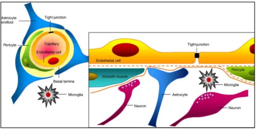
NVU has increasingly been a target of attention from researchers in the area of stroke. Neurons are very delicate cells whose survival depends on other cell types, on a dynamic and complex vascular network in the brain. Blood vessels ensure a precise and proper distribution of nutrients and oxygen for maintaining Central Nervous System homeostasis, coordinating neuronal activity with supporting cells and maintaining an optimal brain microenvironment adequate for neuronal survival by adjusting BBB parameters based on brain physiological needs. Neurons, in combination with astrocytes, pericytes, smooth muscle cells and EC, which are supported by the extracellular matrix, form the NVU and play a key role in the pathophysiology of stroke (8, 15) (Figure 1).
Figure 1 – Representative image of the cytoarchitecture of the NVU. Image from (16).
The angiogenic process
The formation of blood vessels occurs by vasculogenesis (de novo formation) and angiogenesis (formation from pre-existing vessels). In angiogenesis the first step is vessel destabilization and hyperpermeability induced by vascular endothelial growth factor-A (VEGF-A). Then, EC
formation. Afterwards, mesenchymal cells proliferate and migrate to differentiate into pericytes, which are important for vessels integrity and function. Finally, the blood vessel stabilization and the formation on the extracellular matrix depends on the release of growth factors (e.g. fibroblast growth factor, epidermal growth factor, platelet derived growth factor) (17). This process is important to induce vascular repair, which is mainly conducted by
endogenous and circulating endothelial progenitor cells (cEPC). cEPC are bone marrow-derived cells that can be mobilized into circulation when needed (e.g. ischemia) and incorporate damaged or forming vessels. EPC have a high proliferative rate and restorative capacities compared to mature cells. Additionally they also release relevant trophic factors like VEGF-A (18). In fact, in stroke the number of cEPC is increased (19).
Neural stem cells (NSC) and neurogenic niches
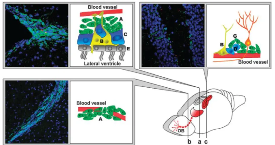
Neurogenesis occurs in two main neurogenic niches in the brain, the subgranular zone (SGZ) of the dentate gyrus of the hippocampal formation (Figure 2c) and the subventricular zone (SVZ) (Figure 2a). The SVZ contains slowly dividing radial astrocyte-like NSC (type B cells; also type B cells in the SGZ). NSC are cells with important characteristics: they are capable to proliferate, to self-renew and are multipotent (20, 21). Proliferating NSC originate transit
amplifying progenitors (type C cells; equivalent to type D cells in the SGZ) that differentiate into neuroblasts (type A cells; equivalent to type G cells in the SGZ). The interface between the ventricules and the neurogenic niche is carried by multiciliated ependymal cells (E). In the healthy rodent brain, neuroblasts migrate along the blood vessels from the lateral ventricle to the olfactory bulb. This migration pathway is known as rostral migratory stream. However, after brain injury such as stroke, neuroblasts break this fixed pattern of migration and re-route to the lesion site where they further differentiate into neurons. In the healthy brain, there is complex cell-to-cell signalling between the endothelium and the neurogenic niches (b) (22).
Impact of angiogenesis and neurogenesis on stroke
Brain angiogenesis and neurogenesis are highly coordinated and interdependent processes after ischemic brain injury. After a stroke, neurogenesis is stimulated and an increased number of neuroblasts (immature neurons) migrate from the neurogenic niche to the damaged brain tissue as part of an endogenous repair system. This process also stimulates and requires active angiogenesis. New neuroblasts are recruited to an area in the peri-infarct cortex, where EC are actively proliferating on the first days after cortical stroke. In the human brain, there are few indications for a migratory pathway of neuroblasts, but there are reports for active neurogenesis upon brain injury. Additionally, EC are known to secrete important trophic and chemotactic factors that recruit and support migrating neuroblasts to the site of injury. Earlier experiences claim that EC-secreted molecules such as erythropoietin, VEGF-A, endothelial nitric oxide synthase not only enhance focal angiogenesis, but also promote neurogenesis in the injured brain (23-25). Thus, it seems likely
that an efficient brain recovery after stroke is dependent on neurovascular plasticity (4, 23, 26-28). Animal studies have in fact shown that promoting post-ischemic angiogenesis can improve
the recovery of neurological function, indicating it is a promising therapeutic target for ischemic stroke.
Experimental models of ischemia
To study the injury caused by stroke there are several in vitro and in vivo models of different degrees of complexity: in vitro experimental setups such as induced glucose deprivation (GD) or oxygen/glucose deprivation (OGD) and animal models that attempt to mimic the ischemic environment (1). No model can mimic human stroke completely because of the disease
heterogeneity. In addition, the available models display variations in size and location of the infarct since animals may present differences in blood vessel distribution and flow. Nevertheless, the type of stroke that occurs most frequently is ischemic and the model that best mimics what occurs in such circumstances is termed middle cerebral artery occlusion (MCAO), which can be transient or permanent. The intensity of the stroke greatly depends on the MCAO period (1, 29). Although in humans most types of events are caused by permanent
ischemia, the use of transient occlusion allows for the evaluation of a neuroprotective and functional effect. In transient focal ischemia models, vessels can be blocked for 30 minutes up to 3-4 hours, followed by prolonged reperfusion; whereas, in permanent focal ischemia, the arterial blockage is maintained throughout an experiment, usually for one or more days. Permanent occlusion leads to extremely low survival rates. The use of an OGD model in this study aims to mimic the loss of cells in the ischemic core (30).
Retinoic acid (RA) signalling
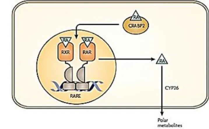
RA, a derivative of vitamin A, is one of the most biologically active molecules belonging to the retinoid family. This element was found to have an important role in development of the mammalian nervous system. RA is obtained from the diet in the form of retinyl esters from animal meat or β-carotene from plants. Inside the cell, the captured retinol is enzymatically converted, first to retinal by the retinol or alcohol dehydrogenases, and then to RA by the retinaldehyde dehydrogenases (RALDH) (31, 32). RA enters the cell nucleus attached to a
cellular retinoic-acid-binding protein 2. After a first connection with RA, RA receptors (RAR) and retinoid X receptors (RXR) heterodimerize and bind to a DNA sequence known as the RA response element, or RARE. This binding activates the transcription of target genes and subsequently RA is catabolized in the cytoplasm by the CYP26 class of P450 enzymes (Figure 3) (31, 33).
Figure 3 - Pathways describing RA signaling and catabolism. Scheme adapted from Maden M., 2007 (33).
The modulatory role of RA in neurogenesis
RA is a key modulator of neurogenesis because it is responsible for the upregulation of proneurogenic genes and negatively regulates genes that inhibit neurogenesis. Previous in
vitro and in vivo studies showed that RA also potentiates the increase of transcription factors
essential for neuronal differentiation, such as Mash1, Neurogenin1(34) and Neurogenin2 (35).
RA has been shown to be capable of stimulating neonatal SVZ and adult hippocampal neurogenesis as well as being capable of inducing the differentiation of progenitor cells into neuronal cells, confirming that this molecule is a good candidate for enhancing post-injury neurogenesis. In an in vivo model of stroke, it was demonstrated that RA reduces infarct volume of rats subjected to MCAO. Moreover, other studies demonstrated that continued oral ingestion of RA enhances SVZ cell proliferation in adult rats (31, 36, 37). Hence, RA is a good
therapeutic agent to stimulate neurogenesis and repair neuronal loss after stroke. The neuroprotective effects of vitamin A and RA may be mediated, at least in part, by the formation of heterodimers with other nuclear receptors (31, 33).
The modulatory role of RA in angiogenesis
The extent of neuronal repair and protection after stroke also depends largely upon the state of the brain vasculature. Of note, RA is known to induce the endothelial secretion of growth factors such as VEGF-A(38) and fibroblast growth factor(39) that modulate neurogenesis.
However, the effect of RA on angiogenesis is controversial. Some studies affirm that it has anti-angiogenic effects while others suggest pro-angiogenic effects. RA can exist as different isoforms trans and cis, each with a unique function. Several studies have shown that all-trans RA (ATRA) and its derivatives modulate angiogenesis. In vitro studies have demonstrated that ATRA is able to induce capillary tube formation in the presence of endogenous VEGF, known to play a central role in angiogenesis via EC. In in vitro and in vivo experiments, ATRA and 9-cis RA, presents pro-angiogenic effects mediated by the functional activation of RAR with consequent production of fibroblast growth factor-2, a component of blood vessels (40-42).
However, some retinoids may inhibit angiogenesis (e.g. fenretinide) and in a context of cancer-associated angiogenesis, ATRA may have an anti-angiogenic effect (43, 44).
Since angiogenesis is fundamental to the repair of stroke-damaged tissue, retinoids may be key to the development of new therapies.
RA-loaded nanoparticles (RA-NP)
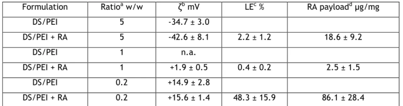
Free RA has limitations, such as low solubility in aqueous solution and fast degradation because is rapidly metabolized by cells. In order to overcome these limitations, RA-NP were developed. This preparation was achieved by electrostatic interaction between two polymers, the polycation polyethylenimine (PEI) and the polyanion dextran sulfate (DS). The nanoparticles used in this project have a ratio of 0.2 (DS/PEI), a favourable zeta-potential and an increased quantity of charged RA (ATRA isoform) per mass of nanoparticle compared to the other formulations tested (Table 1).
Table 1: Characteristics of DS/PEI nanoparticles in the presence and absence of RA
Formulation Ratioa w/w ζb mV LEc % RA payloadd µg/mg
DS/PEI 5 -34.7 ± 3.0 DS/PEI + RA 5 -42.6 ± 8.1 2.2 ± 1.2 18.6 ± 9.2 DS/PEI 1 n.a. DS/PEI + RA 1 +1.9 ± 0.5 0.4 ± 0.2 2.5 ± 1.5 DS/PEI 0.2 +14.9 ± 2.8 DS/PEI + RA 0.2 +15.6 ± 1.4 48.3 ± 15.9 86.1 ± 28.4
aPolyelectrolyte initial weight ratio. The experimental ratio for the 0.2 and 0.2RA formulations was
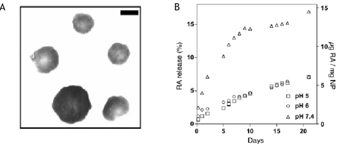
These nanoparticles of approximately 200 nm (Figure 4A) can be internalized via endocytosis, micropinocitose or phagocytosis and after having achieved the cytoplasm there is an efficient and gradual release of RA by desorption, diffusion through the nanoparticle, or nanoparticle erosion. The release profile of RA is determined by pH. The normal pH in cytosol is 7.4. RA-NP exposed to solution with pH=7.4 demonstrates controlled release of RA for 21 days proving the efficiency and efficacy of the formulation (45) (Figure 4B).
Figure 4 – Representative image (A) and release profile (B) of RA-NP. Scale bar 200 nm. Images from (45).
RA-NP have been shown to promote NSC differentiation into functional neurons in vivo presenting up to 2,500-fold higher efficiency. The internalization of RA-NP has no major impact on cell viability and proliferation however proves to be effective in promoting differentiation. RA-NP demonstrate regenerative capacities because they induce neurogenesis in the adult brain and in SVZ cell cultures due to the interaction between RA and its functional receptor, RAR. There are scarce reports for the use on RA on stroke and none for RA-NP, which highlights the innovative component of this project (34, 45).
Aims
Stroke represents a serious medical condition and a significant socioeconomic burden, but current standard of treatment is limited because it only benefits a small number of patients and may cause serious side effects. After a stroke neurogenesis is stimulated and neuroblasts are redirected to the lesioned area. To secure this repair mechanism, blood vessels provide physical support for neuroblast migration and trophic support for neural proliferation and survival. Hence, neuronal recuperation depends on the integrity of the vasculature. Since stroke also affects EC activity it is important to repair vascular function to promote neuronal recuperation and replenishment.
RA is a promoter of neuronal survival, differentiation and a regulator of angiogenesis but free RA has limitations that prevent it from reaching its therapeutical potential in vitro and in
vivo. In order to overcome these limitations it was necessary to develop a formulation that
protects RA while releasing it gradually, RA-NP.
We propose the repair of the vasculature as a means to maximize neurogenesis using the RA-NP as the therapeutic agent in an experimental model of ischemia. To mimic the ischemic environment experiments will be conducted under OGD. More specifically, in these settings, the following parameters will be determined: (1) the modulatory role of RA-NP treatment on vascular activity. EC will be subjected to an in vitro model of OGD to mimic the ischemic condition and the direct effect of RA-NP will be evaluated on proliferation, permeability and network formation and (2) the impact of RA-NP-treated EC secretome (healthy EC versus ischemic EC) on NSC. NSC will be treated with EC-conditioned media (EC-CM), in order to evaluate NSC survival, proliferation and differentiation.
Chapter 2 - Materials and methodology
Endothelial Cells (EC) Cultures
Mouse Hemangioendothelioma Endothelial Cells (EOMA) (kindly provided by Dr. Lino Ferreira, Center for Neuroscience and Cell Biology (CNC, University of Coimbra, Portugal) and Biocant (Innovation Center, Cantanhede, Portugal) were grown in Dulbecco's Modified Eagle Medium (DMEM) (Invitrogen, Barcelona, Spain) supplemented with 10% fetal bovine serum, 100 U/ml penicillin and 100 μg/ml streptomycin (Invitrogen). Cells were kept at 37ºC in a 95% atmospheric air and 5% CO2 humidified atmosphere. Before seeding, number of viable cells
was evaluated counting trypan blue-excluding cells.
Neural Stem Cells (NSC) Cultures
All experiments were performed in accordance with European guidelines (2010/63/EU) for the care and use of laboratory animals. NSC were prepared from 1-3 day old C57BL/6 donor mice as previously described.(46) Briefly, mice were euthanized by decapitation, and the brains
were removed and placed in calcium- and magnesium-free Hank’s Balanced Salt solution supplemented with 100 U/mL penicillin and 100 µg/mL streptomycin (Invitrogen) under sterile conditions. Fragments of SVZ were dissected out of 450 µm-thick coronal brain sections using a McIlwain tissue chopper, and then SVZ was digested in 0.025% trypsin (Invitrogen) and 0.265 mM EDTA (Invitrogen) (10 min, 37 °C), following mechanical dissociation with a P1000 pipet. The cell suspension was diluted in Serum Free Media composed of DMEM/F12 + GlutaMAXTM-I supplemented with 100 U/mL penicillin, 100 µg/mL streptomycin, 1% B27 supplement, 10 ng/mL epidermal growth factor (EGF) and 5 ng/mL basic fibroblast growth factor-2 (all from Invitrogen). Single cells were then plated on uncoated Petri dishes at a density of 3000 cells/cm2 and were allowed to develop in an incubator with 5% CO
2 and 95% atmospheric air
at 37°C.
Cell treatment
EC treatment included the evaluation of death, proliferation, permeability and network formation under physiological conditions and EC death and proliferation under OGD to mimic the ischemic condition. NSC treatment included the evaluation of death, proliferation and differentiation. Cells were plated at a density of 2x10⁴ cells per well in coverslips in 24-well
(EC and NSC respectively) with 3-50 µg/ml RA-NP and with 1-50 µM solubilized RA (free RA). In parallel cells were treated with their respective controls: 3-20 µg/ml blank nanoparticles and DMSO in the same proportion found in the free RA solution.
Collection of conditioned media (CM)
To evaluate the role of RA-NP on EC secretome, EC conditioned media (EC-CM) were collected from healthy or ischemic cells (see the next section). EC-CM was collected from cells incubated for 24 hours in serum-free DMEM medium in the presence of RA-NP, clarified by centrifugation 20 minutes at 14000 rpm and used without dilution on NSC.
OGD experiments
To mimic the ischemic environment the original glucose-containing media was replaced with PBS and cells were maintained in an anaerobic chamber (0.1% O2) for 5 hours. Experimental
conditions will include cells not subjected to OGD (untreated), treated with RA-NP alone, subjected to OGD alone and subjected to OGD and treated with RA-NP.
Scheme 1: Timeline for EC treatments under OGD.
Cell death
Propidium iodide (PI) (3 µg/mL; Sigma-Aldrich, St. Louis, Missouri, USA) was added 10 minutes before the end of the incubation period with RA-NP. Thereafter, cells were fixed with 4% paraformaldehyde (PFA) for 10 minutes, rinsed in PBS, stained with Hoechst-33342 (2 µg/mL; Invitrogen) in PBS for 5 minutes at RT, and mounted in Dakocytomation fluorescent medium (Dakocytomation Inc., California, USA). Photomicrographs of PI uptake labelling were recorded using a digital camera (Axiocam HRC, Carl Zeiss, Jena, Germany) adapted to an Axioskop 2 Plus fluorescent microscope (Carl Zeiss, Jena, Germany).
OGD (PBS; hypoxia)
Reoxygenation (Serum-free media; normoxia)
+ Cell treatments
t = 24hrs EC-CM collection t = 5 hrs
Cell proliferation
5-bromo-2'-deoxyuridine (BrdU; 10 µM; Sigma-Aldrich) was added 4 hours before the end of the incubation period with RA-NP (24 hours for EC and 48 hours for NSC). Cells were fixed in 4% PFA for 10 minutes and rinsed in 0.15 M PBS. BrdU was unmasked following 10 minutes in 1% Triton X-100 (Sigma-Aldrich) at room temperature (RT), and 30 minutes in 1 M HCl at 37°C. Cells were rinsed in PBS, and then incubated in a blocking solution with 3% bovine serum albumin (BSA; Sigma-Aldrich) and 0.3% Triton X-100 in PBS for 30 minutes at RT. Cells were incubated with mouse anti-BrdU (1:50; Invitrogen) in PBS containing 0.3% Triton X-100 and 0.3% BSA overnight at 4°C. Cells were incubated with a secondary antibody anti-mouse Alexa Fluor 546 (1:200; Invitrogen). Cell nuclei were counterstained with 6 µg/mL Hoechst-33342 in PBS for 5 minutes at RT and mounted in Dakocytomation fluorescent medium (Dakocytomation Inc.). Photomicrographs of BrdU-stained cells were recorded using a digital camera (Axiocam HRC, Carl Zeiss) adapted to an Axioskop 2 Plus fluorescent microscope (Carl Zeiss). The number of proliferating cells (BrdU-positive) were counted and expressed as percentage of total cells stained with Hoechst-33342 (Invitrogen).
Immunocytochemistry
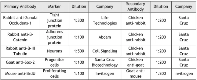
Cells were fixed with 4% PFA (Sigma-Aldrich) for 10 minutes. Then, to permeabilize the cell membrane, they are incubated for 20 minutes with 0.3 % bovine serum albumin solution (Sigma-Aldrich) and 3 % Triton X-100 solution (Sigma-Aldrich) in PBS. After permeabilization, the cells are incubated overnight at 4°C in a primary antibody solution (3% bovine serum albumin). The next day, cells were washed with PBS and incubated for 1 hr at RT with the corresponding secondary antibody. The antibodies used are shown in the Table 2.
Table 2: Antibodies used for immunocytochemistry
Primary Antibody Marker Dilution Company Secondary
Antibody Dilution Company Rabbit anti-Zonula Occludens-1 Tight junction protein 1:300 Life Technologies Chicken anti-rabbit 1:200 Santa Cruz Rabbit anti-β-Catenin Adherens junction protein 1:100 Abcam Chicken anti-rabbit 1:200 Santa Cruz Rabbit anti-β-III
Tubulin Neurons 1:500 Cell Signaling
Chicken
anti-rabbit 1:200
Santa Cruz Goat anti-Sox-2 Progenitor
cells 1:100 Santa Cruz Biotechnology Chicken anti-goat 1:200 Santa Cruz Mouse anti-BrdU Proliferating
cells 1:100 Invitrogen
Goat
Zonula Occludens-1 and β-Catenin were acquired using a confocal microscope (Zeiss LSM 710). Confocal images were obtained, with a Zeiss LSM 710 laser scanning confocal microscope (Carl Zeiss., USA) with 40x objectives.
Tubule formation assay
For in vitro angiogenesis assays, EC were plated in well (3x10⁴ cells/well in a 12-well slide
chamber, Ibidi, Martinsried, Germany) containing a substrate to mimic the extracellular matrix (poly-D-lysine, Sigma-Aldrich). Afterwards, growth medium was replaced with RA-NP treatment for 24h hours. During this period tubule growth was checked to determine the more suitable time point for the formation of tubules. Images were acquired under the magnification of 20x at the Zeiss Axiovert 200 imaging microscope (Axiobserver Z1, Carl Zeiss, Oberkochen, Germany) and analyzed with NIH free software ImageJ, using the Angiogenesis Analyzer program.
Western blotting
Cells were treated with RA-NP for 3 days to evaluate cellular differentiation and then were washed with PBS. Then, cells were incubated with RIPA lysis buffer (0.15 M NaCl, 0.05 M Tris-base, 5 mM ethylene glycol tetraacetic acid, 1% Triton X-100, 0.5% deoxycholic acid, 0.1% sodium dodecyl sulfate, 10mM dichlorodiphenyltrichloroethane containing a cocktail of proteinase inhibitors). Total protein from cell lysates was quantified using the bicinchoninic acid assay (Thermo Scientific,Massachusetts, USA). Then, samples were loaded onto 12% bis-acrylamide gel (Applichem, Darmstadt, Germany) (for β-III tubulin detection). Proteins were separated by Sodium dodecyl sulfate-polyacrylamide gel electrophoresis (120 V) using a running buffer (Tris-glicine with 10% sodium dodecyl sulfate (Acros organics, Geel
Belgium; pH 8.3) and then transferred (300 mA) to polyvinylidene fluoride membranes (GE
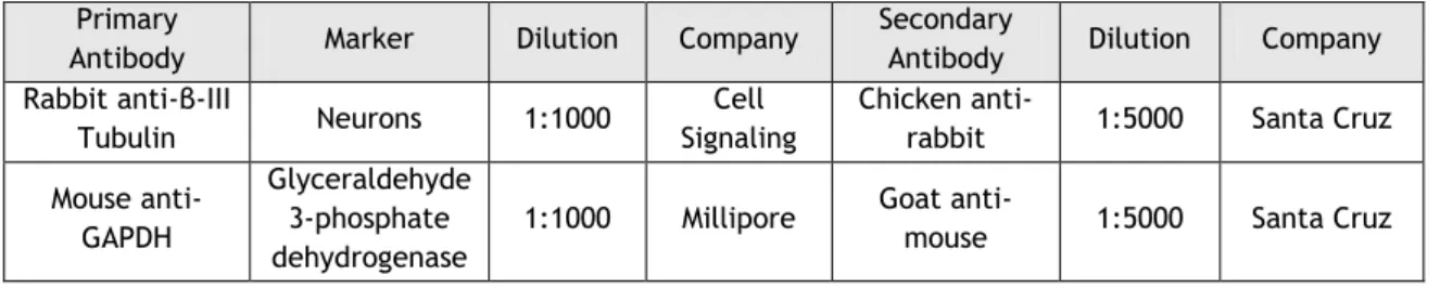
Healthcare, Little Chalfont, UK) for 90 minutes at 4ºC in a solution containing 10 mM CAPS (Sigma-Aldrich) and 20% methanol (Fisher chemicals, Hampton, New Hampshire). Membranes were blocked in 0.1% gelatin (Fluka, St. Louis, Missouri, USA) in Tris-buffer saline containing 0.1 % Tween-20 (Fisher Scientific, Massachusetts, USA) for 15 min, at RT, and then incubated overnight at 4ºC with the primary antibody (Table 3). After, membranes were incubated for 1 hour at RT with secondary antibody (conjugated to horseradish peroxidase) (Table 3). The detection of peroxidase was performed by enhanced chemiluminescence detection and densitometric analyses, using the software ImageLab (Bio-Rad, Hercules, CA, USA).
Table 3: Antibodies used for western blotting Primary
Antibody Marker Dilution Company
Secondary
Antibody Dilution Company Rabbit anti-β-III
Tubulin Neurons 1:1000
Cell Signaling
Chicken
anti-rabbit 1:5000 Santa Cruz Mouse
anti-GAPDH
Glyceraldehyde 3-phosphate dehydrogenase
1:1000 Millipore Goat
anti-mouse 1:5000 Santa Cruz
Statistics
All data are expressed as means ± standard error of mean (SEM). Statistical significance was determined by using Student’s t test, 1-way ANOVA followed by Dunnett’s Test or Bonferroni’s Multiple Comparison Test. Percentages of BrdU, or PI-positive cells were calculated from cell counts of three independent microscopic fields in each coverslip with a 40x objective.
Chapter 3 – Results
This next section will be presented in two separate parts: part I and part II. Part I will focus on the characterization of RA-NP treatment on healthy EC (under physiological conditions) followed by data on ischemic EC (under OGD). Part II will focus on the effect of RA-NP-treated EC secretome on several parameters of NSC activity.
Part I
RA is an important modulator of angiogenesis (40). However, in its free form RA presents some
limitations. To overcome RA low solubility and fast degradation, a polymeric nanoformulation was designed, RA-NP. In this next section, the therapeutic potential of RA-NP will be evaluated on key parameters of vascular activity, namely, cell viability, proliferation, permeability and network formation. Some of these parameters (cell viability and proliferation) will be evaluated on ischemic cells.
Effect of RA-NP treatment on EC death in physiological
conditions
In order to determine RA-NP cytotoxicity in physiological conditions, EC were exposed for 24 hours to treatment and cell death was then assessed by PI uptake (Scheme 1).
Scheme 1: Timeline for PI uptake in EC under physiological conditions.
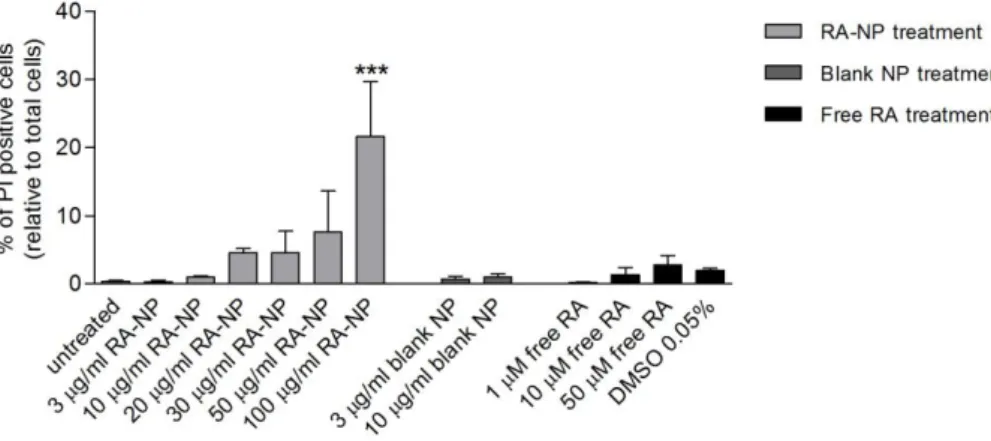
PI (3,8-diamino-5-(3-(diethylmethylammonio)propyl)-6-phenyl phenanthridinium diiodide)) is a common red-fluorescent marker used to detect cells that lost membrane integrity and for this reason, are non-viable. This method is used to detect necrotic cells, in which there is disruption of the plasma membrane and DNA fragmentation. The percentage of basal death for these cells was approximately 0.3% and it was found that up to 10 µg/ml RA-NP and 10 µM free RA do not induce cytotoxicity. The concentrations 3 and 10 µg/ml RA-NP (0.26±0.26%,
Endothelial cell treatment
t = 24hrs Cell death evaluation 10 min before end of treatment
PI uptake t = 0
has very low solubility in aqueous solutions 100% dimethyl sulfoxide (DMSO) was used as RA solvent, meaning that 0.05% DMSO (1.98±0.31%, n=2) is the final concentration in the 50 µM free RA (2.81±1.36%, n=2) solution. Although 0.5% DMSO alone presented some cytotoxicity to cells, this effect was not exacerbated in the RA solution, meaning that toxicity caused this solution was likely from the solvent. The blank formulations (RA-free polymeric nanoparticles) presented a similar effect to their respective loaded formulations (Figure 1).
Figure 1 - Effect of RA-NP treatment on EC viability. In physiological conditions, percentage of PI-positive cells, with RA-NP (3 and 10 µg/ml) and 10 µM free RA treatment did not present cytotoxicity when compared to untreated condition, indicating that these concentrations did not induce EC death. Free RA can induce cytotoxicity due to its solvent DMSO; likewise, blank formulations showed no toxicity as the respective RA-loaded formulations. Data are show as the mean ± SEM (n=2-7). ***P<0.001 using 1-way ANOVA followed by Dunnett's Multiple Comparison Test to compare with untreated condition.
Effect of RA-NP treatment on EC proliferation in physiological
conditions
To evaluate proliferation in physiological conditions, EC were exposed to treatments for 24 hours and after this time, bromodeoxyuridine (BrdU) was added for the remaining 4 hours of treatment (Scheme 2). BrdU (5-bromo-2'-deoxyuridine) is a thymidine analogue that allows the identification of newly synthesized DNA in dividing cells.
Scheme 2: Timeline for BrdU assay in EC under physiological conditions.
t = 0
Endothelial cell treatment
t = 24hrs
Cell proliferation evaluation 4 hrs before end of treatment
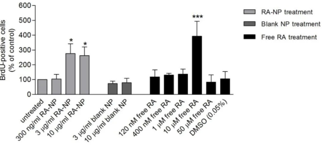
The treatment with 3 and 10 µg/ml RA-NP (276.10±66.21%, n=6, p<0.05; 262.40±58.32%, n=6,
p<0.05) and 10 µM free RA (393.00±101.30%, n=3, p<0.001) induced a significant increase in
EC proliferation comparatively to the untreated condition (100%, absolute value is 6.7%) (Figure 2). These concentrations are within the range of RA-NP and free RA treatment that did not cause cytotoxicity. A higher free RA concentration (50 µM) did not potentiate or sustain proliferation highlighting how RA depends on an optimal/narrow concentration window to drive its effects, as observed by others (47). Lower concentrations did not present
impact on proliferation.
Figure 2 - Effect of RA-NP treatment on EC proliferation. In physiological conditions, the percentage of BrdU-positive cells increased with RA-NP (3 and 10 µg/ml) and 10 µM free RA treatment. Data are expressed as mean ± SEM (n=2-10). *P<0.05; ***P<0.001 using 1-way ANOVA followed by Dunnett's Multiple Comparison Test to compare with untreated condition.
Effect of RA-NP treatment on EC permeability in physiological
conditions
To determine EC permeability with RA-NP treatments in physiological conditions, cells were exposed for 24 hours to 10 µg/ml RA-NP and then immunolabeled for β-Catenin and Zonula occludens-1 (ZO-1), which are part of the intracellular anchorage between EC (Figure 3). β-Catenin expression increased (Figure 3B) after treatment when compared to untreated condition (Figure 3A). ZO-1 expression seemed unaltered by treatment (figures 3C and 3D).
Figure 3 – Effect of RA-NP treatment on EC permeability. A) Representative fluorescence image of β-Catenin immunostaining (red) in control condition (untreated), B) exposed to RA-NP (10 µg/ml) for 24 hours, C) ZO-1 immunostaining (red) in control condition (untreated) and D) exposed to RA-NP (10 µg/ml) for 24 hours (n=2). Scale bars are 20µm.
Effect of RA-NP treatment on EC angiogenesis in physiological
conditions
The formation of new blood vessels from pre-existing ones (angiogenesis) is a key characteristic of EC activity. To analyze this ability, cells were treated with 10 µg/ml RA-NP for 24 hours and then observed over time in order to evaluate the complexity of tubules formed. In these preliminary assays, only one concentration of RA-NP was chosen because experiments conducted in parallel revealed that 3 µg/ml RA-NP did not have a significant effect on ischemic cell repair. These data will be presented on the next section. The more complex network was observed at 24 hours and then was examined with Angiogenesis
Untreated 10 µg/ml RA-NP Untreated
A
B
20 µm 10 µg/ml RA-NP UntreatedD
C
extremities (A, B); number of nodes, identified as pixels that have at least 3 neighboring elements, corresponding to a bifurcation (A, B). Segments (C2, D2) are elements delimited by two junctions (C3, D3); Branches (C4, D4) are elements delimited by a junction and one extremity. Master segments (E3) consist in pieces of tree delimited by two junctions none exclusively implicated with one branch, called master junctions (E2). Master junctions are junctions linking at least three master segments. Finally, meshes (F1) are areas enclosed by segments or master segments (Scheme 3).
Scheme 3 - Detection of the important elements in analyzing the complexity of the tubules formed by cells. Image obtained from (48).
Cells treated with 10 µg/ml RA-NP may increase the complexity of network, since the number of extremities, nodes, branches, junctions, segments and mesh increased, as well the length of branches and segments and area of meshes when compared to control condition (untreated) (figure 4).
Figure 4 – Effect of RA-NP treatment on angiogenesis. Several parameters of network formation were measured, such as A) number of extremities, B) number of nodes, C) number of branches, D) branch length, E) number of junctions, F) number of segments, G) segment length, H) number of meshes and I) mesh area. Data are expressed as mean ± SEM (n=2).
Effect of RA-NP treatment on EC death in OGD conditions
After an ischemic stroke, blood vessels succumb to lack of oxygen and nutrients and may show loss of integrity/activity and ultimately cell death, which reflects negatively on the previously irrigated tissue. To evaluate RA-NP treatment efficiency against ischemia, EC were subjected to 5 hours of oxygen and glucose deprivation in a hypoxic chamber, containing only 0.1% O2, 95% N2 and 5% CO2 and in PBS (to remove all media nutrients). After 5 hours, cells
were treated with the same concentrations of RA-NP already tested in physiological conditions. The treatment consisted of 24 hours and then cell death was evaluated through PI uptake (Scheme 4).
Scheme 4: Timeline for PI uptake in EC under OGD.
The percentage of PI-positive cells increased significantly after insult when compared to the percentage of PI-positive in cells in physiological conditions, which indicated that there was indeed a serious disturbance of EC physiology. The dashed line represents basal death of EC in physiological conditions (≈0.3%). Treatment with 10 µg/ml RA-NP (32.50±11.06%, n=4, p<0.01) and 10 µM free RA (36.07±11.74%, n=5, p<0.01) significantly protected the cells from death after suffering the ischemic insult (Figure 5). In parallel, the concentrations of free RA equivalent to 3 µg/ml RA-NP (120 nM) and to 10 µg/ml RA-NP (400 nM) were tested and had no effect on cell viability. Although the recovery was not full, this treatment restored significantly the survival of cells.
Figure 5 - Effect of RA-NP treatment on EC death, after OGD. Percentage of PI-positive cells decreased
OGD (PBS; hypoxia)
Reoxygenation
(Serum-free media; normoxia) + Cell treatments
t = 24hrs Cell death evaluation t = 5hrs 10 min before end of treatment
PI incorporation t = 0
Effect of RA-NP treatment on EC proliferation in OGD conditions
To analyze the effect of RA-NP treatment in EC proliferation, after OGD, cells were subjected to same protocol used in scheme 5. Then 4 hours before the end of treatment, BrdU was incorporated (Scheme 5).
Scheme 5: Timeline for BrdU assay in EC under OGD.
The dashed line represents the percentage of normal proliferation (≈6.7%) of these cells under basal conditions (Figure 6). None of the tested treatments could restore the basal proliferative EC function, after being subjected to OGD.
Figure 6 - Effect of RA-NP treatment on EC proliferation, after OGD. None of these concentrations recovered the basal proliferative state of EC. Data are expressed as mean ± SEM (n=2-4).
Part II
The brain vasculature provides physical support for NSC survival and secretes molecules that are involved in the proliferation and differentiation of NSC (49). For that reason, conditioned
media were collected from healthy and ischemic EC. In this next section, the effect of RA-NP-treated EC secretome was evaluated on NSC viability, proliferation and differentiation.
OGD (PBS; hypoxia)
Reoxygenation
(Serum-free media; normoxia) + Cell treatments
t = 24hrs Cell proliferation
evaluation t = 5 hrs 4 hrs before end of treatment
BrdU incorporation t = 0
Effect of EC-CM treatment in NSC death
To evaluate the effect of RA-NP-modulated EC secretome on NSC viability, NSC were exposed to EC-CM collected from RA-NP-treated healthy (Figure 8; color light gray) and ischemic EC cells (Figure 8; color dark gray). The treatment follows the protocol shown in Scheme 6.
Scheme 6: Timeline for PI uptake in NSC treated with EC-CM.
EC medium alone induced NSC death (38.54±9.22%, n=7; 21.33±2.89%, n=8; respectively). However, EC-CM from untreated healthy EC significantly decreased NSC death (13.73±1.54, n=6, p<0.05). The same effect was observed with EC-CM from RA-NP-treated healthy EC (6.66±1.84%, n=3, p<0.05). There was also statistical difference between this condition and EC-CM untreated healthy EC which further highlights the effect of RA-NP on EC secretome. EC-CM from untreated ischemic EC induced a similar impact on NSC as EC medium alone, while significantly increasing basal NSC death (44.20±8.37%, n=5, p<0.05). EC-CM from RA-NP-treated ischemic EC significantly decreased NSC death (24.46±7.07%, n=5, p<0.05) when compared to EC-CM from untreated ischemic EC. All of the other media treatments had an effect comparable to EC media alone (Figure 7). An additional control was also performed to determine whether an alteration in pH or in the constitution of the EC media, when subjected to hypoxia, could affect viability. However there was no difference between the two media (n=2).
Figure 7 – Effect of RA-NP-modulated EC secretome on NSC viability. Percentage of PI-positive cells from treatment indicated that EC-CM from RA-NP-treated EC significantly decreased NSC death when compared to EC-CM from untreated condition. Data are expressed as mean ± SEM (n=3-8). *P<0.05using 1-way ANOVA followed by Dunnett’s Multiple Comparison Test to compare with EC media condition;
Neural stem cell treatment
t=48hrs Cell death evaluation 10 min before end of treatment
PI incorporation EC-CM collected t=0
Effect of supernatants treatments on NSC death
EC-CM was centrifuged after collection to remove cell debris and contaminating nanoparticles. To confirm the efficiency of this protocol step an additional control was performed. EC media alone, not exposed to cells, was treated with RA-NP for 48 hours and processed in the same way as EC-CM to obtain the deposition of the RA-NP and its respective supernatant. After, the supernatants were collected for subsequent NSC treatment. The protocol was used as shown in Scheme 7.
Scheme 7: Timeline for PI uptake in NSC treated with supernatants.
These treatments did not protect NSC from EC media-induced death, which demonstrated that the protective effect shown induced by RA-NP-treated EC-CM was derived from pro-survival factors secreted by EC and not by RA-NP in suspension (Figure 8).
Figure 8 - Effect of supernatants on NSC viability. Percentage of PI-positive cells demonstrated that none of these treatments significantly alter cell viability. Data are expressed as mean ± SEM (n=2).
Effect of EC-CM treatments in NSC proliferation
To assess the effect of EC-CM collected from ischemic cells treated with RA-NP, cells were exposed to treatment for 48 hours and then were subjected BrdU incorporation (Scheme 8).
Neural stem cell treatment
t = 48hrs Cell proliferation
evaulation 4 hours before end of treatment
BrdU incorporation BrdU incorporation t = 0
EC-CM collected
Neural stem cell treatment
t = 48hrs Cell death evaluation 10 min before end of treatment
PI incorporation Supernatants collected t = 0
When NSC were exposed to treatments with EC-CM collected from ischemic cells (3µg/ml RA-NP (29.84±8.59%, n=3, p<0.01)) and treatments with EC-CM collected from healthy cells (3µg/ml RA-NP (19.01±1.71%, n=3, p<0.05)), treatments significantly increased cell proliferation when compared to untreated cells (3.48±1.36%, n=3) (Figure 9).
Figure 9 – Effect of EC-CM collected from ischemic EC treated with RA-NP on NSC proliferation. BrdU-positive NSC increased when treated EC-CM collected from cells treated with 3 µg/ml RA-NP. Data are expressed as mean ± SEM (n=3-6). *P<0.05 and **P<0.01 using Student t-test compared to EC media.
Effect of supernatants treatment on NSC proliferation
In order to verify that the effect of EC-CM in NSC proliferation was not due de RA-NP that were suspended in the medium, NSC cells were subjected to RA-NP supernatants. The experimental procedure was the same but occurred without the presence of cells, over 24 hours at 37°C. Then, these suspensions were centrifuged, leading to deposition of the RA-NP. Afterwards, the supernatants were collected for subsequent treatment and evaluation of NSC proliferation (Scheme 9).
Scheme 9: Timeline for BrdU assay in NSC treated with supernatants.
The percentage of BrdU-positive cells indicated that none of supernatants induced NSC proliferation (Figure 10).
Neural stem cell treatment
t = 48hrs Cell proliferation
evaluation 4 hours before end of treatment
BrdU incorporation t = 0
Figure 10 - Effect of EC supernatants collected from ischemic EC treated with RA-NP on NSC proliferation. None of the treatments altered proliferation. Data are expressed as mean ± SEM (n=2).
Effect of EC-CM treatment on NSC differentiation
In order to evaluate effect of treatments on NSC differentiation, NSC were treated with EC-CM from healthy and ischemic cells treated with RA-NP, over 3 days (Scheme 10). To determine the differentiation state of NSC, it was necessary to evaluate the expression of β-III tubulin and Sox-2 by western blotting. β-β-III tubulin is a constitutive protein of microtubules that is responsible for axonal growth, specifically in neurons (50). On the other hand, Sox-2 is a
transcription factor that it is responsible for the maintenance of stem cells state and for that is used to label immature cells (51).
Scheme 10: Timeline for evaluation of NSC differentiation.
All treatments from healthy EC increased the expression of β-III tubulin. However, when NSC were treated with EC-CM from cells collected under OGD, β-III tubulin protein expression only increased in the presence of EC-CM from EC treated with 3 µg/ml RA-NP (Figure 11B) (162.2±29.86%, n=5, p<0.05) and not from untreated EC (78.73±29.22%, n=5) (Figure 11A).
Neural stem cell treatment
t = 3 days
Cell differentiation evaluation t = 0
Figure 11 – Effect of CM collected from ischemic EC treated with RA-NP on NSC differentiation. EC-CM from 3 µg/ml RA-NP-treated EC (OGD) increased β-III tubulin expression in NSC.50kDa β-III tubulin expression normalized to 37kDa GAPDH. Data are expressed as mean ± SEM (n=5) *p<0.05 using Student
t-test to compared with EC media condition, #p<0.05 using Student t-test to compared with untreated
(OGD) condition.Scale bars are 50µm.
To further support the previous result, Sox-2 expression was evaluated. When NSC were treated with EC-CM collected from ischemic RA-NP-treated cells there was a decrease of protein expression (Figure 12D) (9.15±2.05%, n=4, p<0.05) when compared to untreated condition (30.15±5.23%, n=4, p<0.01) (NSC media; 18.50±1.78%, n=4, p<0.05) (Figure 12C). When NSC were treated with EC media there was a small increase in Sox-2 expression compared to NSC media (32.60±6.43%, n=3; 18.50±1.78%, n=4). untreated 3 µg/ml RA-NP
A
B
50 µm β-III tubulin GAPDHFigure 12 - Effect of EC-CM collected from ischemic EC treated with RA-NP on the maintenance of the progenitor cell population. EC-CM from 3 µg/ml RA-NP-treated EC (OGD) decreased significantly Sox-2 expression, in NSC. Data are expressed as mean ± SEM (n=3-4) *p<0.05 and *p<0.01 using 1-way ANOVA followed by Dunnett's Multiple Comparison Test to compare with EC media condition, ###p<0.001 using 1-way ANOVA followed by Bonferroni's Multiple Comparison Test to compare with untreated (OGD) condition. Scale bars are 50µm.
untreated
3 µg/ml RA-NP