Universidade de Lisboa
Faculdade de Ciências
Departamento de Física
Data Acquisition, Curation and Modeling
for Integration of Alzheimer’s Disease
Neuroimaging Data from ADNI in the
Translational Biomedicine Platform
tranSMART
Mestrado Integrado em Engenharia Biomédica e Biofísica
Perfil em Engenharia Clínica e Instrumentação Médica
Vasco de Almeida Jorge Veríssimo
Dissertação orientada por:
Orientador Externo: Professor Doutor Reinhard Schneider
Orientador Interno: Professor Doutor Nuno Matela
“Viver não custa, custa saber viver…”
i
Resumo
Nos dias que correm, as doenças neurodegenerativas afetam milhões de pessoas em todo o mundo, havendo mais de 600 doenças diferentes que incidem sobre o sistema nervoso, sendo as doenças de Alzheimer e Parkinson as mais comuns. Estudos indicam que 60-70% das pessoas que sofrem de distúrbios cerebrais, são casos de Alzheimer. Se olharmos para os Estados Unidos como exemplo, mais de 5 milhões de pessoas sofrem de Alzheimer e pelo menos 500 mil pessoas sofrem de Parkinson.
As doenças neurodegenerativas são doenças caracterizadas pela deterioração progressiva do sistema nervoso, que ocorre quando as suas células, os neurónios, começam a degenerar e a morrer. Isso é preocupante pois os neurónios são a base do funcionamento do sistema nervoso e não se reproduzem, não podendo ser substituídos, o que faz com que as doenças neurodegenerativas sejam, normalmente, incuráveis e debilitantes.
A doença de Alzheimer, descrita pela primeira vez por Alois Alzheimer, é um tipo de demência que afecta o cérebro, provocando problemas ao nível da memória, pensamento e comportamento. Os seus sintomas desenvolvem-se lentamente, em geral, com pioras progressivas que acabam por interferir com as tarefas do dia-a-dia. Esta doença afecta cerca de 44 milhões de pessoas a nível mundial, sendo que este número é esperado aumentar até três vezes mais nos próximos 50 anos. É uma doença geralmente associada à idade, embora haja casos de incidência antecipada, com pacientes nos seus 40s ou 50s anos de idade.
Embora as causas para a doença de Alzheimer não estejam ainda completamente compreendidas, os cientistas acreditam que, para a maioria das pessoas, esta doença resulta duma combinação de factores genéticos, ambientais e de estilo de vida, que vão afectando o cérebro ao longo do tempo.
Por ser a doença neurodegenerativa mais comum no Mundo, a doença de Alzheimer acaba também por ser a doença com maior impacto financeiro nos países desenvolvidos, representando também um enorme peso para os pacientes e para a sua família, tanto a nível económico, como a nível mental e psicológico, visto que esta
ii
devastadora doença, apesar de ter medicação disponível para eventualmente reduzir o efeito dos sintomas, não tem ainda cura.
Com estes dados em mente, podemos verificar a importância de se começar a direcionar a investigação no sentido de melhorar o diagnóstico e terapia clínica destas doenças neurodegenerativas.
Uma das maneiras mais eficazes de alcançar este objetivo é tentar centralizar todo o conhecimento e informação sobre essas doenças e reformar sistemas de classificação de doenças previamente estabelecidos, tais como a CID (Classificação Internacional de Doenças). A doença de Alzheimer é actualmente classificada pelos seus sintomas e severidade, embora seja claro hoje em dia que este sistema não representa as diferentes causas possíveis para a doença, pois os mesmo sintomas podem ser gerados por diferentes causas genéticas ou moleculares. Existe então um reconhecimento geral de que os sistema de classificação tem de mudar, por exemplo para um que utilize uma taxonomia baseada em mecanismos (mechanism-based
taxonomy), a qual se baseia no conhecimento sobre as vias biológicas envolvidas na
etiologia de uma doença, de modo a guiar a classificação das classes e subclasses da doença.
É portanto neste contexto que surge o projeto AETIONOMY, cujo principal objetivo incide no desenvolvimento de uma taxonomia baseada em mecanismos para as doenças de Alzheimer e Parkinson, sendo que o seu maior desafio reside no facto de que, para a maioria das doenças neurodegenerativas, as vias biológicas disfuncionais subjacentes à doença não são conhecidas.
O AETIONOMY pretende identificar e recolher todos os dados disponíveis mundialmente, incluíndo dados clínicos, de imagem ou genéticos, sobre a doença de Alzheimer, com o fim de desenvolver uma estrutura comum que combine todos os dados obtidos, de maneira a ser possível identificar padrões que tornem possível a divisão da doença em sub-grupos de pacientes com causas semelhantes da sua doença, na expectativa de identificar mudanças patofisiológicas durante a doença a nível molecular, que possam conduzir a uma nova taxonomia da doença.
O presente estudo incidiu sobre a doença de Alzheimer, mais especificamente sobre os dados clínicos da ADNI (Alzheimer’s Disease Neuroimaging Initiative), e como processar e tratar estes dados para que seja possível integrá-los numa plataforma de biomedicina translacional chamada tranSMART, cujo intuito é a
iii
criação de novas rotas para a identificação dos mecanismos subjacentes à doença, antes de os organizar e propor uma taxonomia racional da mesma.
Este estudo teve como principal objetivo obter os dados da ADNI, analisá-los e estudar toda a envolvência desta iniciativa, de maneira a conseguir uma melhor perceção do panorama geral da mesma, fazendo de seguida o processamento, modelação e limpeza destes dados clínicos para integração no tranSMART. Ao mesmo tempo procurou-se aprofundar o conhecimento sobre a doença de Alzheimer, inclusive os seus sintomas, possíveis causas e diagnóstico. Recorreu-se neste trabalho a diferentes pacotes de software e linguagens de programação, tais como o OpenRefine e o Python.
Este estudo propõe então uma revisão completa dos conceitos e dados estruturais da ADNI, ao mesmo tempo que obtém e armazena localmente todos os dados, valores e variáveis utilizados no âmbito desse mesmo projecto. Foi também criado um software para a transformação dos dados da ADNI, com o objectivo de os integrar numa plataforma de biomedicina translacional. No entanto, primeiro é indispensável compreender os princípios de tratamento de dados para o carregamento de dados no tranSMART para poder ser possível a integração dos dados da ADNI, fazendo com que estejam disponíveis para outros projectos a nível mundial, tal como o AETIONOMY e o eTRIKS, por exemplo.
Com este estudo tornou-se possível adicionar os dados da ADNI à base de dados do tranSMART, de forma a torná-los disponíveis para terceiros e outros projetos, ajudando então a tão desejada centralização do conhecimento sobre esta doença, o que representa um passo importante na direção dos objectivos traçados por este projecto, ajudando na recolha de dados, e, sendo a ADNI uma das maiores iniciativas de estudo clínico sobre Alzheimer alguma vez feita, o seu carregamento no traSMART é crucial para uma melhor compreensão da doença e dos seus mecanismos.
A dissertação encontra-se dividida em 5 capítulos. O primeiro consiste numa introdução ao tema da dissertação e aos seus objetivos, e no segundo são descritos os principais conceitos inerentes ao trabalho desenvolvido --- como por exemplo conceitos sobre doenças neurodegenerativas, mais especificamente a doença de
Alzheimer --- analisando ao pormenor as suas causas, as diferentes fases,
iv
também temas e conceitos relacionados com a bioinformática por detrás deste estudo, especificando também algumas das plataformas e pacotes de software utilizados.
O terceiro capítulo descreve os materiais e metodologias utilizadas, explicando pormenorizadamente todos os passos e processos involvidos neste projecto, em cada uma das quatro principais fases dos mesmo (aquisição, tratamento, modelação e integração dos dados da ADNI), sendo que no quarto capítulo se encontram os resultados obtidos após a conclusão da parte prática deste estudo, apresentando também uma discussão sobre os mesmos, à medida que vão sendo demonstrados.
No quinto e último capítulo encontram-se a Conclusão e Trabalho Futuro, ondese apresentam as ideias finais deste estudo, juntamente com sugestões de trabalho futuro que poderá dar seguimento a este estudo.
Existe ainda uma secção de Anexos, onde se encontram os dicionários criados para este projecto e o script de Python criado para a modelação dos dados.
Palavras-chave: Doenças Neurodegenerativas, Doença de Alzheimer,
v
Abstract
Neurodegenerative diseases affect millions of people worldwide, and Alzheimer’s disease (AD) is the most common type. For example in America, more than five million people are living with AD, although some estimates are much higher. With that in mind, it is more and more important to start working towards the improvement of these neurodegenerative diseases’ diagnosis and clinical therapy.
One way to achieve that goal is to centralize all the knowledge on those diseases and reform established disease classification systems, such as ICD [International Classification of Disease], using a “mechanism-based taxonomy”, based upon the knowledge about the biological pathways involved in the aetiology of a disease to guide the classification of disease classes and subclasses, and that is the main goal of the AETIONOMY project, with its greatest challenge lying on the fact that, for most neurodegenerative diseases, the dysfunctional biological pathways underlying the disease are not known.
This project will focus on the Alzheimer’s Disease Neuroimaging Initiative (ADNI) Clinical Data, and how to properly curate this clinical data and process it for integration in a translational biomedicine platform (tranSMART), analyzing and studying its relevance, as well as having a better understanding of this initiative’s global overview, with the purpose of defining new routes towards the identification of the underlying disease mechanisms, before organizing the latter, as well as proposing a rational disease taxonomy.
This study made it possible to add the ADNI clinical data to the tranSMART’s database, making it available to third-part projects and thus helping the centralization of the current knowledge about this disease.
Key-words: Neurodegenerative Diseases, Alzheimer’s Disease (AD),
vii
Acknowledgements
First I would have to extend a special thank you to Professor Reinhard Schneider, for devising an amazing project for my internship and allowing me the possibility of working besides him and his fantastic group, to whom I’m also grateful for all the help.
Second, I would like to thank Dr. Adriano Barbosa, for guiding me throughout this entire year, helping me and counseling me about everything that I needed, making me comfortable since my first day and always making time for me when I needed his help. More than my supervisor, you were my friend, inside and outside the LCSB.
I would also like to thank my internal supervisor at FCUL, Professor Nuno Matela, for his promptness in helping me every time I asked, always demonstrating full availability.
I cannot name everyone involved in this project and in the BioCore group because there are too many, but I have to mention also Dr. Venkata Satagopam and Dr. Wei Gu for all the help and support along my internship, and especially during the Datathon in Boston; I also have to mention sweet Aishu who worked beside me in this project, and without whom I would never have been able to finish it.
Even when you are working in an amazing Research Center like the LCSB, with challenging and interesting projects, surrounded by brilliant scientists and incredible people, it is always difficult to live abroad, to leave your country, your friends, your family, your home… It is equally important to find, in your new country, a new group of friends, a new family, a new place you can call home. That is why I can say for sure that I was blessed with luck to have found it all here in Luxembourg. The discomfort of being away from home was eased by meeting the most incredible group of friends which I can call my family, the “Happy Wednesday” group! I am not going to name you one by one because you are too many, but I want to thank you all for being a part of my life and for making this staying in Luxembourg so incredibly awesome for me! Sometimes I even forget I am not actually home… You guys are amazing! Thank you for everything!
viii
As I once read, “the journey is what brings you happy, not the destination”, so I also have to thank all my childhood friends, all my friends from high school, all my friends from college, all my friends that in one way or another managed to change my life, all my friends who helped me grow as an academic, as a person, as a human being, to all my friends that came into my life and left a mark, to all my friends that made me who I am now.
Last but not least, nay last and most important, I have to thank my family! And for them, “thank you” cannot even remotely describe the way I feel… There are not enough words to express how thankful I am to have them and how lucky I am to have chanced in such a wonderful family.
To my mother, whose unconditional love and affection for me and whose unshaken care for my well-being and happiness have always made me feel safe and knowing I would never be alone. Forgive me for my “undeserving” behavior sometimes and thank you for being who you are! I am who I am nowadays largely because of you, because of the amazing person that you are and fought for me to be. To my brother, who has always taken care of me since I was born, lecturing me when he had to and advising me even when I didn’t want to listen. You have been there for my best and worst moments, celebrate with me when I am happy, comforting me when I am sad, congratulate me on my successes and pick me up on my failures. Everyone can have brothers or sisters, but I have more than that. In you I have a best friend, a partner for life, with me no matter what! Thank you for being who you are.
To my father, with whom I embarked on this great adventure in Luxembourg. A son couldn’t possibly ask for a better father! You are my role model, my hero, my idol. You make me want to be better every day, and you wisely guide me not only with your precious advices which I treasure, but by leading by example. You know that actions speak louder than words and following your footsteps have made me a better man than I thought I could ever be. Thank you for never giving up on me and for always believing in me. I’ll be happy if I ever make you feel half as proud of me as I am of you.
ix
Contents
Resumo ... i Abstract ... v Acknowledgements ... vii Contents ... ix List of Figures ... xi List of Abbreviations ... xv 1. Introduction ... 11.1 Thesis Objectives and Outline ... 1
2. Background... 3
2.1 Neurodegenerative Diseases ... 3
2.2 Alzheimer’s Disease ... 4
2.2.1 Symptoms, Characteristics ... 6
2.2.2 Stages ... 9
2.2.2.1 Preclinical Alzheimer’s Disease ... 10
2.2.2.2 Mild Cognitive Impairment ... 10
2.2.2.3 Mild Dementia ... 11 2.2.2.4 Moderate Dementia ... 11 2.2.2.5 Severe Dementia ... 12 2.2.3 Causes ... 12 2.2.3.1 Genetic Hypothesis ... 14 2.2.3.2 Cholinergic Hypothesis ... 17 2.2.3.3 Amyloid Hypothesis ... 19 2.2.3.4 Tau Hypothesis ... 21 2.2.4 Pathophysiology ... 22 2.2.4.1 Neuropathology ... 22 2.2.4.2 Biochemistry ... 23 2.2.5 Diagnosis ... 24 2.2.5.1 Criteria ... 24
2.2.5.2 Tests and Techniques ... 25
2.2.5.3 Early Diagnosis... 27
x 2.3 Clinical Trials ... 29 2.4 AETIONOMY ... 30 2.5 ADNI Project ... 31 2.6 Translational Bioinformatics ... 33 2.7 Big Data ... 35
2.8 Data Curation (OpenRefine) ... 36
2.9 Data Integration (tranSMART)... 38
3. Materials and Methods ... 41
3.1 ADNI Data ... 41
3.2 OpenRefine Features ... 45
3.3 Python features ... 51
3.4 tranSMART Features ... 55
4. Results and Discussion ... 59
4.1 Data Acquisition ... 59
4.2 Data Curation ... 62
4.3 Data Modeling ... 67
4.4 Data Integration ... 71
5. Conclusion and Future Work ... 81
Bibliography ... 83
xi
List of Figures
Figure 2. 1 – Brain image illustrating different kinds of effects on the brain of different kinds of NDs.
(http://www.cannabisoils.ca/wp-content/uploads/2014/11/neurodegenerative-diseases.jpg) ... 3 Figure 2. 2 – Microscope image comparing AD cells and healthy cells.
(https://www.alz.org/braintour/plaques_tangles.asp) ... 4 Figure 2. 3 – Brain atrophy in advanced AD in comparison with a normal brain.
(http://alzheimerdisease.tv/wp-content/uploads/2014/11/alzheimers-disease-term.jpg) ... 5 Figure 2. 4 – Growth in dementia cases expectation by 2050.
(http://www.fic.nih.gov/News/GlobalHealthMatters/PublishingImages/fogarty-nih-infographic-global-growth-dementia-cases-by-region-by-2050.jpg) ... 6 Figure 2. 5 – Brain sections mostly affected by AD.
(http://www.medicalook.com/diseases_images/alzheimers_disease.jpg)... 7 Figure 2. 6 – Image showing the different effects of AD on the brain from the healthy phase to severe AD. (http://sierram.web.unc.edu/files/2011/04/Alzheimers-disease.jpg) ... 9 Figure 2. 7 – Illustration of a group of plaques. (https://www.alz.org/braintour/plaques.asp) 13 Figure 2. 8 – Microscope image of the formation of tangles in brain cells.
(https://www.alz.org/braintour/tangles.asp) ... 14 Figure 2. 9 – Molecular mechanism involved in the genetic hypothesis of AD.31 ... 15 Figure 2. 10 – Schematic diagram of a neuron representing (A) alternations in
neurotransmission in AD and (B) the hypothetical mode of action of AChE inhibitors.
(http://jnnp.bmj.com/content/66/2/137/F1.large.jpg)... 18 Figure 2. 11 – The amyloid hypothesis.
(http://www.nature.com/nrd/journal/v10/n9/fig_tab/nrd3505_F1.html) ... 20 Figure 2. 12 – Representation of the microtubule disintegration on tau hypothesis.
(http://www.rsc.org/images/b906101k-350-FOR-TRIDION_tcm18-156498.jpg) ... 21 Figure 2. 13 – Image representing synapses, brain cell communications.
(http://www.alz.org/research/) ... 28 Figure 2. 14 – Clinical trial phases. ... 30 Figure 2. 15 – ADNI study timeline. (http://adni.loni.usc.edu/about/) ... 33 Figure 2. 16 – Schematic representation of Translation Bioinformatics’ focus and goal.
(https://www.dbmi.columbia.edu/wp-content/uploads/2013/09/Translational_Bioinformatics_Infographic.jpg) ... 34 Figure 2. 17 – The convergence of multiple positive changes has created a tipping point for innovation, making room for a new view of big data in the healthcare industry.
(http://www.pharmatalents.es/assets/files/Big_Data_Revolution.pdf) ... 35 Figure 2. 18 – This platform specializes in turning messy data into clean data.
(http://www.dailymo.de/wp-content/uploads/openrefine.jpg) ... 37 Figure 2. 19 – Screenshot representing the tranSMART web platform. ... 39 Figure 2. 20 – TranSMART Architecture: schematic representation of curation and semantic layer interfacing between data providers and data warehouse.
xii
Figure 3. 1 – Data request process. ... 41
Figure 3. 2 – Representation of the downloaded clinical data files. ... 41
Figure 3. 3 – Screenshot showing an example (PTDOBYY) of the labels’ inconspicuous acronyms. ... 44
Figure 3. 4 – Screenshot showing an example of the messy data in numerical codes (1 and 2) and simplistic abbreviations (sc and f). ... 46
Figure 3. 5 – Screenshot where it is shown an example of the manually inputted data. ... 46
Figure 3. 6 – Screenshot showing the text facet created, and the changes made after, substituting the numbers by their meanings, in order to change the values in every row at the same time. ... 47
Figure 3. 7 – Screenshot showing an example of the manually inputted values inconsistency. 48 Figure 3. 8 – Screenshot showing the text facet clustering, using the Key Collision method. ... 49
Figure 3. 9 - Screenshot showing the text facet clustering, using the Nearest Neighbor method. ... 49
Figure 3. 10 – Screenshot showing the changes on the VISCODE column values. ... 50
Figure 3. 11 – Screenshot showing a portion of the code automatically generated by OpenRefine when changes are made. ... 50
Figure 3. 12 – Screenshot showing that thousands of changes were made on different files just by using the generated code on every file. ... 51
Figure 3. 13 – Representation of a standard column mapping file. ... 52
Figure 3. 14 – Flowchart representing the Python script created to module the column mapping files. ... 53
Figure 3. 15 – Workflow of the clinical data loading into tranSMART. ... 55
Figure 3. 16 – Screenshot showing a previously curated Clinical File. ... 55
Figure 3. 17 – Screenshot representing a previously created Column Mapping File. ... 56
Figure 3. 18 – Screenshot showing the Loading Script for tranSMART. ... 57
Figure 3. 19 – Screenshot showing the logs from the loading process. ... 57
Figure 3. 20 – Table showing how the Column Mapping File should be represented when loading multiple files. ... 57
Figure 4. 1 – List of the pre-selected Clinical Files. ... 59
Figure 4. 2 – Screenshot showing part of the SDD created. (see file in its full extent on Appendix A) ... 60
Figure 4. 3 - Screenshot showing a zoomed image of the SDD, where there can be seen the division of the files into groups and sub-groups, with the original files’ names. ... 60
Figure 4. 4 - Screenshot of an example of one of the assessment schedules created on the SDD (ADNI 1 – Normal Subjects), with the tests performed, their short description, when they were performed and to which file they are associated with. (see file on its full extent on Appendix B) ... 61
Figure 4. 5 - Screenshot showing part of the Labels Dictionary created, with the labels’ names and their meanings. ... 61
Figure 4. 6 – Screenshot displaying the list of the clinical files loaded into OpenRefine... 63
Figure 4. 7 – Screenshot representing the before and after of the curation of the numerical codes and abbreviations. ... 64
xiii
Figure 4. 8 – Screenshot showing the text facets created and edited to the right values, the
meanings of the numbers, and its effect on the actual file. ... 65
Figure 4. 9 - Screenshot showing the considerable differences between the number of choices before and after the clustering merge was applied. ... 65
Figure 4. 10 – Screenshot showing the effects of the clustering merge applied. ... 66
Figure 4. 11 - Screenshot showing the different file exporting options available. ... 67
Figure 4. 12 – Screenshot showing the successfully cleaned and curated Clinical Files. ... 67
Figure 4. 13 – Python script part 1 ... 68
Figure 4. 14 – Python script part 2 ... 68
Figure 4. 15 – Python script part 3 ... 68
Figure 4. 16 – Python script part 4 ... 69
Figure 4. 17 – Python script part 5 ... 69
Figure 4. 18 – Python script part 6 ... 69
Figure 4. 19 – Python script part 7(a) ... 70
Figure 4. 20 – Python script part 7(b)... 70
Figure 4. 21 – List of the Column mapping files created after the script was ran. ... 70
Figure 4. 22 – Screenshot showing an example of a Column mapping file created with the Python script, ready to be loaded into tranSMART. ... 71
Figure 4. 23 – Screenshot showing the ADNI data successfully loaded into tranSMART, with a tree-structured representation. ... 72
Figure 4. 24 – Analyze tab. ... 73
Figure 4. 25 – Navigating through studies. ... 73
Figure 4. 26 - Selecting cohorts. ... 74
Figure 4. 27 – Numerical constraint selection. ... 74
Figure 4. 28 - Drag and drop more features to summary statistics. ... 75
Figure 4. 29 – Boxplot: Disease stage vs Age. ... 75
Figure 4. 30 – Plots generated on the analysis. ... 76
Figure 4. 31 – The selected cohorts can be exported from the export lab. ... 77
Figure 4. 32 - General tranSMART presentation, with the ADNI dataset already loaded. ... 77
Figure 4. 33 - Comparison of the MMSE scores between females and males. ... 78
Figure 4. 34 - Comparison of the CDR scores between females and males... 78
Figure 4. 35 - Comparison of the MMSE scores from female and male subjects, through 3 different visits (Month 12, 18 and 24). ... 79
Figure A. 1 – Study Data Dictionary part 1/6 ... 92
Figure A. 2 – Study Data Dictionary part 2/6 ... 93
Figure A. 3 – Study Data Dictionary part 3/6 ... 94
Figure A. 4 – Study Data Dictionary part 4/6 ... 95
Figure A. 5 – Study Data Dictionary part 5/6 ... 96
Figure A. 6 – Study Data Dictionary part 6/6 ... 97
Figure B. 1 – Assessment Schedule for ADNI 1 Normal subjects. ... 100
Figure B. 2 – Assessment Schedule for ADNI 1 MCI subjects. ... 101
Figure B. 3 – Assessment Schedule for ADNI 1 AD subjects. ... 102
Figure B. 4 – Assessment Schedule for ADNI GO EMCI subjects. ... 103
xiv
Figure B. 6 – Assessment Schedule for ADNI 2 new CN, SMC, EMCI and LMCI. ... 105 Figure B. 7 – Assessment Schedule for ADNI 2 new AD subjects. ... 106 Figure B. 8 – Assessment Schedule for ADNI 2 follow-up CN and LMCI (ADNI1) and EMCI (ADNI GO) ... 107
Figure C. 1 – Python Script part 1/2 ... 110 Figure C. 2 – Python Script part 2/2 ... 111
xv
List of Abbreviations
AA Alzheimer's Association ACh Acetylcholine AChE Acetylcholinesterase AD Alzheimer's DiseaseADAS-cog Alzheimer's Disease Assessment Scale - cognitive subscale
ADNI Alzheimer's Disease Neuroimaging Initiative
ADNI 1 Alzheimer’s Disease Neuroimaging Initiative 1
ADNI 2 Alzheimer’s Disease Neuroimaging Initiative 2
ADNI-GO Alzheimer’s Disease Neuroimaging Initiative Grand Opportunity
APA American Psychiatric Association
APOE Apolipoprotein E
APP Amyloid Precursor Protein
ARDA Alzheimer's Disease and Related Disorders Association
Aβ Beta-Petide
BIN1 Bridging Integrator 1
CCI Cognitive Change Index
CCI-I Informant-rated Cognitive Change Index
CCI-S Self-rated Cognitive Change Index
CDR Clinical Dementia Rating
ChE Cholinesterase
CLU Clusterin
CN Cognitively Normal
xvi
CSF Cerebrospinal Fluid
CT Computed Tomography
DSM-IV-TR Diagnostic and Statistical Manual of Mental Disorders
EMCI Early Mild Cognitive Impairment
FAD Familial Alzheimer's Disease
GWAS Genome-Wide Association Study
HD Huntington's Disease
ICD Intenational Classification of Disease
IDA Image Data Archive
LD Labels Dictionary
LMCI Late Mild Cognitive Impairment
LONI Laboratory of Neuroimaging
MCI Mild Cognitive Impairment
MMSE Mini Mental State Exam
MoCA Montreal Cognitive Assessment
MRI Magnetic Resonance Imaging
ND Neurodegenerative Disease
NFT Neurofibrillary Tangle
NIA National Institute of Aging
NIH National Institute of Health
NINCDS Neurological and Communicative Disorders and Stroke
PD Parkinson's Disease
PET Positron Emission Tomography
PHF Paired Helical Filament
PICALM Phosphatidylinositol Binding Clathrin Assembly Protein
PS-1 Presenilin-1
xvii
SDD Study Data Dictionary
SMC Significant Memory Concern
SPECT Single-Photon Emission Computed Tomography
1
CHAPTER 1
1. Introduction
1.1 Thesis Objectives and Outline
In the present days, neurodegenerative diseases affect millions of people worldwide, and Alzheimer’s disease (AD) and Parkinson’s disease (PD) are the most common types. For example in America, more than five million people are living with AD, and at least 500,000 live with Parkinson's disease, although some estimates are much higher. With that in mind, it is more and more important to start working towards the improvement of these neurodegenerative diseases’ diagnosis and clinical therapy.
One way to achieve that goal is to centralize all the knowledge on those diseases and reform established disease classification systems, such as ICD [International Classification of Disease], using a “mechanism-based taxonomy”, based upon the knowledge about the biological pathways involved in the aetiology of a disease to guide the classification of disease classes and subclasses.
The AETIONOMY project goal is to develop the above-mentioned “mechanism-based taxonomy” for Alzheimer’s and Parkinson’s disease and its biggest challenge lies on the fact that for most neurodegenerative diseases, the dysfunctional biological pathways underlying the disease are not known.
This project will focus on the Alzheimer’s disease, more specifically the Alzheimer’s Disease Neuroimaging Initiative (ADNI) Clinical Data, and how to properly curate this clinical data and process it for integration in a translational biomedicine platform (tranSMART), with the purpose of defining new routes towards
2
the identification of the underlying disease mechanisms, before organizing the later, as well as proposing a rational disease taxonomy.
The aim of this study was thus to obtain the ADNI data, analyze it and study its relevance and have a better understanding of this initiative’s global overview, followed by processing, modeling and curating the clinical data for integration in tranSMART, using different software packages and programming languages, like OpenRefine and Python; whilst getting a deepened knowledge about AD, including its symptoms, causes and diagnosis.
In this project we propose a complete overview about the concepts and data structure of the ADNI project while retrieving and storing in a local data warehouse all the variables and values used in the scope of the ADNI project. We also intend to create a software for the ADNI data transformation aiming the inclusion of this dataset in a translational bioinformatics environment, but first is extremely necessary to understand the curating principles for data upload in the software tranSMART so we can be able to actually integrate the ADNI data, and its respective descriptors, on the tranSMART platform, and make this dataset available to third-part projects such as AETIONOMY and eTRIKS.
This work made it possible to add the ADNI clinical data to the tranSMART’s database, making it available to third-part projects and thus helping the centralization of the current knowledge about this disease.
This dissertation is organized in the following way. Chapter 2 provides background information about neurodegenerative diseases, with special emphasis on AD, clinical research and current projects like AETIONOMY and ADNI, and also about translational bioinformatics and data curation. Chapter 3 describes the data, software and methodologies used and Chapter 4 presents the results obtained and a discussion about those results. Chapter 5 summarizes and discusses its findings and contributions, future work arising from this study and concluding remarks.
3
CHAPTER 2
2. Background
2.1 Neurodegenerative Diseases
Brain disorders are among the most serious health problems facing modern society, wherein many of these disorders become more common with advancing age. The number of neurodegenerative diseases (ND) is growing inexorably as the population ages, with enormous economic and human costs.1
A neurodegenerative disease is an umbrella term for a range of conditions which primarily affect the neurons in the human brain. There are more than 600 disorders that affect the nervous system, including Parkinson’s (PD), Alzheimer’s (AD), and Huntington’s disease (HD), and this disorders can cause problems with movement (called ataxias), or mental functioning (called dementias), wherein dementias are responsible for the greatest burden of disease with Alzheimer’s representing approximately 60-70% of cases.2
Figure 2. 1 – Brain image illustrating different kinds of effects on the brain of different kinds of NDs. (http://www.cannabisoils.ca/wp-content/uploads/2014/11/neurodegenerative-diseases.jpg)
Neurodegenerative diseases are defined as hereditary and sporadic conditions which are characterized by progressive nervous system dysfunction that occur when
4
nervous system cells (neurons) in the brain and spinal cord begin to deteriorate progressively, degenerating and/or dying.3
These neurons are the building blocks of the nervous system and they normally don’t reproduce or replace themselves, so when they become damaged or die they cannot be replaced by the body, therefore neurodegenerative diseases are normally incurable and debilitating.4,5
Figure 2. 2 – Microscope image comparing AD cells and healthy cells. (https://www.alz.org/braintour/plaques_tangles.asp)
As neurons deteriorate, an individual may first experience relatively mild symptoms — problems with coordination or remembering names. But as huge numbers of neurons die, symptoms progressively worsen. In some cases, patients lose the ability to walk independently, think clearly, or generally function in the world. Ultimately, many of these diseases are fatal.6
This project will focus specifically on Alzheimer’s disease.
2.2 Alzheimer’s Disease
Alzheimer's disease, named after the doctor who first described it (the German Alois Alzheimer), is a type of dementia (group of brain disorders that results in the loss of intellectual and social skills) that affects the brain causing problems with
5
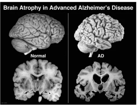
memory, thinking and behavior. Symptoms usually develop slowly and get worse over time, becoming severe enough to interfere with daily tasks.7
Figure 2. 3 – Brain atrophy in advanced AD in comparison with a normal brain. (http://alzheimerdisease.tv/wp-content/uploads/2014/11/alzheimers-disease-term.jpg)
Dementia affects 44 million people globally, and that figure is set to rise to 135 million by 2050, mostly due to the ageing of the population, and Alzheimer's is the most common form of dementia, a general term for memory loss and other intellectual abilities serious enough to interfere with daily life. This disease accounts for 60-70% of dementia cases, as mentioned before, and is not a normal part of aging, although the greatest known risk factor is increasing age, and the majority of people with Alzheimer's are 65 and older. But Alzheimer's is not just a disease of old age. Up to 5 percent of people with the disease have early onset Alzheimer's (also known as early-onset), which often appears when someone is in their 40s or 50s. In 2010 dementia resulted in about 486,000 deaths and there were between 21 and 35 million people worldwide with AD, being one of the most financially costly diseases in developed countries.8
6
Figure 2. 4 – Growth in dementia cases expectation by 2050.
(http://www.fic.nih.gov/News/GlobalHealthMatters/PublishingImages/fogarty-nih-infographic-global-growth-dementia-cases-by-region-by-2050.jpg)
In Alzheimer’s disease, the brain cells themselves degenerate and die, causing a steady decline in memory and mental function, and although the disease medications and management strategies may temporarily improve symptoms, helping people with AD to maximize function and maintain independence, there is no cure yet for this devastating disease, and caring for patients as their disease progresses represents an immense burden for family members, care givers, and health and social care systems.9
2.2.1 Symptoms, Characteristics
Alzheimer's dementia symptoms gradually worsen over a number of years, eventually affecting most areas of your brain, including those important in memory, thinking, judgement, language, problem-solving, personality and movement. In its early stages, memory loss is mild, but with late-stage Alzheimer's, individuals lose the ability to carry on a conversation and respond to their environment. Those with Alzheimer's live an average of eight years after their symptoms become noticeable to others, but survival can range from 4 to 20 years, depending on age and other health conditions.10
7
Figure 2. 5 – Brain sections mostly affected by AD.
(http://www.medicalook.com/diseases_images/alzheimers_disease.jpg)
Alzheimer's has no current cure, but treatments for symptoms are available and research continues. Although current Alzheimer's treatments cannot stop Alzheimer's from progressing, they can temporarily slow the worsening of dementia symptoms and improve quality of life for those with Alzheimer's and their caregivers. Today, there is a worldwide effort under way to find better ways to treat the disease, delay its onset, and prevent it from developing.11
The most common early symptom of Alzheimer's is difficulty remembering newly learned information because Alzheimer's changes typically begin in the part of the brain that affects learning. As Alzheimer's advances through the brain it leads to increasingly severe symptoms, including disorientation, mood and behavior changes; deepening confusion about events, time and place; unfounded suspicions about family, friends and professional caregivers; more serious memory loss and behavior changes; and difficulty speaking, swallowing and walking.12
People with memory loss or other possible signs of Alzheimer’s may find it hard to recognize they have a problem. Signs of dementia may be more obvious to family members or friends.13
As previously mentioned, brain changes associated with AD normally lead to growing trouble with:
8
Memory: memory loss associated to AD varies from normal day-to-day memory lapses because it persists and worsens, affecting people’s ability to function at work and at home. Events like repeating statements and questions over and over and forgetting conversations and people’s names, or routinely misplace possessions are normally sign of the presence of AD.
Disorientation and misinterpreting spatial relationships: getting lost in familiar places, may also be a sign of AD, as the disease disrupts the brain’s ability to interpret what people see, making it difficult to understand the surroundings, and also making them lose their sense of what day or even season it is.
Speaking and writing: another sign of AD is the declination of the ability to read, write or find the right words to identify objects or express thoughts.
Thinking and reasoning: Alzheimer’s disease also causes difficulty concentrating and thinking, eventually progressing to inability to recognize and deal with numbers, making it challenging, if not impossible, to manage finances or keep track of bills.
Making judgements and decisions: people with AD end up having trouble responding effectively to everyday problems, like unexpected driving situations or even food burning on the stove.
Planning and performing familiar tasks: losing the ability to complete routine activities or perform basic tasks such as dressing and bathing is also a known symptom of AD.
Changes in personality and behavior: last but not least, brain changes that occur in AD can also affect the way people with AD act or feel, from depression, anxiety or mood swings, to changes in sleeping habits, loss of inhibitions or even delusions.
The rate of progression for AD varies widely. On average, people with AD live 8 to 10 years after diagnosis, but some survive as long as 25 years. Pneumonia is a common cause of death because impaired swallowing allows food or beverages to enter the lungs, where an infection can begin. Other common causes of death include complications from urinary tract infections and falls.11
9
Information, skills and habits learned early in life are among the last abilities to be lost as the disease progresses, which is why one of the keys to maintaining a high quality of life, even when in a moderate phase of the disease, is to capitalize on those abilities and skills.14
2.2.2 Stages
There are several different ways to divide and qualify Alzheimer’s disease because these stages are rough guides based on averages and generalizations and the disease is a continuous process, meaning developed symptoms can vary from person to person.
Figure 2. 6 – Image showing the different effects of AD on the brain from the healthy phase to severe AD. (http://sierram.web.unc.edu/files/2011/04/Alzheimers-disease.jpg)
This project considers five different stages associated with AD: Preclinical Alzheimer’s Disease, Mild Cognitive Impairment, Mild Dementia, Moderate Dementia and Severe Dementia.15
10
2.2.2.1 Preclinical Alzheimer’s Disease
Alzheimer’s disease begins deep in the brain, in the entorhinal cortex, a brain region that is near the hippocampus and has direct connections to it. Healthy neurons in this region begin to work less efficiently, lose their ability to communicate, and ultimately die.16
AD’s first symptoms are often mistakenly attributed to ageing or stress, because the first symptoms are usually related to memory lapses, due to the fact that the process of brain atrophy gradually spreads to the hippocampus, the brain region that plays a major role in learning and is involved in converting short-term memories to long-term memories. Detailed neuropsychological testing can reveal mild cognitive difficulties up to eight years before a person fulfills the clinical criteria for diagnosis of AD, so scientists believe that these brain changes begin before any clinically detectable signs or symptoms of forgetfulness appear. These early symptoms can affect the most complex daily living activities.12
At some point, the damage occurring in the brain begins to show itself in very early clinical signs and symptoms. Much research is being done to identify these early changes, which may be useful in predicting dementia or AD. Subtle problems with the executive functions of attentiveness, planning, flexibility, and abstract thinking, or impairments in semantic memory (memory of meanings, and concept relationships) can also be symptomatic of the early stages of AD.13
2.2.2.2 Mild Cognitive Impairment
People with mild cognitive impairment (MCI) have mild changes in their memory and thinking ability and manifest deficits which are subtle. These changes aren't significant enough to affect work yet but might be noted by people who are closely associated with them. People with MCI may have memory lapses when it comes to information that is usually easily remembered, such as conversations, recent events or appointments, having also trouble making sound decisions and judging the amount of time needed for a task, or the steps needed to complete it.17
Not everyone with mild cognitive impairment has Alzheimer's disease and the same procedures used to identify preclinical Alzheimer's disease can help determine whether MCI is due to Alzheimer's disease or something else.18
11
2.2.2.3 Mild Dementia
Alzheimer's disease diagnosis can be made with considerable accuracy in this mild dementia stage, when it becomes clear to family and doctors that a person is having significant trouble with memory and thinking.
As AD spreads through the brain, the number of plaques and tangles grows, shrinkage progresses, and more and more of the cerebral cortex is affected. Memory loss continues and changes in other cognitive abilities begin to emerge. In a small percentage, difficulties with language, executive functions, perception (agnosia), or execution of movements (apraxia) are more prominent than memory problems.19
Language problems are mainly characterized by a shrinking vocabulary and decreased word fluency, leading to a general impoverishment of oral and written language and the dominant mood at this stage is apathy, as the subjects often seem less responsive than previously. As the disease progresses, the patients can often continue to perform many tasks independently, but may need assistance or supervision with the most cognitively demanding activities.20
In the absence of complicating medical pathology, the diagnosis of AD can be made with considerable certainty from the beginning of this stage, Studies indicate that the duration of this stage of mild dementia is a mean of approximately 2 years.21
2.2.2.4 Moderate Dementia
Moderate Dementia is typically the longest stage and can last for many years, during which people grow more confused and forgetful, due to the progressive deterioration, and begin to need help with daily activities and self-care. By this stage, AD damage has spread to the areas of the cerebral cortex that control language, reasoning, sensory processing and conscious thought.
Speech difficulties become evident due to an inability to recall vocabulary, which leads to frequent incorrect word substitutions (paraphasias). Reading and writing skills are also progressively lost. Complex motor sequences become less coordinated as time passes and AD progresses, so the risk of falling increases. During this phase, memory problems worsen, and the person may fail to recognize close relatives. Long-term memory, which was previously intact, becomes impaired. Basically, at this stage, deficits are of sufficient magnitude so patients can no longer manage on their own in the community.21–23
12
Behavioral and neuropsychiatric changes become more prevalent and manifestations of wandering and irritability, leading to crying, become common. Subjects also lose insight of their disease process and limitations (anosognosia) and urinary incontinence can develop. These symptoms create stress for relatives and care givers, which can be reduced by moving the person from home care to other long-term care facilities.11
2.2.2.5 Severe Dementia
With Severe Dementia, mental function continues to decline and the disease has a growing impact on movement and physical capabilities of the patients, as the plaques and tangles are widespread throughout the brain and most areas of the brain have shrunk further. At this stage of dementia, people cannot recognize family and loved ones and they are completely dependent upon caregivers. Language is reduced to simple phrases or even single words, eventually leading to complete loss of speech. Although aggressiveness can still be present, extreme apathy and exhaustion are much more common symptoms.10,24
Confused about past and present and generally incapacitated, the patients will not be able to perform even the simplest tasks independently, leading to muscle mass and mobility deteriorate to the point where they are bedridden and unable to feed themselves. The cause of death is usually an external factor, such as infection of pressure ulcers or pneumonia, not the disease itself.7
2.2.3 Causes
Although the causes of Alzheimer’s disease are not yet fully understood, like all types of dementia, Alzheimer's is caused by brain cell death. It is a neurodegenerative disease, which means there is progressive brain cell death that happens over a course of time.
Scientists believe that for most people, Alzheimer's disease results from a combination of genetic, lifestyle and environmental factors that affect the brain over time.25
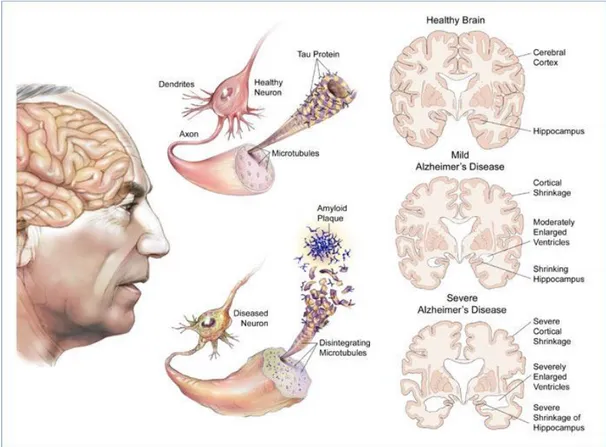
While they cannot be seen or tested in the living brain affected by Alzheimer's disease, postmortem/autopsy will always show tiny inclusions in the nerve tissue, called plaques and tangles:
13
Plaques: plaques are abnormal clusters of protein fragments, build up between nerve cells. These clumps of a protein called beta-amyloid may damage and destroy brain cells in several ways, including interfering with cell-to-cell communication. Although the ultimate cause of brain-cell death in Alzheimer's isn't known, the collection of beta-amyloid on the outside of brain cells is a prime suspect.10,26
Figure 2. 7 – Illustration of a group of plaques. (https://www.alz.org/braintour/plaques.asp)
Tangles: dead and dying nerve cells contain tangles, which are made up of twisted strands of another protein, and they destroy a vital cell transport system made of proteins. In healthy areas, the transport system is organized in orderly parallel strands, helped by a protein called tau, but in areas where tangles are forming, tau collapses into twisted strands (tangles) so the tracks can no longer stay straight, falling apart and disintegrating, thus the nutrients can no longer move through the cells, that eventually die.27
14
Figure 2. 8 – Microscope image of the formation of tangles in brain cells. (https://www.alz.org/braintour/tangles.asp)
The cause for most Alzheimer's cases is still mostly unknown except for less than 5% of cases where genetic differences have been identified. Several competing hypotheses exist trying to explain the cause of the disease, the most important being the Genetic, the Cholinergic, the Amyloid and the Tau Hypotheses.
2.2.3.1 Genetic Hypothesis
The more the scientists learn about this devastating disease, the more they realize that genes play an important role in its development. Research conducted and funded by the National Institute on Aging (NIA) at the National Institutes of Health (NIH) and others is advancing the field of AD genetics.
Some diseases are caused by a genetic mutation, or permanent change in one or more specific genes. If a person inherits from a parent a genetic mutation that causes a certain disease, then he or she will usually get the disease. In other diseases, a genetic variant may occur. This change in a gene can sometimes cause a disease directly. More often, it acts to increase or decrease a person's risk of developing a disease or condition. When a genetic variant increases disease risk but does not directly cause a disease, it is called a genetic risk factor.28–30
There are two types of Alzheimer's: early-onset and late-onset. Both types have a genetic component.
15
Early-onset Alzheimer's disease occurs in people age 30 to 60. It is rare, representing less than 5 percent of all people who have Alzheimer's. Some cases of early-onset Alzheimer's have no known cause, but most cases are inherited, a type known as familial Alzheimer's disease (FAD), caused by any one of a number of different single-gene mutations on chromosomes 21, 14, and 1. Each of these mutations causes abnormal proteins to be formed. Mutations on chromosome 21 cause the formation of abnormal amyloid precursor protein (APP). A mutation on chromosome 14 causes abnormal presenilin-1 (PS-1) to be made, and a mutation on chromosome 1 leads to abnormal presenilin-2 (PS-2).25
Figure 2. 9 – Molecular mechanism involved in the genetic hypothesis of AD.31
Scientists know that each of these mutations plays a role in the breakdown of APP, a protein whose precise function is not yet known. This breakdown is part of a process that generates harmful forms of amyloid plaques, a hallmark of the disease. A child whose mother or father carries a genetic mutation for FAD has a 50/50 chance of inheriting that mutation. If the mutation is in fact inherited, the child almost surely will develop FAD.32
On the other hand, most cases of Alzheimer's are the late-onset form, which develops after age 60. The causes of late-onset Alzheimer's are not yet completely understood, but they likely include a combination of genetic, environmental, and lifestyle factors that influence a person's risk for developing the disease.33
The single-gene mutations directly responsible for early-onset Alzheimer's disease do not seem to be involved in late-onset Alzheimer's. Researchers have not
16
found a specific gene that causes the late-onset form of the disease. However, one genetic risk factor does appear to increase a person's risk of developing the disease. This increased risk is related to the apolipoprotein E (APOE) gene found on chromosome 19. APOE contains the instructions for making a protein that helps carry cholesterol and other types of fat in the bloodstream. APOE comes in several different forms, or alleles. There’s APOE ε2, which is relatively rare and may provide some protection against the disease; APOE ε3, which is the most common allele, and is believed to play a neutral role in the disease (neither decreasing nor increasing risk); and APOE ε4, which is present in about 25 to 30 percent of the population and in about 40 percent of all people with late-onset Alzheimer's. People who develop Alzheimer's are more likely to have an APOE ε4 allele than people who do not develop the disease.34,35
Dozens of studies have confirmed that the APOE ε4 allele increases the risk of developing Alzheimer's, but how that happens is not yet understood. These studies also help explain some of the variation in the age at which AD develops, as people who inherit one or two APOE ε4 alleles tend to develop the disease at an earlier age than those who do not have any APOE ε4 alleles. So APOE ε4 is called a risk-factor gene because it increases a person's risk of developing the disease. However, inheriting an APOE ε4 allele does not mean that a person will definitely develop Alzheimer's.8,30
Using a relatively new approach called genome-wide association study (GWAS), researchers have identified a number of genes in addition to APOE ε4 that may increase a person's risk for late-onset Alzheimer's, including BIN1 (bridging integrator 1), CLU (clusterin), PICALM (phosphatidylinositol binding clathrin assembly protein), and CR1 (complement receptor 1). Finding genetic risk factors like these helps scientists better understand how AD develops and identify possible treatments to study.8,28
Although a blood test can identify which APOE alleles a person has, it cannot predict who will or will not develop AD. It is unlikely that genetic testing will ever be able to predict the disease with 100 percent accuracy because too many other factors may influence its development and progression.
17
2.2.3.2 Cholinergic Hypothesis
The cholinergic hypothesis, the oldest hypothesis, was proposed when the researchers began to focus on neurotransmitter imbalances as a cause of AD. Hypothesis on which most currently available drug therapies are based, but it has not maintained widespread support, largely because medications intended to treat acetylcholine (ACh) deficiency have not been very effective.36
This hypothesis proposes that a dysfunction of acetylcholine-containing neurons (involved in both memory and learning) in the brain contributes substantially to the cognitive decline observed in those with advanced age and AD.
A variety of studies in humans indicate that basal forebrain and rostral forebrain cholinergic pathways including converging projections to the thalamus serve important functional roles in conscious awareness, attention, working memory, and a number of additional mnemonic processes. Furthermore, studies of the brains of advanced AD patients frequently showed damage or abnormalities in these pathways, which correlates with the level of cognitive decline.
A prediction of the cholinergic hypothesis is that drugs that potentiate central cholinergic function should improve cognition and perhaps even some of the behavioral problems experienced with Alzheimer’s disease. There are a number of approaches to the treatment of the cholinergic deficit in Alzheimer’s disease, most of which have initially focused on the replacement of ACh precursors (choline or lecithin) but these agents failed to increase central cholinergic activity. Other studies have investigated the use of cholinesterase (ChE) inhibitors that reduce the hydrolysis of ACh.2,36–39
18
Figure 2. 10 – Schematic diagram of a neuron representing (A) alternations in neurotransmission in AD and (B) the hypothetical mode of action of AChE inhibitors. (http://jnnp.bmj.com/content/66/2/137/F1.large.jpg)
As shown on the Figure 2.10 (A), about the proposed neurochemical changes in AD, it is hypothesized that these changes give rise to the clinical symptoms of AD and contribute to the spread of pathology. Explaining the processes demonstrated:
1. Reduced cortical cholinergic innervation;
2. Reduced corticocortical glutamatergic neurotransmission due to neuron or synapse loss;
19
4. Shift of tau to the hyperphosphoryalted state—precursor of neurofibrillary tangles;
5. Reduced secretion of soluble APP;
6. Increased production of β-amyloid protein; 7. Decreased glutamate production.
On the figure 2.10 (B), there is a display of the rectification processes of neurotransmission with cholinesterase inhibitors:
1. Acetylcholinesterase (AChE) inhibitors reduce the breakdown of endogenously released acetylcholine (ACh), resulting in greater activation of postsynaptic ACh receptors;
2. Reduced phosphorylation of tau;
3. Secretion of sAPP returned towards normal; 4. Reduced β-amyloid production;
5. Glutamatergic neurotransmission returns towards normal, possibly due to activation of muscarinic and nicotinic receptors.
While some of the assumptions at the basis of its original formulation are disputable in the light of recent developments, the cholinergic hypothesis has, however, constituted an invaluable stimulus to better understand not only the anatomy and the biochemistry of the cholinergic systems of brain connections but also its developmental biology, its complex relationships with trophic factors and its role in cognitive functions.36
2.2.3.3 Amyloid Hypothesis
Amyloid precursor protein (APP) is a protein found widely throughout the body. The amyloid hypothesis is that a fault with the processing of APP in the brain leads to the production of a short fragment of APP known as beta-amyloid. The hypothesis thus postulates that extracellular amyloid beta-peptide (Aβ) deposits are the fundamental cause of the disease, causing the neurodegeneration in AD by forming accumulated clumps, called plaques, in brain tissue, leading to damage to neurons which in turn triggers inflammatory responses as the brain attempts to repair itself. It is also thought to cause the formation of tangles, made up of a protein
20
called tau, that also contribute to the damage to brain cells which causes the symptoms of dementia, but that is thought to be a fault with the over production of Aβ or with the mechanism that usually clears it from the brain, or both.40
Support for this postulate comes from the location of the gene for the amyloid precursor protein (APP) on chromosome 21, together with the fact that people with trisomy 21 (Down Syndrome) who have an extra gene copy almost universally exhibit AD by 40 years of age. Also, a specific isoform of apolipoprotein, APOE4, is a major genetic risk factor for AD.41,42
Figure 2. 11 – The amyloid hypothesis. (http://www.nature.com/nrd/journal/v10/n9/fig_tab/nrd3505_F1.html)
Later on, this theory was updated, suggesting that a close relative of the beta-amyloid protein, and not necessarily the beta-beta-amyloid itself, may be a major culprit in the disease. The theory holds that an amyloid-related mechanism that prunes neuronal connections in the brain in the fast-growth phase of early life may be triggered by ageing-related processes in later life to cause the neuronal withering of Alzheimer's disease, with Aβ playing a complementary role, by depressing synaptic function.43,44
21
2.2.3.4 Tau Hypothesis
The tau hypothesis proposes that tau protein abnormalities initiate the disease cascade. According to this hypothesis, excessive or abnormal phosphorylation of tau results in the transformation of normal adult tau into PHF-tau (paired helical filament) and intracellular neurofibrillary tangles (NFT). When this occurs, the microtubules disintegrate, destroying the structure of the cell's cytoskeleton which collapses the neuron's transport system. This may result first in malfunctions in biochemical communication between neurons and later in the death of the cells.27
Mutations that alter function and isoform expression of tau lead to hyperphosphorylation. The process of tau aggregation in the absence of mutations is not known but might result from increased phosphorylation, protease action or exposure to polyanions, such as glycosaminoglycans.27,40
Figure 2. 12 – Representation of the microtubule disintegration on tau hypothesis. (http://www.rsc.org/images/b906101k-350-FOR-TRIDION_tcm18-156498.jpg)
Tau hypothesis also doesn’t explain everything, however. The formation of tangles is often observed in the brain of people as young as thirty years of age.
22
Apparently, there are some processes and mechanisms that can eliminate the tangles or prevent them from affecting the brain functions.40
2.2.4 Pathophysiology
2.2.4.1 Neuropathology
Neuropathology is the branch of medicine concerned with diseases of the nervous system tissue, and is a subspecialty of anatomic, neurology and neurosurgery.
As to Alzheimer’s disease, microscopic changes in the brain begin long before the first signs of memory loss.
People with AD have a shortage of some important chemicals in their brain. These chemical messengers help to transmit signals around the brain. When there is a shortage of them, the signals are not transmitted as effectively.
The brain has 100 billion nerve cells (neurons). Each nerve cell connects with many others to form communication networks. Groups of nerve cells have special jobs. Some are involved in thinking, learning and remembering. Others help us see, hear and smell. To do their work, brain cells operate like tiny factories. They receive supplies, generate energy, construct equipment and get rid of waste. Cells also process and store information and communicate with other cells. Keeping everything running requires coordination as well as large amounts of fuel and oxygen.7,46
Scientists believe AD prevents parts of a cell's factory from running well. They are not sure where the trouble starts. But just like a real factory, backups and breakdowns in one system cause problems in other areas. As damage spreads, cells lose their ability to do their jobs and, eventually die, causing irreversible changes in the brain.
The neuropathological hallmarks of Alzheimer disease include “positive” lesions such as abundant amyloid plaques and neurofibrillary tangles, neuropil threads, and dystrophic neurites containing hyperphosphorylated tau, that are accompanied by astrogliosis and microglial cell activation, and “negative” lesions such as neuronal and synaptic loss in the cerebral cortex and certain subcortical regions. This loss results in gross atrophy of the affected regions, including
23
degeneration in the temporal lobe and parietal lobe, and parts of the frontal cortex and cingulate gyrus.7,16
Degeneration is also present in brainstem nuclei like the locus coeruleus. Studies using Magnetic Resonance Imaging (MRI) and Positron Emission Tomography (PET) have documented reductions in the size of specific brain regions in people with AD as they progressed from mild cognitive impairment to AD, and in comparison with similar images from healthy older adults.7,24
Scientists do not know exactly what role plaques and tangles play in AD. Most experts believe they somehow play a critical role in blocking communication among nerve cells and disrupting processes that cells need to survive.
Although all these neuropathological characteristics are useful diagnostic markers, the cognitive impairment in patients with AD is closely associated with the progressive degeneration of the limbic system, neocortical regions, and the basal forebrain. This neurodegenerative process is characterized by early damage to the synapses with retrograde degeneration of the axons and eventual atrophy of the dendritic tree and perikaryon. Indeed, the loss of synapses in the neocortex and limbic system is the best correlate of the cognitive impairment in patients with AD.7,16
In summary, the concept of neurodegeneration in AD has been expanded from the idea of general neuronal loss and astrogliosis to include earlier alterations such as synaptic and dendritic injury and disturbances in the process of adult neurogenesis, circuitry dysfunction, and aberrant innervation. All of these factors are important to understand the neuropathology behind AD.
2.2.4.2 Biochemistry
Biochemistry, intrinsically, is the chemistry of living things. It deals with the chemical compounds and processes occurring in organisms and studies the chemical characteristics and reactions of a particular living organism or biological substance. In regard to Alzheimer's disease, its biochemistry is yet not well understood, but AD has already been identified as a protein misfolding disease (proteopathy), caused by plaque accumulation of abnormally folded amyloid beta protein, and tau protein in the brain.47,48
24
AD is also considered a tauopathy due to abnormal aggregation of the tau protein. Every neuron has a cytoskeleton, an internal support structure partly made up of structures called microtubules which act like tracks, guiding nutrients and molecules from the body of the cell to the ends of the axon and back. These microtubules are stabilized by tau when phosphorylated, and is therefore called a microtubule-associated protein. In AD, tau undergoes chemical changes, becoming hyper phosphorylated, beginning to pair with other threads, creating neurofibrillary tangles and disintegrating the neuron's transport system.47–49
2.2.5 Diagnosis
There are several different methods and tools used to help determine whether a person who is having memory problems has or not AD. Doctors are usually able to diagnose AD based on the person's medical history, history from relatives, and behavioral observations, as well as by carrying out standard medical tests or performing brain scans like Computed Tomography (CT), MRI or PET, or even by conducting tests of memory, problem solving, attention, counting and language.
The presence of characteristic neurological and neuropsychological features and the absence of alternative conditions is supportive. Advanced medical imaging with CT or MRI, and with Single-Photon Emission Computed Tomography (SPECT) or PET can be used to help exclude other cerebral pathology or subtypes of dementia. Moreover, it may predict conversion from prodromal stages (like MCI) to AD.
Medical organizations have created diagnostic criteria to ease and standardize the diagnostic process for practicing physicians. The diagnosis can be confirmed with very high accuracy post-mortem when brain material is available and can be examined histologically.
2.2.5.1 Criteria
The National Institute of Neurological and Communicative Disorders and Stroke (NINCDS) and the Alzheimer's Disease and Related Disorders Association (ADRDA), now known as the Alzheimer's Association, established the most commonly used NINCDS-ADRDA Alzheimer's Criteria for diagnosis in 1984, extensively updated in 2007. These criteria require that the presence of cognitive impairment, and a suspected dementia syndrome, be confirmed by neuropsychological testing for a clinical diagnosis of possible or probable AD, and a