Analgesic action of laser therapy (LLLT) in an animal model
Daniel Humberto Pozza 1, Patrícia Wehmeyer Fregapani 2, João Batista Blessmann Weber 3, Marília Gerhardt de Oliveira 4,
Marcos André Matos de Oliveira 5, Nelson Ribeiro Neto 6, João Batista de Macedo Sobrinho 7
(1) Laser Technology, Universidade Federal da Paraíba (UFPB) and Universidade Federal da Bahia (UFBA), Post Doctorate Intern, Histology and Embryology Institute, School of Medicine, Universidade do Porto, Portugal
(2) Universidade Luterana do Brasil (Ulbra), Canoas, Brazil
(3) Pediatric Dentistry, School of Dentistry of Bauru, Universidade de São Paulo (USP); PhD, Dentistry, Pontifícia Universidade Católica do Rio Grande do Sul (PUCRS), Porto Alegre, Brazil. Adjunct Professor, School of Dentistry, PUCRS, Porto Alegre, Brazil
(4) Maxillofacial Trauma and Surgery, PUCRS, Porto Alegre, Brazil. PhD, Dentistry, PUCRS, Porto Alegre, Brazil. Researcher, Brazilian Council for Scientific and Technological Development (CNPq). Full Professor, PUCRS, Porto Alegre, Brazil
(5) Maxillofacial Trauma and Surgery, Universidade Federal de Pernambuco (UFPE). PhD, Immunology, UFBA. Professor, Co-ordinator, Specialization Course in Implantology, Associação Educativa do Brasil (SOEBRAS), Faculdade Unidas do Norte de Minas (FUNORTE); Professor, Anatomy, Universidade Metropolitana de Educação e Cultura (UNIME); Head of the Service of Maxillofacial Trauma and Surgery, Hospital Agenor Paiva
(6) Specialists, Maxillofacial Trauma and Surgery; Specialist, Implantology; PhD, Laser Technology, UFPB and UFBA. Professor, Specialization Course in Implantology, Soebras, Funorte
(7) Specialist, Master, Maxillofacial Trauma and Surgery, Université Pierre et Marie Curie VI, Paris, France. Specialist, Temporoman-dibular Dysfunction and Orofacial Pain, Federal Dentistry Board. PhD, Laser Technology, UFPB and UFBA. Coordinator, Specia-lization Course in Maxillofacial Trauma and Surgery, Temporomandibular Dysfunction and Orofacial Pain, Soebras, Funorte
Correspondence:
Dr. Marília Gerhardt de Oliveira, PhD Av. Cel. Lucas de Oliveira, 1841, conj. 203 90460.001 - Porto Alegre, RS, Brazil E-mail: [email protected] Received: 19/12/2007
Accepted: 02/04/2008
Pozza DH, Fregapani PW, Weber JBB, Oliveira MG, Oliveira MAM, Ribeiro-Neto N, Macedo-Sobrinho JB. Analgesic action of laser the-rapy (LLLT) in an animal model. Med Oral Patol Oral Cir Bucal. 2008 Oct1;13(10):E648-52.
© Medicina Oral S. L. C.I.F. B 96689336 - ISSN 1698-6946
http://www.medicinaoral.com/medoralfree01/v13i10/medoralv13i10p648.pdf
Abstract
Objectives: To evaluate the analgesic effect of laser therapy on healthy tissue of mice. Study design: Forty-five animals were divided in three groups of 15: A – infrared laser irradiation (830 nm, Kondortech®, São Carlos, SP, Brazil); B – red laser irradiation (660 nm, Kondortech®, São Carlos, SP, Brazil); C – sham irradiation with laser unit off. After laser application, the mice remained immobilized for the injection of 30 µl of 2% formalin in the plantar pad of the irradiated hind paw. The time that the mouse kept the hind paw lifted was measured at 5 min intervals for 30 minutes. Results: Results showed statistically significant differences comparing the control group with the infrared laser group at 5, 20, 25 and 30 accumulated minutes, and with the red laser group at all time points. The analysis of partial times, at each 5 minutes, showed statistically significant differences between the control and the laser groups up to 20 minutes. Conclusions: Laser therapy had an analgesic effect and red laser had the best results.
Key words: Low-level laser therapy, lasers, analgesia, pain measurement, mice.
Indexed in:
-Index Medicus / MEDLINE / PubMed -EMBASE, Excerpta Medica -SCOPUS
-Indice Médico Español -IBECS
Med Oral Patol Oral Cir Bucal. 2008 Oct1;13(10):E648-52. LLLT in an animal model Med Oral Patol Oral Cir Bucal. 2008 Oct1;13(10):E648-52. LLLT in an animal model Med Oral Patol Oral Cir Bucal. 2008 Oct1;13(10):E648-52. LLLT in an animal model Med Oral Patol Oral Cir Bucal. 2008 Oct1;13(10):E648-52. LLLT in an animal model
Introduction
Several studies (1-5) have found that laser is an effective short-term method to relieve chronic muscle pain be-cause of the anti-inflammatory effect of the reduction of prostaglandin synthesis, inhibition of prostacyclin, and increase in blood flow, peripheral nerve action and analgesic effects. Chow et al. (6) evaluated 90 patients and found that laser treatment was effective and painless, had a low incidence of adverse effects and was relatively easy to be administered.
The increase of blood flow after laser therapy is an im-portant factor in pain relief because it increases oxyge-nation, lymphatic drainage, the activity of neutrophils, macrophages, fibroblasts, and the metabolism of damaged or defective cells (7-10).
Wavelength and dose are important factors in laser the-rapy, and their variation may inhibit or stimulate certain effects, such as ATP synthesis (11,12).
Ebert and Roberts (13) found that HeNe laser used at 7 J/ cm2 increased sciatic nerve action potential in frogs and reduced nerve conduction, particularly when infrared wavelengths were used.
The injection of formalin solution in the hind paw of rats or mice produces two phases of nociceptive behavior. The first phase, in the first 5 minutes, results from direct chemical activation of nociceptive peripheral afferent fibers, which promotes the release of pro-inflammatory peptides, such as bradykinin and substance P, and triggers the noxious stimulus. The second phase, between 15 and 30 minutes, results from the release of inflammation me-diators, such as histamine, serotonin, prostaglandin and bradykinin, or the hypersensitization of the spinal cord in the first phase, although such mechanisms have not been fully explained yet (14-18).
The purpose of this study was to evaluate the analgesic action of red (660 nm) and infrared (830 nm) laser thera-py applied before the injection of 2% formalin in healthy tissue of the plantar pad of the hind paw of mice.
Material and Methods
This study was conducted in accordance with the ethical principles for the care and use of animals in research, as well as the norms for animal vivisection for teaching and research purposes. The study project was evaluated and approved by the Committee on Science and Ethics of the School of Dentistry of Pontifícia Universidade Católica do Rio Grande do Sul, Porto Alegre, Brazil.
Forty-five male Swiss mice weighing from 37 to 41 g and clinically healthy were used in this study. The animals were fed a solid diet and ad libitum water, which were discontinued on the experimental day, when 15 animals were randomly selected for each of the 3 groups.
In group A, and infrared laser diode (Kondortech®,
plantar pad of the animals by contact. Irradiation dose was 10 J/cm2 in a total of 334 s.
In group B, a red laser diode (Kondortech®, São Carlos, SP, Brazil; 660 nm, 30 mW) operating in continuous wave was applied to irradiation points on the plantar pad by contact. Irradiation dose was 10 J/cm2 in a total of 334 s.
The diameter of the laser beam was 3 mm and it was applied to a point in the center of the plantar pad of mice.
In group C, the control group, the laser pointer was placed on the same point as in the other two groups, that is, on the plantar pad, and kept there for the same amount of time, but the unit was kept off.
In all groups, animals were kept immobile by head restraint during laser application.
Immediately after laser therapy, 30 µl of sterile 2% for-malin was injected in the subcutaneous tissue of the same plantar pad with a 0.5 ml insulin syringe and a 0.33 mm needle. After the removal of the needle, the animals were placed in individual 20 cm2 boxes, 40 cm high, with a mirror on the floor and clear glass panes on the sides to facilitate visualization of the animal paws.
One observer and one measurement evaluator, previously calibrated by simulation of the procedures, measured the time that each mouse kept the right hind paw lifted. Two stopwatches were used for the measurements: the first was started each time the animal lifted the hind paw and sto-pped when the hind paw reached full contact with the mi-rrored floor of the box, and the moments when the mouse kept only the lateral portion of the paw in touch with the floor were also classified as lifted hind paw; the second stopwatch worked for 30 non-interrupted minutes, and at each 5 minutes the person controlling this stopwatch told the observer the time so that accumulated partial values were recorded. The accumulated partial times in which the animal kept the hind paw lifted were obtained in the first 5 minutes, as well as at the accumulated 10, 15, 20, 25 and 30 subsequent minutes. Observers were calibrated by simula-tion of the procedures. Two mice were injected to test for how long they lifted their hind paw. After the procedures, both mice were killed by barbiturate overdose.
Results
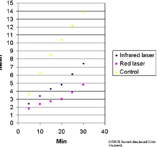
The results of the Kruskal-Wallis test revealed a statis-tically significant difference between groups. The results of each animal were used to calculate means according to the times evaluated (Figure 1), and then analyzed two by two to check differences.
Results of the post-hoc Dunn test revealed statistically significant differences when group A was compared with group C at the first 5 minutes and at the accumulated 20, 25 and 30 minutes, and when group B was compared with group C at all time points evaluated.
Group Description Time Mean Standard Deviation
A Infrared Laser 10min-5min 0.96 0.68
B Red Laser 10min-5min 0.59 0.41
C Control 10min-5min 2.66 1.88
A Infrared Laser 15min-10min 0.88 0.62
B Red Laser 15min-10min 0.35 0.25
C Control 15min-10min 2.24 1.58
A Infrared Laser 20min-15min 0.56 0.40
B Red Laser 20min-15min 0.26 0.18
C Control 20min-15min 1.89 1.34
A Infrared Laser 25min-20min 1.30 0.92
B Red Laser 25min-20min 0.86 0.61
C Control 25min-20min 1.76 1.24
A Infrared Laser 30min-25min 1.33 0.94
B Red Laser 30min-25min 0.95 0.67
C Control 30min-25min 1.66 1.18
Table 1. Descriptive analysis of mean differences of accumulated times in minutes.
SOURCE: Research data, Instat® 3.0 for Windows®
Fig. 1. Descriptive analysis of mean time in minutes that each mouse kept the right hind paw lifted according to time points evaluated.
Med Oral Patol Oral Cir Bucal. 2008 Oct1;13(10):E648-52. LLLT in an animal model Med Oral Patol Oral Cir Bucal. 2008 Oct1;13(10):E648-52. LLLT in an animal model Med Oral Patol Oral Cir Bucal. 2008 Oct1;13(10):E648-52. LLLT in an animal model Med Oral Patol Oral Cir Bucal. 2008 Oct1;13(10):E648-52. LLLT in an animal model
was obtained for each 5 minutes after the initial phase. The results of the Kruskal-Wallis test revealed statistica-lly significant differences when the following values were subtracted: 10 minutes from 5 minutes; 15 from 10; and 20 from 15. The results of each animal were used to calculate means according to the time point evaluated (Table 1), and then paired to check differences.
Results of the post-hoc Dunn test revealed statistically significant differences when group A was compared with group C at the periods between 15 and 10 minutes and between 20 and 15 minutes, and when group B was compared with group C at the periods between 10 and 5 minutes, between 15 and 10 minutes, and between 20 and 15 minutes.
Laser irradiation of the plantar pad of mice before injection of 2% formalin had an analgesic effect when compared with mock irradiation. The best results were obtained with the use of red laser (660 nm).
Discussion
The formalin used in this study was a 2% solution because, according to Rosland et al. (19), Abbott et al. (20), and Saddi and Abbott (21), solutions at a concentration lower than 2% may not be efficient in inducing pain responses in mouse tissue.
The laser dose used was 10 J/cm2 (11,12). Moore et al. (12) found positive results with this dose for cell growth when using a red light wavelength, and for fibroblast inhibition when using an infrared wavelength. In this study, we ob-tained better results with red laser. Ebert and Roberts (13) used 7 J/cm2 and found good results only with infrared laser. However, they performed irradiation directly on the nervous tissue, with no interference of the cell substrates found in in vivo studies, such as in our study. Other tissue factors should also be taken into consideration, such as the thickness of animal tissue, the degree of optical interferen-ce of endogenous pigments and the constant blood flow. When not accurately applied, high laser doses may cause damage to tissues, as reported by Hawkins and Abrahamse (11). Conversely, low doses may not cause biological effects on irradiated tissues, as reported by Núñez et al. (10) in a study that evaluated the effects of HeNe laser on the microcirculation of rats using only 1 J/cm2.
Laser therapy promotes an increase in the synthesis and release of endorphins, as well as a decrease in the release of nociceptive receptors, such as bradykinin and sero-tonin (1,22). However, these mechanisms were probably not implicated in pain reduction, the objective of this study, because of the single application and the short evaluation time (30 minutes). Another fact that supports this hypothesis is that irradiated animals showed greater analgesia in the initial phase of pain evaluation, between 5 and 20 minutes.
mice. Such possibility is supported by studies (23,24) that found a stabilization of the cell membranes that regulate the transmission of the nervous impulse. Such regulation inhibits depolarization by an increase in ATP synthesis, which promotes a significant increase in nerve latency. As sensory nerve conduction velocity is reduced, pain relief is observed (7,13,25-27).
Another fundamental factor in pain relief in irradiated animals, when compared with the control group, was the vascular effects of therapeutic laser, particularly that of red laser. The increase of blood flow increases oxygenation, lymphatic drainage, activity of neutrophils, macrophages and fibroblasts, and the metabolism of defective or dama-ged cells, and is responsible for pain relief after the first minutes of tissue irradiation (3-5,7,8).
Red laser produces a photochemical effect that is faster because it acts directly on mitochondria. Our results of the red laser group were better than those found for the control group. Statistically significant differences were found as early as the first 5 minutes and extended to the subsequent accumulated periods of time. Infrared laser produces a photophysical or photoelectrical effect and is slower because it acts indirectly on mitochondria, changes its membrane potential and promotes its stimulation. In our infrared group, we found statistically significant di-fferences in the first 5 minutes, no significant didi-fferences at the accumulated 10 and 15 minutes, and significant differences again at the final 20, 25 and 30 minutes. Such findings indicate better results for the infrared wavelength laser at the later time points.
The individual analysis of animal behavior at each 5 mi-nutes between the accumulated times showed statistically significant differences for red laser at the initial time points up to 20 minutes. For infrared laser, there was, again, an interruption between 10 and 5 minutes, and also statisti-cally significant differences at the subsequent time points, which, similarly to results of the red laser group, was not observed at 20 minutes.
The observation of animal behavior showed a new pain peak at about 20 minutes after the injection of formalin. Formalin has a second pain phase between 12 and 40 minutes after injection according to Saddi and Abbott (21), between 15 and 20 minutes according to Shibata et al. (14), between 20 and 30 minutes according to Jett and Michelson (15), Abbadie et al. (16) and Taylor et al. (17). In our study, the second pain peak was observed at 20 minutes for the animals in the laser groups. In the control group, the mice also started to lift the right hind paw, which indicated pain, at 20 minutes, but they kept the study hind paw up for a shorter time. This may have been caused by the greater pain of these animals in the initial phases, which made them less susceptible to pain at the final time points of the evaluation. Another possibility
Further studies applied to dental clinical conditions are warranted to evaluate the results of this method, as laser seems to be useful before infiltrative anesthesia, administration of medication, biopsies, or procedures that require the use of needles. Topical anesthesia and several maneuvers are used to mitigate the sensation of discomfort experienced by the patient. Therapeutic laser may reduce such discomfort due to its effect on healthy tissue, as found in this study.
References
1. Gur A, Karakoc M, Cevik R, Nas K, Sarac AJ, Karakoc M. Efficacy of low power laser therapy and exercise on pain and functions in chronic low back pain. Lasers Surg Med. 2003;32(3):233-8.
2. Gur A, Cosut A, Sarac AJ, Cevik R, Nas K, Uyar A. Efficacy of different therapy regimes of low-power laser in painful osteoarthritis of the knee: a double-blind and randomized-controlled trial. Lasers Surg Med. 2003;33(5):330-8.
3. Hakgüder A, Birtane M, Gürcan S, Kokino S, Turan FN. Efficacy of low level laser therapy in myofascial pain syndrome: an algometric and thermographic evaluation. Lasers Surg Med. 2003;33(5):339-43. 4. Gur A, Sarac AJ, Cevik R, Altindag O, Sarac S. Efficacy of 904 nm gallium arsenide low level laser therapy in the management of chronic myofascial pain in the neck: a double-blind and randomize-controlled trial. Lasers Surg Med. 2004;35(3):229-35.
5. Ilbuldu E, Cakmak A, Disci R, Aydin R. Comparison of laser, dry needling, and placebo laser treatments in myofascial pain syndrome. Photomed Laser Surg. 2004 Aug;22(4):306-11.
6. Chow RT, Heller GZ, Barnsley L. The effect of 300 mW, 830 nm laser on chronic neck pain: a double-blind, randomized, placebo-controlled study. Pain. 2006 Sep;124(1-2):201-10.
7. Maegawa Y, Itoh T, Hosokawa T, Yaegashi K, Nishi M. Effects of near-infrared low-level laser irradiation on microcirculation. Lasers Surg Med. 2000;27(5):427-37.
8. Schaffer M, Bonel H, Sroka R, Schaffer PM, Busch M, Reiser M, et al. Effects of 780 nm diode laser irradiation on blood microcirculation: preliminary findings on time-dependent T1-weighted contrast-enhanced magnetic resonance imaging (MRI). J Photochem Photobiol B. 2000 Jan;54(1):55-60.
9. Schindl A, Heinze G, Schindl M, Pernerstorfer-Schön H, Schindl L. Systemic effects of low-intensity laser irradiation on skin microcircu-lation in patients with diabetic microangiopathy. Microvasc Res. 2002 Sep;64(2):240-6.
10. Núñez SC, Nogueira GE, Ribeiro MS, Garcez AS, Lage-Marques JL. He-Ne laser effects on blood microcirculation during wound healing: a method of in vivo study through laser Doppler flowmetry. Lasers Surg Med. 2004;35(5):363-8.
11. Hawkins D, Abrahamse H. Biological effects of helium-neon laser irradiation on normal and wounded human skin fibroblasts. Photomed Laser Surg. 2005 Jun;23(3):251-9.
12. Moore P, Ridgway TD, Higbee RG, Howard EW, Lucroy MD. Effect of wavelength on low-intensity laser irradiation-stimulated cell proliferation in vitro. Lasers Surg Med. 2005 Jan;36(1):8-12.
13. Ebert DW, Roberts C. In vitro frog sciatic nerve as a peripheral nerve model for studies of the mechanism of action of low energy lasers: Part one. Lasers Surg Med. 1997;21(1):32-41.
14. Shibata M, Ohkubo T, Takahashi H, Inoki R. Modified formalin test: characteristic biphasic pain response. Pain. 1989 Sep;38(3):347-52. 15. Jett MF, Michelson S. The formalin test in rat: validation of an automated system. Pain. 1996 Jan;64(1):19-25.
16. Abbadie C, Taylor BK, Peterson MA, Basbaum AI. Differential contribution of the two phases of the formalin test to the pattern of c-fos expression in the rat spinal cord: studies with remifentanil and lidocaine. Pain. 1997 Jan;69(1-2):101-10.
17. Taylor BK, Peterson MA, Roderick RE, Tate J, Green PG, Levine JO, et al. Opioid inhibition of formalin-induced changes in plasma extrava-sation and local blood flow in rats. Pain. 2000 Feb;84(2-3):263-70.
18. Granados-Soto V, Arguelles CF, Alvarez-Leefmans FJ. Peripheral and central antinociceptive action of Na+-K+-2Cl- cotransporter blockers on formalin-induced nociception in rats. Pain. 2005 Mar;114(1-2):231-8.
19. Rosland JH, Tjølsen A, Maehle B, Hole K. The formalin test in mice: effect of formalin concentration. Pain. 1990 Aug;42(2):235-42. 20. Abbott FV, Ocvirk R, Najafee R, Franklin KB. Improving the effi-ciency of the formalin test. Pain. 1999 Dec;83(3):561-9.
21. Saddi G, Abbott FV. The formalin test in the mouse: a parametric analysis of scoring properties. Pain. 2000 Dec 15;89(1):53-63. 22. Walker J. Relief from chronic pain by low power laser irradiation. Neurosci Lett. 1983 Dec 30;43(2-3):339-44.
23. Iijima K, Shimoyama N, Shimoyama M, Mizuguchi T. Evaluation of analgesic effect of low-power He:Ne laser on postherpetic neuralgia using VAS and modified McGill pain questionnaire. J Clin Laser Med Surg. 1991 Apr;9(2):121-6.
24. Palmgren N, Jensen GF, Kaae K, Windelin M, Colov HC. Low-power laser therapy in rheumatoid arthritis. Lasers Med Sci. 1989 Sep;4(3):193-6.
25. Snyder-Mackler L, Bork CE. Effect of helium-neon laser irradiation on peripheral sensory nerve latency. Phys Ther. 1988 Feb;68(2):223-5. 26. Cambier D, Blom K, Witvrouw E, Ollevier G, De Muynck M, Vanderstraeten G. The influence of low intensity infrared laser irradia-tion on conducirradia-tion characteristics of peripheral nerve: a randomised, controlled, double blind study on the sural nerve. Lasers Med Sci. 2000 Sep;15(3):195-200.
27. Greco M, Vacca RA, Moro L, Perlino E, Petragallo VA, Marra E, et al. Helium-Neon laser irradiation of hepatocytes can trigger increase of the mitochondrial membrane potential and can stimulate c-fos expression in a Ca2+-dependent manner. Lasers Surg Med. 2001;29(5):433-41.
Conflicts of Interest
No conflict of interest. The authors declare that they have no commercial interests and have not been paid by the company that manufactures the lasers used in this study.