342 / J of IMAB. 2012, vol. 18, book 3 /
CHANGES IN THE IMMUNOLOGIC MARKERS OF
COLLAGEN TYPE IV DEGRADATION IN
SUBJECTS WITH METABOLIC SYNDROME
Edward Mekenyan, Nadya Stancheva, Snejana Tisheva
First Cardiological Clinic UMHAT “G. Stranski” - Pleven, Medical University Pleven, Bulgaria.
Journal of IMAB - Annual Proceeding (Scientific Papers) 2012, vol. 18, book 3
ABSTRACT:
Background: Collagen is the major protein component of the vessels. Collagen type IV is found exclusively in the basal membrane and doesn’t form individual fibers, but instead is presented as a polygonal amorphous matrix that is associated with laminin and other matrix macromolecules to form the unique matrix basal membrane. Under the influence of the risk factors characterizing the metabolic syndrome, a variety of basal memrane degrading enzymes are activated. This leads to an early changes in vascular wall and accelerates the vascular aging The early manifestation of the metabolic syndrome in younger people in the modern society, leads to earlier manifestation of the complications of early vessels aging. Loss of elasticity is a key component in the pathogenesis of cardiovascular complications.
Materials and methods: A study is conducted on 62 subjects with metabolic syndrome without vascular complications and 42 controls. The main objective of the study was to compare the imunological markers of Collagen typr IV degradation in both groups and to assess their relationship with the risk factors characterizing the metabolic syndrome.
Results: When comparing the levels of Anti Coll IV Ab IgG in the control group and subjects with metabolic syndrome (respectively 0.28 + / - 0.08 and 0.40 + / - 0.11) a statistically significantly higher levels of Anti Coll IV Ab IgG were determined in the group with metabolic syndrome, F = 30.299, p = 0.000, In the whole sample Anti Col IV Ab IgG showed negative correlation with HDL with a correlation Spearman coefficient r = 0,26, and p = 0,02. The antibodies showed positive correlation with the diastolic pressure (DP), blood sugar (Gluc), total cholesterol (Tchol), triglycerides (Tg) and LDL. The positive corelations were with Pearson correlation coefficient as follows: DP - r = 0,22, p = 0,04; Gluc – r=0,27, p=0,01; TChol – r=0,30, p=0.005; Tg – r=0,34, p=0,002; LDL – r=0,32, p=0,002.
Conclusion: It is proved that the ACol IVAb IgG and are significantly elevated in the subjects with metabolic
syndrome without manifested cardiovascular complications compared with the control group and there is a strong correlation between the Ab and the risc factors.
Key words: Metabolic syndrome, AEAb IgG, ATEAb IgG, risk factor
BACKGROUND:
Collagen is the major protein component of the vessels. Collagen type IV is found exclusively in the basal membrane and doesn’t form individual fibers, but instead is presented as a polygonal amorphous matrix that is associated with laminin and other matrix macromolecules to form the unique matrix basal membrane. Under the influence of the risk factors characterizing the metabolic syndrome, a variety of basal memrane degrading enzymes are activated. This leads to an early changes in vascular wall and accelerates the vascular aging The early manifestation of the metabolic syndrome in younger people in the modern society, leads to earlier manifestation of the complications of early vessels aging. Loss of elasticity is a key component in the pathogenesis of cardiovascular complications.
MATERIALS AND METHODS:
A study is conducted on 62 subjects with metabolic syndrome without vascular complications and 42 controls. The main objective of the study was to compare the imunological markers of Collagen typr IV degradation in both groups and to assess their relationship with the risk factors characterizing the metabolic syndrome.
RESULTS:
When comparing the levels of Anti Coll IV Ab IgG in the control group and subjects with metabolic syndrome (respectively 0.28 + / - 0.08 and 0.40 + / - 0.11) a statistically significantly higher levels of Anti Coll IV Ab IgG were determined in the group with metabolic syndrome, F = 30.299, p = 0.000, Figure 1.
/ J of IMAB. 2012, vol. 18, book 3 / 343
Fig. 1. Comparison of the levels of Col IV Ab IgG in the control group and subjects with metabolic syndrome
No significant difference was found when comparing the medians of Anti Col IV Ab IgM in healthy subjects and metabolics respectively 0.41 and 0,39, p> 0.05.
In all tested subjects ACollIVAb IgG showed weak positive but statistically significant correlation with the levels of diastolic blood pressure with Pearson’s r = 0,22, and p = 0,04. Regression analysis best describes this relationship which is linear, r = 0,22, p = 0,04, FigureACol IVAb IgG
Fig. 3. Correlation analysis between ACollIVAb IgG
Fig. 2. Correlation analysis between AColl IVAb IgG and levels of DBP in the whole group
There is a weak positive statistically significant correlation between ACollIVAb IgG levels and blood sugar levels with a Pearson correlation coefficient of r = 0,27, and p = 0,01. Regression analysis best describes this relationship which is linear, r = 0,27, p = 0,01, Fig. 3.
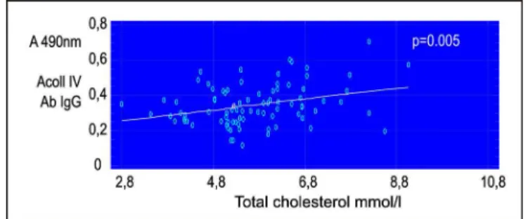
Fig. 4. Correlation analysis between ACollIVAb IgG and total cholesterol levels in the whole group
There is a moderate positive correlation between the ACollIVAb IgG levels and the triglycerides levels, with a Pearson correlation coefficient of r = 0,34, p = 0,002. Regression analysis best describes this relationship which is linear, r = 0,34, p = 0,002, fig.5
and glucose levels in the whole group
A moderate positive correlation was determined between the levels of ACollIVAb IgG and total cholesterol levels, with a Pearson correlation coefficient r = 0,30, p = 0,005. Regression analysis best describes this relationship which is linear, r = 0,30, p = 0,005, Fig.4
Fig. 5. Correlation analysis between ACollIVAb IgG and triglyceride levels in the the whole group
344 / J of IMAB. 2012, vol. 18, book 3 /
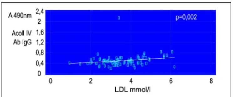
Fig. 6. Correlation analysis between AColIVAb IgG and levels of LDL in the whole group
There is a moderate negative correlation between the levels of ACollIVAb IgG and the levels of HDL-C with a correlation coefficient of Spearman ñ = -0,26, and p = 0,02. Regression analysis best describes this relationship which is linear, ñ = -0,26, p = 0,02, fig.7ACol IVAb IgG
.
CONCLUSION:
Collagen type IV is found mainly in the basal membrane. With the launch of endothelial dysfunction in the early stages of the atherosclerotic process, the anti-inflammatory endothelial protective function is depleted, allowing more aggressive intervention of the humoral and hemostatic components of the blood, followed by damage and accelerated degeneration of the basal membrane and collagen type IV. Our results reveal statistically significant higher levels of ACollIVAb IgG in the group of subjects with metabolic syndrome compared with the control group, while a significant difference was not detected in ACollIVAb IgM in both groups. When assessing the correlation between the antibody levels and the studied risk factors (components of the metabolic syndrome) in both groups were found statistically significant linear correlations between the levels of IgG ACollIVAb with diastolic blood pressure and the serum levels of blood sugar, total cholesterol, triglycerides, LDL, HDL.
Therefore, even in the early stages of the atherosclerotic process there are changes in the metabolism of collagen type IV wich is a main components constituting the basal membranes. Anticolagen type IV antibodies of class IgG, characterising the secondary immune response are elevated in the long-term chronic inflammation characterizing the metabolic syndrome and atherosclerosis, respectively, and characterize vascular aging process, whereas antibodies of class IgM, in serum during the initial response in the early stages of vascular aging are already depleted.
Fig. 7. Correlation analysis between AColIVAb IgG and levels of HDL in the whole group
Address for correspondence: Dr. Edward Mekenyan
First Cardiological Clinic, UMHAT “G. Stranski” - Pleven 8A, Georgi Kochev str., Pleven, Bulgaria
Phone: 00359 64 886 140
Email: [email protected]; 1. Liotta LA, Tryggvason K,
Garbisa S, Hart I, Foltz CM, Shafie S. Metastatic potential correlates with enzymatic degradation of basement membrane collagen. Nature. 1980 Mar 6;284 (5751):67-8. [PubMed]
2. Kilo C, Vogler N, Williamson JR: Muscle capillary basement membrane changes related to aging and to diabetes mellitus. Diabetes. 1972 Aug;21(8):881-905. [PubMed]
3. Williamson JR, Kilo C. Current status of capillary basement-membrane disease in diabetes mellitus. Diabetes.
REFERENCES:
1977 Jan;26(1):65-73. [PubMed] 4. Williamson JR, Kilo C. Capillary basement membranes in diabetes.
Diabetes. 1983 May;32(Suppl 2):96-100. [PubMed]
5. Hollertz O. Sulphur: the vulnerable factor X in atherosclerosis.
Med Hypotheses. 2002 Jul;59(1):35-38. [PubMed]
6. Weber S, Dolz R, Timpl R, Fessler JH, Engel J. Reductive cleavage and reformation of the interchain and intrachain disulfide bonds in the globular hexameric domain NC1
involved in network assembly of basement membrane collagen (type IV).
Eur J Biochem. 1988 Aug 1;175(2):229-236. [PubMed] [CrossRef]
7. Najjar SS, Scuteri A, Lakatta EG. Arterial aging: is it an immutable cardiovascular risk factor?
Hypertension. 2005 Sep;46(3):454-62. Epub 2005 Aug 15. [PubMed] [CrossRef]